This article contains AI generated content.
· UpdatedTibial Torsion Genes And Biomarkers - 5 Genes And 6 Biomarkers To Track
Introduction
Living with tibial torsion — whether you discovered it as a child whose feet turned inward, as a teenager with unexplained knee pain, or as an adult who finally connected the dots between gait mechanics and chronic joint stress — means navigating a condition that rarely gets the attention it deserves. Most people are told it either resolves on its own or that little can be done beyond orthotics and physiotherapy. That is not wrong, exactly. It is just incomplete.
What rarely comes up in clinical settings is the biological environment surrounding the tibia. Bone is a living tissue. Collagen in tendons and periosteum turns over constantly. Inflammation rises and falls with lifestyle choices. Growth factor signaling shapes how joints hold up under load across decades. These factors matter enormously for tibial torsion — not because they define the condition, but because they determine how well or poorly your body manages it over time.
Generic advice to eat well and exercise more does not address the specific vulnerabilities that tibial torsion creates in the musculoskeletal system. Understanding your particular biomarker profile and genetic predispositions allows for a far more targeted approach — one where you know what to monitor, what to support, and what specific interventions are most likely to move the needle for your biology.
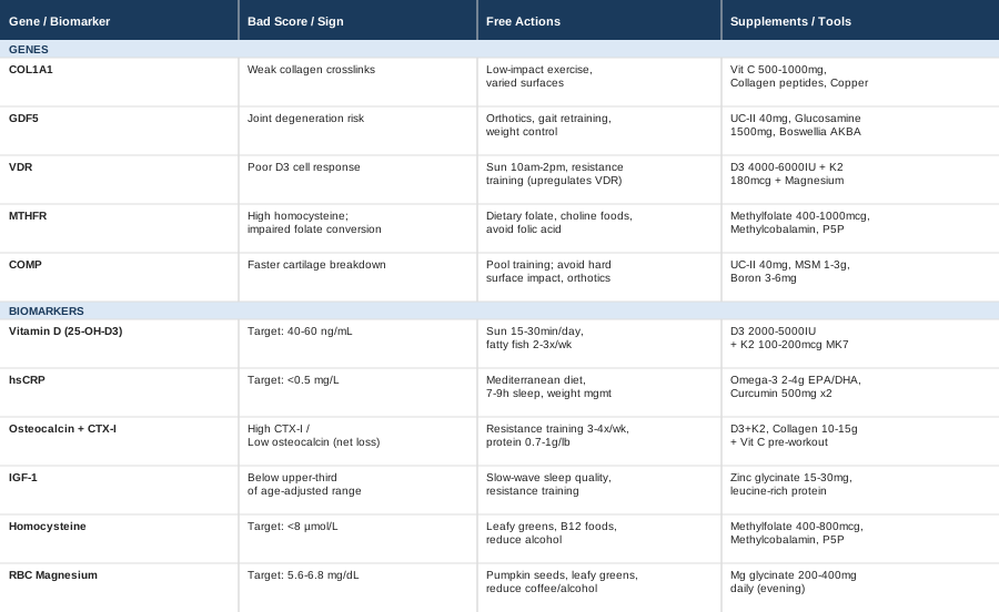
This article takes two angles on that question. The first examines six biomarkers that reflect the biological conditions most relevant to tibial torsion management: bone turnover, inflammation, connective tissue integrity, growth factor signaling, and mineral status. The second explores five genetic variants that influence musculoskeletal development and connective tissue quality — along with concrete plans for compensating when those genes are working against you. Together, they offer a more useful foundation than any one-size-fits-all recommendation could provide.
6 Biomarkers to Track When You Have Tibial Torsion
Tibial torsion is structural at its core, but structure does not exist in a biological vacuum. Bone remodels in response to hormonal signals, mechanical load, inflammatory status, and nutritional availability. The joints downstream — the knees, ankles, and hips — wear at different rates depending on the quality of the cartilage, tendons, and ligaments that absorb the rotational mismatch. The biomarkers below are not diagnostic tools for tibial torsion itself; they are windows into the biological terrain that determines how well your musculoskeletal system holds up over time. For most people, this terrain has never been assessed in any depth.
Biomarker 1: Vitamin D (25-Hydroxyvitamin D3)
Why It Matters
Vitamin D is the master regulator of calcium metabolism and bone mineralization. During skeletal development, inadequate vitamin D impairs the quality of bone matrix and can contribute to abnormal bone geometry. In adults, low vitamin D accelerates bone resorption and weakens the structural integrity of load-bearing bones including the tibia. Beyond bone, vitamin D regulates hundreds of genes involved in inflammation, immune function, and muscle performance — all of which affect how the body compensates for a rotational malalignment.
Research consistently links vitamin D insufficiency with increased musculoskeletal pain, reduced muscle strength, and impaired bone density. For someone with tibial torsion who already places asymmetric stress on the knees, ankles, and hips, insufficient vitamin D compounds the mechanical risk.
How to Measure It
The test is a 25-hydroxyvitamin D blood draw (25-OH-D3). Cost ranges from $30 to $60 through standard labs. It is sometimes covered by insurance with a physician's order. Peter Attia recommends targeting 40–60 ng/mL rather than simply hitting the lab's lower normal threshold of 20 ng/mL, which many researchers consider inadequate for musculoskeletal outcomes. Re-test every three months when adjusting supplementation.
If the Score Is Bad: The Plan Without Supplements
Aim for 15–30 minutes of midday sun exposure with arms and legs uncovered, at least four to five days per week. Include fatty fish (salmon, mackerel, sardines) two to three times per week, along with egg yolks and fortified dairy regularly. For most people in northern latitudes or with predominantly indoor lifestyles, food and sun alone are rarely sufficient to move deficient levels into an optimal range. They nonetheless form a meaningful base.
If the Score Is Bad: The Plan With Supplements or Equipment
Vitamin D3 combined with vitamin K2-MK7 is the standard protocol. K2 ensures that calcium mobilized by D3 is routed into bone rather than arterial tissue. A typical starting dose is 2000–5000 IU D3 with 100–200 mcg K2-MK7 daily, taken with the largest fat-containing meal of the day. For those testing below 20 ng/mL, 5000–8000 IU may be appropriate initially, with re-testing at three months.
Cycling and side effects: Vitamin D3 is appropriate for long-term use at these doses for most adults. Hypercalcemia is rare below 10,000 IU/day when K2 is included. Do not supplement without baseline testing. The goal is optimization, not maximum dosing.
Biomarker 2: High-Sensitivity C-Reactive Protein (hsCRP)
Why It Matters
Chronic low-grade inflammation is one of the most underappreciated drivers of musculoskeletal degradation. In people with tibial torsion, compensatory gait mechanics create repetitive asymmetric stress on the knees, hips, and ankles. Over years, this drives localized inflammation that slowly degrades cartilage, tendon, and bone quality. High-sensitivity CRP is the most accessible systemic marker for this kind of background inflammatory activity.
Elevated hsCRP also signals that the body is in a state of ongoing tissue damage that is outpacing repair — the worst possible biological context for managing a structural condition over the long term. Keeping inflammation controlled is one of the most consequential things a person with tibial torsion can monitor and address.
How to Measure It
A standard hsCRP blood test costs $15–$40 at commercial labs. No fasting is required, though it can be temporarily elevated by illness, injury, or recent intense exercise. Test when healthy and rested. Optimal target: below 0.5 mg/L. Values above 3 mg/L indicate significant inflammation warranting investigation.
If the Score Is Bad: The Plan Without Supplements
The Mediterranean dietary pattern has the strongest evidence for reducing hsCRP: olive oil as the primary fat, two or more servings of fatty fish weekly, abundant vegetables and legumes, minimal ultra-processed food. Equally important: seven to nine hours of sleep per night (sleep deprivation raises CRP significantly), stress management, and maintaining a healthy body weight — visceral fat is a major driver of elevated CRP. Eliminating the most obvious inflammatory inputs — alcohol, smoking, refined seed oils, chronic sleep debt — often moves the needle faster than adding supplements.
If the Score Is Bad: The Plan With Supplements or Equipment
Omega-3 fatty acids (EPA and DHA combined) are the most evidence-supported anti-inflammatory supplement for reducing hsCRP. A dose of 2–4 grams of combined EPA/DHA daily is used in clinical trials demonstrating CRP reductions. Curcumin — formulated as phosphatidylcholine-bound or BCM-95 — at 500 mg twice daily has demonstrated anti-inflammatory effects in multiple randomized trials, particularly for joint inflammation. Magnesium glycinate at 300–400 mg daily also helps reduce inflammatory markers through multiple pathways.
Cycling and side effects: Omega-3s are appropriate for long-term use. Curcumin can be taken continuously. Re-test hsCRP at three months to assess response. At high doses, omega-3s can thin blood — relevant for those on anticoagulants.
Biomarker 3: Bone Turnover Markers — Osteocalcin and CTX-I
Why They Matter
Bone is continuously being broken down by osteoclasts and rebuilt by osteoblasts in a process called remodeling. In tibial torsion, abnormal rotational mechanics place asymmetric mechanical stress on the tibia, potentially disrupting the balance of this cycle. Osteocalcin reflects active bone formation; CTX-I (C-terminal telopeptide of type I collagen) reflects bone resorption. Together, they reveal whether the tibia's structural environment is trending toward strength or fragility.
An imbalance — high CTX-I with low osteocalcin — indicates net bone loss. For a bone already under abnormal mechanical load, this worsens structural integrity in ways that compound over years. Osteocalcin also functions as a metabolic hormone with downstream effects on insulin sensitivity and inflammation, making it relevant beyond bone health alone.
How to Measure Them
Both are blood tests drawn fasting in the morning — bone markers fluctuate with meals and circadian rhythm. Osteocalcin costs $30–$60; CTX-I costs $40–$80. Functional medicine panels often combine both. Standard endocrinologists and rheumatologists order these for metabolic bone disease; a GP referral or functional medicine provider can request them specifically.
If the Score Is Bad: The Plan Without Supplements
Weight-bearing exercise is the single most powerful stimulus for osteocalcin production. Resistance training three to four times per week, with progressive lower-limb loading (calibrated to individual pain and alignment), directly upregulates osteoblast activity. Adequate dietary protein (0.7–1g per pound of body weight) is essential for bone matrix synthesis. Improving sleep quality has measurable effects, as the majority of bone formation occurs during slow-wave sleep.
If the Score Is Bad: The Plan With Supplements or Equipment
Vitamin D3 combined with K2 is foundational for improving the osteocalcin/CTX-I balance. Collagen peptides at 10–15g daily (taken with vitamin C, 30–60 minutes before exercise) have shown in randomized trials to stimulate bone formation markers. Calcium from food is strongly preferable to supplemental calcium; if supplementing, calcium citrate is better absorbed than carbonate and less associated with the cardiovascular risks flagged in some calcium supplement research.
Cycling and side effects: Re-test bone markers at six months — they change slowly. Calcium supplements above 1000 mg daily have been associated with cardiovascular risk in some study populations; food sources remain the safer primary strategy.
Biomarker 4: IGF-1 (Insulin-Like Growth Factor 1)
Why It Matters
IGF-1 is the primary downstream mediator of growth hormone signaling in bone and cartilage. It stimulates osteoblast proliferation, collagen synthesis, and cartilage matrix production. While growth hormone drives skeletal growth during development, IGF-1 remains essential throughout adult life for maintaining bone mineral density and connective tissue repair capacity. In tibial torsion, the quality of cartilage in the knee and ankle joints — structures absorbing most of the misalignment's compensatory stress — depends partly on sustained IGF-1 signaling.
Low IGF-1 is associated with accelerated joint space narrowing, reduced bone density, and slower recovery from microtrauma. Peter Attia has highlighted IGF-1 as one of the more underappreciated longevity and musculoskeletal markers in aging adults. It tends to decline gradually with age, and by middle adulthood many people are well below the upper portion of the reference range where optimal musculoskeletal function is supported.
How to Measure It
A serum IGF-1 test costs $50–$90. Drawn in the morning, fasting. Normal adult reference ranges typically span 50–350 ng/mL depending on age and sex. For musculoskeletal optimization, the upper third of the age-adjusted reference range is generally considered favorable.
If the Score Is Bad: The Plan Without Supplements
Sleep is the most powerful lever for IGF-1. The primary growth hormone pulse occurs during slow-wave sleep in the first half of the night. Any intervention that improves sleep architecture — consistent sleep schedule, dark and cool room, limiting alcohol — directly supports this anabolic window. Resistance training three to four times per week also stimulates the GH/IGF-1 axis. Adequate caloric intake matters too: chronic caloric restriction significantly suppresses IGF-1. Reducing visceral fat improves growth hormone sensitivity and downstream IGF-1 production.
If the Score Is Bad: The Plan With Supplements or Equipment
Zinc glycinate or zinc picolinate at 15–30 mg daily supports GH axis function and is one of the more evidence-based micronutrient interventions for maintaining IGF-1. Vitamin D and magnesium also influence IGF-1 signaling. Adequate dietary protein — particularly leucine-rich animal proteins — is a primary driver of IGF-1 production. Do not consider exogenous GH or IGF-1 without full clinical evaluation; the risks are significant outside supervised medical contexts.
Cycling and side effects: Re-test IGF-1 at three to six months. Zinc above 40 mg daily can deplete copper; balance with 1–2 mg copper or use a zinc:copper formula at a 10:1 ratio.
Biomarker 5: Homocysteine
Why It Matters
Homocysteine is an amino acid intermediate produced during methionine metabolism. When the methylation cycle is impaired — due to dietary deficiencies or genetic variants like MTHFR — homocysteine accumulates and damages collagen crosslinks throughout the body. This weakens bone matrix, tendons, and ligaments at a structural level. Elevated homocysteine is one of the clearest and most underrecognized signs of connective tissue vulnerability in routine bloodwork.
Thomas Dayspring and Peter Attia have both identified elevated homocysteine as both a cardiovascular and structural health marker of genuine concern. For tibial torsion specifically, the tendons and ligaments of the knee and ankle must handle abnormal rotational loads — their collagen integrity directly affects long-term joint outcomes. McLean and colleagues published work in JAMA (2004) showing that elevated homocysteine was independently associated with increased fracture risk in older adults, and the mechanisms involve direct damage to bone collagen quality.
How to Measure It
Plasma homocysteine is a standard fasting blood test. Cost: $25–$50. Optimal target: below 8 µmol/L. Values above 12 µmol/L correlate with measurable collagen damage and significantly increased structural risk. Many labs consider anything below 15 µmol/L "normal," which functional medicine practitioners widely consider too permissive.
If the Score Is Bad: The Plan Without Supplements
Folate-rich foods (leafy greens, lentils, beans) and adequate dietary B12 from animal foods (meat, fish, eggs, dairy) form the dietary foundation. Reducing alcohol intake directly lowers homocysteine. Improving gut health — where B12 absorption depends on intrinsic factor — is particularly relevant for adults over 50.
If the Score Is Bad: The Plan With Supplements or Equipment
The methylation triad: methylfolate (5-MTHF), methylcobalamin (B12), and pyridoxal-5-phosphate (P5P, active B6). These three vitamins clear homocysteine through the methionine cycle. Methylfolate 400–800 mcg daily, methylcobalamin 500–1000 mcg daily, P5P 25–50 mg daily. Critical point: if you carry MTHFR gene variants (discussed in the genetics section below), standard folic acid does not work — only methylated forms will clear homocysteine effectively. Re-test at three months.
Side effects and cycling: High-dose B6 above 200 mg daily can cause peripheral neuropathy; the doses above are well within safety margins. Some individuals with MTHFR who begin methylfolate experience temporary anxiety or overstimulation as methylation capacity increases — start at 200 mcg and titrate up over two to four weeks. Long-term supplementation is typically appropriate.
Biomarker 6: RBC Magnesium
Why It Matters
Magnesium participates in over 300 enzymatic reactions including vitamin D activation, bone mineral synthesis, muscle contraction and relaxation, and anti-inflammatory signaling. Standard serum magnesium tests are nearly useless for detecting true deficiency because serum levels are maintained by drawing from bone and muscle — normal serum values can coexist with severe intracellular depletion. RBC magnesium is the only clinically meaningful way to assess true magnesium status.
In tibial torsion, the compensatory muscle tension patterns — particularly in the IT band, tibial muscles, and hip external rotators — place chronic demand on magnesium-dependent muscle relaxation pathways. Deficiency creates a self-reinforcing cycle: chronic muscle tightness, poor sleep, elevated inflammatory markers, and compromised bone remodeling. Addressing it is often one of the highest-return interventions available.
How to Measure It
RBC magnesium costs $30–$70 through specialty or functional medicine labs (SpectraCell, LabCorp, Genova Diagnostics). Request it by name — a standard metabolic panel will only measure serum magnesium, which is not what you want. Optimal range: 5.6–6.8 mg/dL. Most people who test for the first time will find themselves in the 4.2–5.0 mg/dL range despite normal serum values.
If the Score Is Bad: The Plan Without Supplements
Top dietary sources: pumpkin seeds (one of the richest plant sources), dark leafy greens, almonds, avocados, black beans, and dark chocolate (70%+). Reduce coffee and alcohol, both of which significantly increase urinary magnesium excretion. Chronic psychological stress depletes magnesium rapidly through cortisol-driven mechanisms — stress management is a legitimate dietary strategy here.
If the Score Is Bad: The Plan With Supplements or Equipment
Magnesium glycinate or magnesium malate are the preferred forms — well-absorbed, well-tolerated, with minimal laxative effect at effective doses. Dose: 200–400 mg elemental magnesium daily, taken in the evening, which also supports sleep quality and parasympathetic recovery. Avoid magnesium oxide, which has poor bioavailability despite being commonly sold.
Cycling and side effects: Long-term supplementation is appropriate for most adults given the widespread prevalence of deficiency. Loose stools indicate the dose is too high or the form should be changed; reduce to 150–200 mg and adjust. Not suitable at high doses for those with impaired kidney function.
Biomarkers tell you what is happening in the body right now. But understanding why some individuals are more prone to tibial torsion — and why certain interventions work better for specific people — requires a look at the underlying genetic architecture. The following section covers five genetic variants that influence musculoskeletal development, connective tissue quality, and the biological pathways the biomarkers above are measuring.
What the Genetics of Tibial Torsion Reveal — 5 Variants Worth Knowing
Tibial torsion has a recognized heritable component. It clusters in families, and first-degree relatives of affected individuals have elevated rates of rotational limb anomalies. Several genes involved in collagen architecture, growth factor signaling, and vitamin D metabolism are particularly relevant. This section draws on the emerging framework popularized by practitioners like Gary Brecka, who has emphasized that identifying genetic liabilities allows for precise compensation strategies rather than one-size-fits-all protocols.
None of this is deterministic. Genetics sets susceptibility; lifestyle, environment, and targeted interventions shape how those genes express. The goal is knowing which levers are worth pulling for your specific profile.
Gene 1: COL1A1 — The Collagen Architecture Gene
What It Affects
COL1A1 encodes the alpha-1 chain of type I collagen, the most abundant structural protein in bone, tendon, ligament, and periosteum. The Sp1 polymorphism (rs1800012) in COL1A1 is one of the best-studied variants affecting bone mineral density and connective tissue strength. Carriers of unfavorable alleles tend to have weaker collagen crosslinking, reduced bone matrix stiffness, and higher susceptibility to stress fractures and ligament injuries. Grant and colleagues published early foundational work on this in The Lancet (1996), establishing the link between COL1A1 Sp1 variants and reduced bone density.
In tibial torsion, where abnormal rotational forces are distributed asymmetrically along the tibia, COL1A1 variants can amplify structural vulnerability in both the bone itself and the surrounding soft tissues.
If the Gene Is Bad: The Plan Without Supplements
Prioritize low-impact weight-bearing exercise — walking on varied and natural surfaces, controlled resistance training — over high-impact activities like running on hard pavement or plyometric training. Swimming and cycling maintain cardiovascular fitness without repetitive tibial impact. Alignment-based movement practices (yoga, Pilates, or supervised gait retraining) help distribute force more evenly along the length of the tibia and reduce localized stress concentration.
If the Gene Is Bad: The Plan With Supplements or Equipment
Vitamin C at 500–1000 mg daily is an essential cofactor for lysyl hydroxylase and prolyl hydroxylase — the enzymes that synthesize and crosslink collagen. Without adequate ascorbate, even a normal COL1A1 gene cannot produce optimally crosslinked collagen. Collagen peptides at 10–15g daily (with vitamin C, 30–60 minutes before exercise) have shown in randomized trials to increase collagen synthesis markers in connective tissue. Copper at 1–2 mg daily fuels lysyl oxidase, which creates mature collagen crosslinks. Lysine at 1g daily provides the precursor amino acid for collagen hydroxylation.
Cycling and side effects: Collagen peptides can be cycled in three-month courses with one-month breaks. Vitamin C is appropriate long-term. Doses above 2g of vitamin C daily may cause loose stools. Copper above 3 mg daily without zinc can disrupt the zinc:copper balance.
Gene 2: GDF5 — The Joint Blueprint Gene
What It Affects
Growth Differentiation Factor 5 is a protein central to joint formation, skeletal patterning, and limb geometry during embryonic development. The rs143384 polymorphism in GDF5 is one of the most replicated genetic associations with osteoarthritis in large genome-wide association studies — most notably identified in research by Miyamoto and colleagues (2007) and replicated across multiple ethnic populations. GDF5 variants have been implicated in altered limb rotational geometry during development, making them directly relevant to tibial torsion susceptibility.
For adults with tibial torsion who carry GDF5 risk variants, the primary concern shifts toward progressive joint degeneration at the knees and ankles — the joints bearing the greatest compensatory burden from the rotational malalignment.
If the Gene Is Bad: The Plan Without Supplements
Custom orthotics designed to correct tibial rotation forces at the foot are the most direct non-supplement intervention. They reduce asymmetric stress transmitted to the knee joint. Gait retraining with a physiotherapist or biomechanist — modifying foot strike pattern and knee tracking during walking and running — has strong evidence for reducing cartilage wear in lower limb malalignment conditions. Maintaining a healthy body weight is non-negotiable: each additional kilogram of body weight multiplies force on the knee by approximately three to four times during normal walking.
If the Gene Is Bad: The Plan With Supplements or Equipment
Undenatured type II collagen (UC-II) at 40 mg daily works through oral tolerization mechanisms to support cartilage matrix integrity — a distinct and evidence-supported mechanism separate from structural collagen supplementation. Glucosamine sulfate (1500 mg daily) and chondroitin sulfate (1200 mg daily) have mixed but generally positive evidence for slowing cartilage degradation in established joint disease; the evidence is weaker for prevention but may be worthwhile in genetically high-risk individuals. Boswellia serrata extract (AKBA fraction, 100–200 mg daily) has reasonable randomized trial evidence for reducing synovial joint inflammation.
Cycling and side effects: Three- to six-month courses of glucosamine and chondroitin with four-week breaks. Glucosamine is generally well-tolerated; some concern for blood sugar effects in individuals with diabetes. Boswellia may cause mild GI discomfort at higher doses.
Gene 3: VDR — The Vitamin D Receptor Gene
What It Affects
The VDR gene encodes the vitamin D receptor, which mediates virtually all genomic effects of active vitamin D in the cell nucleus. Key polymorphisms — including FokI, BsmI, ApaI, and TaqI — alter receptor binding affinity and transcriptional efficiency. Certain variant combinations can significantly reduce the biological effectiveness of circulating vitamin D, meaning a person may have acceptable blood levels of 25-OH-D but poor cellular response at the receptor level.
Multiple studies have documented associations between VDR polymorphisms and bone mineral density outcomes across different populations. The VDR gene also affects muscle function, immune regulation, and inflammatory control — all directly relevant to the musculoskeletal environment of tibial torsion. Standard vitamin D supplementation advice may be insufficient for VDR variant carriers; the approach must be calibrated differently.
If the Gene Is Bad: The Plan Without Supplements
Maximize sun exposure during peak UVB hours (10am–2pm), when radiation intensity is sufficient to trigger meaningful skin vitamin D synthesis. Maintain a diet consistently rich in vitamin D food sources. Physical loading — specifically the mechanical strain of resistance training — upregulates VDR expression in bone cells independently of circulating vitamin D levels, providing partial compensation for reduced receptor sensitivity.
If the Gene Is Bad: The Plan With Supplements or Equipment
VDR variants that reduce receptor sensitivity may require higher-dose supplementation to achieve equivalent biological effect. Target blood levels of 50–70 ng/mL rather than the lower end of normal (20–30 ng/mL). This typically requires D3 at 4000–6000 IU daily with 180–200 mcg K2-MK7. Magnesium is essential because it activates the enzymes (CYP2R1 and CYP27B1) that convert inactive 25-OH-D to the active 1,25-dihydroxyvitamin D form. Re-test 25-OH-D every three months during dose adjustment, and monitor serum calcium alongside.
Side effects: Hypercalcemia is the primary risk at very high doses (above 10,000 IU daily). At the doses above with K2 included, risk is low for most adults, but individual monitoring remains essential.
Gene 4: MTHFR — The Methylation Gene
What It Affects
MTHFR (methylenetetrahydrofolate reductase) encodes a central enzyme in the methylation cycle — involved in DNA repair, neurotransmitter synthesis, and homocysteine clearance. The two most clinically significant variants are C677T and A1298C. These reduce enzyme efficiency by 30–70%, impairing folate conversion and causing homocysteine accumulation. Frosst and colleagues identified the C677T variant as thermolabile with significantly reduced enzyme activity in foundational research published in Nature Genetics (1995), and subsequent research has shown clear bone density implications.
Elevated homocysteine from MTHFR variants directly damages collagen crosslinks in bone matrix and connective tissue — the same mechanism described in the homocysteine biomarker section. Gary Brecka has placed MTHFR at the center of his genetic health optimization framework, noting that a substantial proportion of the population carries at least one copy of these variants without ever knowing it. The downstream consequences for connective tissue and bone quality are specific and addressable.
If the Gene Is Bad: The Plan Without Supplements
Prioritize dietary folate (not folic acid) from leafy greens, legumes, and liver — foods that provide natural folates not requiring full MTHFR conversion. Avoid synthetic folic acid in fortified foods and standard supplements: people with MTHFR variants convert it poorly and may accumulate unmetabolized folic acid. Prioritize choline-rich foods (eggs, liver, fish roe), which fuel an alternative methylation pathway (the betaine pathway) independent of MTHFR.
If the Gene Is Bad: The Plan With Supplements or Equipment
Methylated B vitamins are essential, not optional, with MTHFR variants. Methylfolate (5-MTHF) 400–1000 mcg daily, methylcobalamin 500–2000 mcg daily, pyridoxal-5-phosphate (P5P) 25–50 mg daily. Always check labels — standard multivitamins containing folic acid work against you if you have these variants. Do not substitute standard folic acid at any dose.
Important practical note: Some individuals beginning methylfolate experience temporary anxiety, irritability, or vivid dreams as methylation capacity upregulates. Start at 200 mcg and titrate up over two to four weeks. Monitor homocysteine as the outcome marker — it is the most direct reflection of whether methylation is actually improving. Cycling: Long-term supplementation is typically required as genetic variants do not change.
Gene 5: COMP — The Cartilage Matrix Gene
What It Affects
COMP (Cartilage Oligomeric Matrix Protein) is a glycoprotein that maintains the structural cohesion of cartilage, tendon, and ligament extracellular matrix. It acts as a molecular bridge between collagen fibrils and other matrix components. Pathogenic variants cause rare but severe skeletal dysplasias. More common polymorphisms may subtly reduce cartilage resilience and alter joint geometry during development — relevant to understanding why some individuals with tibial torsion experience faster secondary joint degeneration than others.
Serum COMP also functions as a measurable biomarker for active cartilage turnover: elevated levels in blood indicate significant cartilage breakdown and can guide intervention monitoring in parallel with the genetic context.
If the Gene Is Bad: The Plan Without Supplements
Reduce high-impact activity on hard surfaces: avoid pavement running, high-intensity plyometrics, and sports involving rapid directional changes unless lower-extremity biomechanics have been fully corrected. Pool-based training (swimming, aqua jogging) provides cardiovascular conditioning without cartilage compression. Properly fitted, motion-control footwear and custom orthotics reduce the force translated to knee and ankle cartilage with every step. Healthy body weight is the highest single-impact variable for knee cartilage preservation.
If the Gene Is Bad: The Plan With Supplements or Equipment
Undenatured type II collagen (UC-II) at 40 mg daily is the most targeted intervention for cartilage matrix support. MSM (methylsulfonylmethane) at 1–3 g daily provides sulfur essential for proteoglycan synthesis and has modest randomized trial evidence for reducing joint pain and improving cartilage markers. Boron at 3–6 mg daily supports glycosaminoglycan production in cartilage matrix and has a favorable safety profile. Custom orthotics — discussed here as equipment — may be the most impactful non-supplement intervention by directly reducing the abnormal forces reaching the cartilage layer.
Cycling and side effects: UC-II can be taken long-term. Cycle MSM three months on, one month off and re-evaluate. High-dose MSM may cause mild headache or loose stools in some individuals; reduce and adjust.
The biomarker and genetic angles together provide a science-informed map of individual risk and resilience. What they do not capture alone is the broader framework of movement science, sleep biology, and recovery physiology that turns that map into daily practice. That is where the following section becomes useful.
The Huberman Lab Framework for Bone and Structural Health — 10 Things That May Change How You Approach Tibial Torsion
The Huberman Lab podcast has become one of the most rigorously referenced science communication platforms in health, synthesizing research on sleep, exercise, nutrition, and neurobiology in ways that are directly applicable to structural conditions like tibial torsion. Across multiple episodes on exercise science, bone density, connective tissue recovery, and the autonomic nervous system, Andrew Huberman has outlined principles that challenge some of the passive management approaches conventional medicine tends to take with skeletal malalignment.
1. Mechanical Load Is the Primary Language of Bone
Bone responds primarily to mechanical compression and tension — these signals trigger osteoblasts to activate and mineralize matrix. Without adequate load, bone quality declines regardless of calcium or supplement intake. Weight-bearing exercise must continue for people with tibial torsion, even if loads are distributed carefully. Resistance training and walking on varied surfaces provide the mechanical diversity bone tissue requires.
2. Slow-Wave Sleep Is When Bone Builds
The largest daily pulse of growth hormone occurs during deep slow-wave sleep, primarily in the first half of the night. This GH pulse drives IGF-1 production and osteoblast activity overnight. Sleep architecture is arguably the most underappreciated bone health variable available to anyone without a prescription. Alcohol, late-night eating, and irregular sleep timing all suppress this anabolic window.
3. Cortisol Directly Inhibits Bone Formation
Chronic psychological stress chronically elevates cortisol, which directly inhibits osteoblast function and accelerates bone resorption. Stress management has measurable effects on bone turnover markers — specifically osteocalcin and CTX-I — that make it a genuinely biological rather than merely psychological recommendation. Breathing protocols, cold exposure, and adequate recovery time are all relevant here.
4. Zone 2 Cardio Preserves Bone Without Overloading It
Low-to-moderate aerobic exercise at 60–70% maximum heart rate improves vascular supply to bone without the repetitive impact stress of higher-intensity running. For tibial torsion, cycling, swimming, and brisk walking provide this circulatory stimulus while controlling abnormal torsional loading on the tibia itself.
5. Proprioceptive Training Rewires the Compensatory Neuromuscular System
Huberman has discussed at length how the nervous system — not just muscle and bone — drives movement patterns. In tibial torsion, compensatory gait patterns involve learned, neurologically-encoded adaptations. Single-leg balance work, unstable surface training, and gait-focused movement practice can progressively rewire the neuromuscular patterns that amplify downstream joint stress.
6. Vitamin D and Sex Hormone Signaling Overlap in Bone
Vitamin D and sex hormones (testosterone, estrogen) use overlapping receptor and signaling machinery in bone tissue. Low vitamin D impairs not just calcium metabolism but also the anabolic effects of sex hormones on bone. This is one reason why the effective dose of vitamin D for musculoskeletal outcomes may need to exceed the commonly recommended minimum thresholds.
7. Anti-Inflammatory Nutrition Is Load-Bearing for Joint Longevity
Research highlighted repeatedly in the Huberman Lab shows that omega-3 fatty acids, polyphenol-rich foods, and Mediterranean-style dietary patterns produce measurable reductions in systemic inflammatory markers. For a structural condition managed over years or decades, cumulative inflammatory damage to cartilage and periarticular tissue is a genuine long-term threat. Dietary quality is not a soft recommendation — it is among the highest-leverage daily inputs available.
8. Collagen Synthesis Has an Exercise Timing Window
Research covered in the Huberman Lab highlights that consuming gelatin or collagen peptides with vitamin C approximately 30–60 minutes before connective-tissue-loading exercise significantly increases collagen synthesis compared to supplementing at other times of day. This pre-exercise timing window is one of the most actionable and underused findings in connective tissue science — and it requires no additional product, just better timing of what you are likely already taking.
9. Heat Therapy Reaches Connective Tissue That Exercise Does Not
Regular sauna use (15–20 minutes at 80–90°C, three to four times per week) has been associated with increased heat shock protein production, improved vascular supply to connective tissue, and GH pulses independent of exercise. For joint health specifically, heat increases synovial fluid viscosity and improves cartilage nutrient delivery. Sauna is one of the few passive interventions with documented physiological effects on connective tissue that are distinct from those produced by exercise.
10. Small Consistent Inputs Compound Over Decades
One of the most consistent themes across Huberman Lab episodes is biological non-linearity: small, consistently applied inputs — adequate sleep, daily movement, anti-inflammatory diet, optimized vitamin D and magnesium — compound over years into dramatically different structural outcomes. For tibial torsion, no single intervention produces transformation. The goal is damage reduction and functional preservation through compounding small advantages. Starting that curve early matters more than any individual protocol.
Biomarkers, genetics, and frameworks like the Huberman approach all address different levels of the same problem. The following complementary modalities add hands-on, clinically-grounded options for those looking to take a broader approach to management.
Complementary Approaches Supported by Human Evidence
Yoga
Yoga combines controlled stretch and load across joints in a way that is directly relevant to the muscular imbalances characteristic of tibial torsion. Internal tibial torsion typically drives tightness in the hip internal rotators and medial hamstrings while weakening the hip external rotators and gluteus medius — a pattern that targeted yoga practice can address through both strengthening and lengthening. The mind-body component also trains proprioceptive awareness, which helps individuals consciously adjust compensatory movement patterns.
Multiple systematic reviews support yoga's effectiveness for chronic musculoskeletal pain and functional movement limitations. A randomized controlled trial published in the Journal of Back and Musculoskeletal Rehabilitation found that a 12-week yoga program significantly reduced lower extremity pain and improved functional alignment in adults with lower limb biomechanical conditions. Evidence specific to tibial torsion is limited, but the anatomical rationale is strong.
Focus on postures that externally rotate the hip and strengthen the hip abductors: Warrior II, Extended Side Angle, Pigeon Pose, Goddess Pose, and lateral lunge variations. Avoid deep knee-flexed internal rotation postures (Virasana may be contraindicated without guidance). Practice three to five times per week for 30–45 minutes. Work with a yoga therapist initially to align cues with your specific rotational pattern — generic classes will not address tibial mechanics specifically.
Biofeedback
Biofeedback provides real-time physiological signals — muscle activation patterns, force distribution, joint angles — that allow individuals to consciously modify movement mechanics they would otherwise perform automatically. For tibial torsion, EMG biofeedback targeting the tibialis anterior, peroneal group, and hip external rotators can help retrain abnormal neuromuscular compensation patterns that develop around the rotational malalignment.
A study published in Gait and Posture (2019) demonstrated that biofeedback-assisted gait retraining significantly reduced peak knee adduction moments in adults with lower limb malalignment — a directly relevant outcome for the kind of asymmetric knee stress that tibial torsion creates. The standard protocol involves 6–8 weeks of supervised sessions, transitioning to an independent maintenance program.
Seek a physiotherapist or rehabilitation specialist trained in gait analysis and biofeedback technology. Pressure plate gait analysis combined with real-time EMG is the most informative combination. Cost for a full protocol typically runs $500–$1500 self-pay. Wearable biofeedback devices (pressure insoles, IMU-based gait monitors) can extend the retraining work outside clinic sessions and reduce ongoing cost.
Massage Therapy
The muscular compensation patterns that develop around tibial torsion — tightness in the TFL (tensor fascia latae), IT band, medial tibial compartment, and calf — respond to manual therapy. Massage reduces myofascial tension that would otherwise translate directly into abnormal joint loading, improves local circulation to connective tissue, and helps break pain-tension feedback cycles that amplify the structural problem over time.
A Cochrane systematic review on massage therapy for musculoskeletal pain found consistent evidence for short-term pain reduction and functional improvement. No study targets tibial torsion specifically — the research gap is real and worth acknowledging. The evidence base for massage in mechanically adjacent conditions (chronic knee pain, gait dysfunction, IT band syndrome) is reasonably strong and anatomically transferable.
Deep tissue massage targeting the IT band, lateral tibial compartment, calf complex, and hip external rotators monthly or biweekly provides the most targeted benefit. Self-myofascial release with a foam roller daily (5–10 minutes targeting TFL/IT band and posterior calf) extends the benefit between sessions. For a more specific approach, Active Release Technique (ART) practitioners specialize in treating the chronic soft tissue tethering that develops around rotational deformities and may produce more durable results than general massage.
Tai Chi
Tai chi combines slow, deliberate rotational movements with balance training and coordinated breathing. For tibial torsion, where balance, proprioception, and controlled rotational load distribution are the central physical challenges, tai chi addresses multiple problems simultaneously. The emphasis on weight shifting and lower limb positioning encourages conscious movement mechanics that challenge the compensatory patterns tibial torsion creates.
A meta-analysis published in PLoS ONE (2016, incorporating 17 randomized controlled trials and over 1500 participants) found that tai chi significantly improved balance, proprioception, and lower extremity muscle strength. Separate studies have documented reduced knee pain and improved functional mobility in populations with lower extremity biomechanical issues. Evidence specific to tibial torsion is absent, but the proprioceptive and balance mechanisms are well-established.
Yang-style tai chi for beginners, practiced for 20–30 minutes daily or 45 minutes three times weekly, is a practical starting protocol. In-person classes are strongly preferred initially — incorrect rotational patterns in self-taught tai chi could theoretically load the tibia poorly. After 8–12 weeks with qualified instruction, independent practice becomes effective. Tai chi has an excellent safety profile, no meaningful side effects, and carries cardiovascular and neurological benefits beyond the structural ones discussed here.
Conclusion
Tibial torsion is a structural reality that no biomarker panel or supplement can undo. But the quality of your bone, the integrity of your connective tissue, the level of systemic inflammation your joints are navigating daily, and the gene variants shaping all of the above — these are measurable, addressable, and consequential. They are the difference between a managed condition and a progressively degenerating one.
The six biomarkers covered here — vitamin D, hsCRP, bone turnover markers, IGF-1, homocysteine, and RBC magnesium — give you a real-time picture of the biological terrain surrounding your tibia and the joints compensating for it. The five genetic variants — COL1A1, GDF5, VDR, MTHFR, and COMP — identify which specific systems carry additional risk and why targeted interventions matter more for some people than others. The complementary approaches and movement principles reinforce the biological foundation with practical, human-evidence-supported tools.
The clearest next step is not to implement everything at once. Start with two or three biomarker tests — vitamin D, hsCRP, and homocysteine are the most accessible and immediately actionable. Review what comes back and build from there. A functional medicine physician, sports physiotherapist, or musculoskeletal specialist familiar with biomechanical assessment can help you connect the data to a plan that fits your specific anatomy, genetics, and lifestyle. Better information, applied consistently, is the most honest form of progress available.
Musculoskeletal: Bone Conditions Joint Conditions Muscle Conditions Tendon & Ligament Conditions
Autoimmune: Connective Tissue Conditions