This article contains AI generated content.
· UpdatedTibial Tuberosity Avulsion Fracture — 6 Genes And 7 Biomarkers To Track
Introduction
If you or someone you care for has experienced a tibial tuberosity avulsion fracture, you already know how disorienting the recovery process can feel. The standard medical guidance — rest, immobilization, possibly surgery, then rehab — covers the mechanics, but it rarely explains why healing goes smoothly for some people and feels painfully slow for others. That gap between the textbook and your actual experience is real, and it deserves a better answer.
What most recovery plans miss is that the bone-tendon junction where this injury occurs is an extraordinarily complex biological interface. The tibial tuberosity anchors the patellar tendon to the tibia, and the strength of that junction depends on a layered system of collagen architecture, bone mineral density, local inflammation control, and hormonal signaling. Generic protocols do not account for any of this individual variability. Two athletes at the same age with the same fracture type can have wildly different biological terrains underneath.
That is where a more targeted approach becomes genuinely useful. Tracking specific biomarkers in your blood can tell you whether your bone is actively remodeling, whether your inflammatory load is interfering with healing, and whether key hormones and growth factors are at levels that support tissue repair. Understanding your genetic profile can reveal structural vulnerabilities in collagen and bone metabolism that predispose you to this kind of injury in the first place — and point toward specific compensating strategies.
This article covers both angles. The main section focuses on seven blood biomarkers that experienced sports medicine physicians and metabolic health researchers like Peter Attia and Thomas Dayspring have consistently highlighted as meaningful for bone and connective tissue health. A second section covers six genes with strong relevance to tibial tuberosity avulsion fractures. After that, you will find a summary of a landmark podcast episode that reframes tendon and bone recovery in a way most rehabilitation protocols have not yet caught up with, followed by complementary modalities with meaningful clinical evidence. Better information does not guarantee a faster recovery, but it does narrow the gap between guessing and knowing.
7 Biomarkers Worth Tracking During Recovery
Measuring these biomarkers is not about replacing your orthopedic surgeon's guidance. It is about giving you and your care team a clearer biological picture. Many of these can be ordered through a standard blood draw, though some require a specialty lab. Costs listed reflect typical US out-of-pocket estimates; insurance coverage varies significantly.
1. 25-OH Vitamin D
Why it matters: Vitamin D is not just a supplement fad — it is a steroid hormone precursor that directly regulates calcium absorption, phosphate metabolism, and the activity of osteoblasts (bone-building cells). At the tibial tuberosity, where bone is undergoing rapid repair after an avulsion, adequate 25-OH vitamin D levels are essential. Research consistently shows that low vitamin D status is associated with impaired fracture healing, reduced callus formation, and increased risk of re-injury. A large meta-analysis published through NCBI showed that individuals with vitamin D deficiency had significantly slower recovery from musculoskeletal injuries compared to those in the sufficient range.
How to measure it: A standard serum 25-hydroxyvitamin D test. Available through any primary care physician or direct-to-consumer labs. Cost range: $30–$60 standalone; often included in metabolic panels. Optimal range for bone healing is generally considered 40–60 ng/mL by most sports medicine specialists, not the often-cited clinical minimum of 20 ng/mL.
If the score is low, the plan without supplements: Increase safe sun exposure to 15–30 minutes on large skin areas (arms, legs, back) at solar noon, three to five times per week. This is most effective between spring and early autumn at mid-latitudes. Dietary sources include fatty fish (salmon, sardines, mackerel), egg yolks, and UV-treated mushrooms. Consistent daily outdoor time combined with a diet rich in these foods can meaningfully raise levels over 8–12 weeks. For someone in recovery, supervised outdoor rehabilitation sessions naturally integrate sun exposure.
If the score is low, the plan with supplements or equipment: Vitamin D3 supplementation at 2,000–5,000 IU daily is the standard starting point, always paired with vitamin K2 (MK-7 form, 100–200 mcg/day) to direct calcium toward bone rather than arterial walls. Take with the largest meal of the day containing fat for optimal absorption. Cycling: no established need to cycle vitamin D3 itself, but retest every 12 weeks to adjust dosing. Side effects at these doses are rare; toxicity risk begins above sustained 10,000 IU/day without monitoring. A UV-B lamp (such as a Sperti lamp) is a practical equipment alternative for those with limited sun access in winter months.
2. Parathyroid Hormone (PTH)
Why it matters: PTH is the body's primary regulator of calcium homeostasis, and it directly controls bone remodeling activity. When vitamin D is low or calcium intake is insufficient, PTH rises to pull calcium from bone — the exact opposite of what you need during fracture repair. Chronically elevated PTH (secondary hyperparathyroidism) accelerates bone resorption and can undermine even well-managed rehabilitation. Testing PTH alongside vitamin D gives you a complete picture of calcium regulation. Peter Attia has emphasized this pairing repeatedly as a key component of bone health evaluation.
How to measure it: Serum intact PTH, measured with a morning fasting blood draw. Cost range: $40–$80. Normal range is typically 15–65 pg/mL; in the context of bone healing, you want it in the lower half of that range, indicating calcium balance is adequate.
If the score is high, the plan without supplements: Prioritize dietary calcium from food sources: dairy, leafy greens (kale, bok choy), canned fish with bones (sardines, salmon), and fortified foods. Target 1,000–1,200 mg daily from food. Reduce dietary factors that increase calcium excretion, particularly high sodium intake and excess caffeine. Optimize vitamin D first, as correcting deficiency often normalizes PTH without additional intervention.
If the score is high, the plan with supplements or equipment: If dietary calcium is genuinely insufficient (common in athletes with restricted diets), calcium supplementation at 500 mg twice daily with meals (calcium citrate form for better absorption with or without food) may be warranted. Do not exceed 2,500 mg total daily calcium from all sources. Monitor PTH retesting at 8–12 weeks. Note: calcium carbonate requires stomach acid for absorption; calcium citrate is preferred for most people. Side effects: constipation at higher doses; pair with magnesium to counterbalance.
3. CTX-1 (C-Terminal Telopeptide of Type I Collagen)
Why it matters: CTX-1 is a direct marker of bone resorption — specifically, it measures how much type I collagen is being broken down as osteoclasts remodel bone. After a tibial tuberosity avulsion fracture, you want a controlled remodeling environment: enough resorption to clear damaged tissue, but not so much that it outpaces new bone formation. Elevated CTX-1 signals excessive breakdown and can indicate that systemic factors (low estrogen, high cortisol, low vitamin D, or nutritional deficits) are tipping the balance away from healing. Thomas Dayspring has highlighted CTX-1 as one of the most underused bone health tests in clinical practice.
How to measure it: Fasting morning blood draw is critical — CTX-1 has significant diurnal variation, with levels highest in the morning after overnight fasting. Available through specialty labs (LabCorp, Quest). Cost range: $80–$150. Optimal value for most adults is below 0.3 ng/mL; values above 0.5 ng/mL suggest high turnover requiring investigation.
If the score is high, the plan without supplements: Increase load-bearing activity appropriate to your recovery stage — mechanical loading is a potent suppressor of osteoclast activity. Even partial weight-bearing with crutches, if cleared by your surgeon, sends anabolic signals to bone. Ensure total caloric intake is adequate; caloric restriction is a major driver of elevated CTX-1. Reduce cortisol-elevating behaviors: poor sleep, overtraining, and chronic psychological stress all directly increase bone resorption via the HPA axis.
If the score is high, the plan with supplements or equipment: Ensure vitamin D and calcium are optimized first, as both are primary regulators of CTX-1. If bone loss is confirmed, bisphosphonates or other prescription interventions are a physician decision, not a self-managed one. From a supplementation standpoint, omega-3 fatty acids (2–3 g combined EPA+DHA daily) have demonstrated modest anti-resorptive effects in several human trials. Collagen peptide supplementation (10 g/day) has emerging evidence for supporting bone matrix quality. No cycling required for either; monitor CTX-1 every 3 months.
4. P1NP (Procollagen Type I N-Terminal Propeptide)
Why it matters: Where CTX-1 measures breakdown, P1NP measures bone formation. It reflects osteoblast activity and the production of new type I collagen — the structural backbone of bone. After a tibial tuberosity avulsion fracture, you want P1NP to be actively elevated, signaling robust new bone matrix synthesis. Low P1NP in the context of a healing fracture suggests insufficient anabolic drive, which may reflect nutritional deficits, hormonal insufficiency, or inadequate mechanical loading. The CTX-1/P1NP ratio is particularly informative: a favorable healing environment shows rising P1NP and controlled CTX-1.
How to measure it: Serum P1NP, available through specialty labs. Cost range: $80–$150. Reference ranges vary by lab; for active bone healing, you want values in the upper-normal range, typically above 50 mcg/L. Best ordered alongside CTX-1 for a complete remodeling picture.
If the score is low, the plan without supplements: Adequate protein intake is the most direct dietary driver of P1NP — procollagen synthesis requires sufficient amino acid supply, particularly glycine, proline, and hydroxyproline. Target 1.6–2.2 g of protein per kilogram of body weight daily during fracture recovery. This is above standard recommendations and is supported by bone remodeling research. Resistance training of adjacent muscle groups (non-weight-bearing if required) preserves anabolic hormone signaling even during immobilization.
If the score is low, the plan with supplements or equipment: Collagen peptide hydrolysate (10–15 g/day, taken with vitamin C 50 mg to support hydroxylation) has randomized controlled trial evidence for increasing P1NP and supporting connective tissue repair. Take 30–60 minutes before loading activity when possible. Vitamin C (500 mg/day) separately if dietary intake is low. IGF-1 stimulating strategies such as compound exercise and quality sleep naturally upregulate P1NP; ensure sleep is genuinely prioritized. No cycling necessary for collagen peptides; vitamin C at these doses has no meaningful side effects.
5. IGF-1 (Insulin-Like Growth Factor 1)
Why it matters: IGF-1 is the primary mediator of growth hormone's anabolic effects on bone and connective tissue. It directly stimulates osteoblast proliferation, collagen synthesis, and bone mineral apposition. In adolescents — the demographic most affected by tibial tuberosity avulsion fractures — IGF-1 naturally spikes during growth spurts, which is precisely why this injury is so common in this age group: the bone-tendon junction cannot always keep up with the rapid growth. However, in adult patients with delayed healing, low IGF-1 may signal that the anabolic environment is insufficient to drive repair. Peter Attia regularly tracks IGF-1 as a longevity and musculoskeletal health marker.
How to measure it: Serum IGF-1, standard blood draw. Cost range: $60–$120. Reference ranges are age- and sex-adjusted. For bone healing, values in the upper half of the age-adjusted normal range are generally favorable. Low values warrant investigation of growth hormone axis function, nutritional adequacy, and sleep quality.
If the score is low, the plan without supplements: Sleep is the single most powerful modifiable driver of IGF-1 — the majority of growth hormone pulsatile release occurs during slow-wave sleep. Consistently achieving 7.5–9 hours of high-quality sleep, with good sleep architecture, can meaningfully raise IGF-1 over 4–8 weeks. Resistance exercise involving large muscle groups (leg press, deadlifts once cleared) acutely raises growth hormone and subsequently IGF-1. Intermittent fasting protocols beyond 16 hours have mixed effects on IGF-1 and are generally not recommended during active fracture healing.
If the score is low, the plan with supplements or equipment: No over-the-counter supplement reliably raises IGF-1 to clinically meaningful levels; this is a domain where addressing root causes (sleep, training, nutrition) is the primary tool. Zinc (15–25 mg/day) supports growth hormone secretion and should be checked if dietary intake is low. Protein adequacy is essential. Growth hormone therapy is a physician-managed intervention outside the scope of self-management. Retest IGF-1 every 3 months while tracking lifestyle changes. Zinc cycling: 5 days on, 2 days off, or 3 months on with a copper supplement (2 mg) to prevent copper depletion.
6. High-Sensitivity C-Reactive Protein (hs-CRP)
Why it matters: Inflammation is a necessary part of fracture healing — but only the early, acute phase. When systemic inflammation becomes chronically elevated (hs-CRP above 1–2 mg/L), it disrupts osteoblast activity, impairs collagen synthesis, and extends the resorption phase. For tibial tuberosity injuries specifically, chronic low-grade inflammation can slow the bridging of the avulsed fragment and compromise the eventual strength of the repaired junction. Elevated hs-CRP in an otherwise healthy young patient often points to dietary patterns, gut health, overtraining, or inadequate recovery.
How to measure it: Serum hs-CRP, widely available through standard blood panels. Cost range: $20–$40. Target for healing: below 1.0 mg/L. Values above 3.0 mg/L warrant investigation of source.
If the score is high, the plan without supplements: An anti-inflammatory dietary pattern is the highest-leverage non-supplement intervention: emphasize vegetables, fatty fish, olive oil, berries, and nuts; reduce ultra-processed foods, refined oils high in omega-6 (soybean, corn, canola in excess), and added sugars. Prioritize sleep and reduce training load if overtraining is suspected. Regular moderate aerobic exercise (distinct from overtraining) is one of the most reliably anti-inflammatory lifestyle interventions.
If the score is high, the plan with supplements or equipment: Omega-3 fatty acids (2–4 g EPA+DHA daily) have robust human evidence for reducing hs-CRP. A 2020 meta-analysis in NCBI confirmed omega-3 supplementation significantly reduces CRP in adults. Curcumin with piperine (500 mg curcumin twice daily with 5 mg piperine for absorption) also has meaningful evidence. Take omega-3 with food; cycling not necessary. Side effects: omega-3 at high doses may slightly thin blood — avoid the week before surgery. Curcumin occasionally causes GI discomfort; take with food.
7. Serum Magnesium (or RBC Magnesium)
Why it matters: Magnesium is a cofactor in over 300 enzymatic reactions, including several that are central to bone mineralization, collagen crosslinking, and muscle-tendon contractility. Approximately 60% of the body's magnesium resides in bone. Standard serum magnesium tests are an imperfect proxy — the body tightly defends serum levels at the expense of tissue stores — which is why red blood cell (RBC) magnesium is considered more clinically meaningful. Athletes and active individuals tend to lose more magnesium through sweat, and subclinical deficiency is surprisingly common. For tibial tuberosity healing, magnesium directly supports osteoblast function and reduces the neuromuscular irritability that can stress the recovering junction.
How to measure it: Serum magnesium: $15–$30, widely available. RBC magnesium: $40–$80, through specialty labs like LabCorp or Quest. Optimal serum level: above 2.0 mg/dL; RBC magnesium optimal: above 5.5 mg/dL.
If the score is low, the plan without supplements: Dietary magnesium sources include dark leafy greens (spinach, Swiss chard), pumpkin seeds, almonds, black beans, dark chocolate (70%+), and avocado. Cooking in water reduces magnesium content; steaming or roasting preserves it better. Reducing alcohol and excess caffeine decreases urinary magnesium losses.
If the score is low, the plan with supplements or equipment: Magnesium glycinate or magnesium malate (200–400 mg elemental magnesium at night) are the best-tolerated forms with good bioavailability. Magnesium oxide is cheaper but poorly absorbed. Take at bedtime — it also supports sleep quality, which independently supports bone healing. Cycling: not required for glycinate form at these doses. Side effects: loose stools at higher doses; glycinate form is gentler on GI tract. Transdermal magnesium sprays or Epsom salt soaks offer an alternative delivery route with limited but emerging evidence.
6 Genes That May Shape Your Vulnerability and Recovery
Genetic testing for musculoskeletal health is still an emerging field, and most of the associations below come from population-level studies rather than direct therapeutic trials. That said, knowing your genotype can shift the probability estimates for what interventions are most likely to help. Platforms like 23andMe, AncestryDNA, or clinical genetic testing through a physician can provide the raw data; third-party tools like Genetic Genie or SelfDecode can help interpret it.
COL1A1 — The Foundation of Bone and Tendon Strength
What this gene affects: COL1A1 encodes the alpha-1 chain of type I collagen, which constitutes roughly 90% of the organic matrix in bone and is the primary structural protein in tendons. A well-studied variant (rs1800012, also called the Sp1 site polymorphism) in the COL1A1 gene has been associated with reduced collagen quality, lower bone mineral density, and increased susceptibility to stress fractures and tendon injuries in multiple human cohort studies. The tibial tuberosity avulsion mechanism — a sudden tensile overload at the bone-tendon junction — is precisely the scenario where collagen quality matters most.
If the gene is unfavorable, the plan without supplements: Emphasize mechanical loading protocols that progressively stress collagen architecture — heavy, slow resistance training once healed is the gold standard. Keith Baar's research at UC Davis demonstrates that tendons and ligaments respond to mechanical load by increasing collagen synthesis and crosslinking. Eccentric exercises (slow lowering phase) create more collagen-stimulating mechanotransduction than concentric phases alone. Sleep 8+ hours: growth hormone during sleep is the primary driver of collagen turnover.
If the gene is unfavorable, the plan with supplements or equipment: Collagen peptide hydrolysate (15 g) taken with 50 mg vitamin C, 60 minutes before loading exercise, has randomized trial evidence for increasing collagen synthesis in tendons. A 2017 RCT published in the American Journal of Clinical Nutrition showed this protocol doubled collagen synthesis markers compared to placebo. Frequency: daily, no cycling necessary. Side effects: minimal; rare GI discomfort. Avoid high-dose vitamin C (above 2 g) as it may paradoxically impair collagen crosslinking at excessive levels.
COL5A1 — Tendon Mechanical Properties
What this gene affects: COL5A1 encodes type V collagen, which regulates the diameter of collagen fibrils in tendons and ligaments. Variants in COL5A1 (particularly the BstUI RFLP and rs12722 SNPs) have been consistently associated with Achilles tendinopathy, anterior cruciate ligament rupture, and other soft tissue injuries in human studies. Collagen fibril diameter directly affects how much tensile load a tendon can handle before microtearing — making this gene particularly relevant to patellar tendon integrity at the tibial tuberosity.
If the gene is unfavorable, the plan without supplements: Prioritize tendon-specific loading programs during rehabilitation — isometric holds (70% of maximum voluntary contraction, held for 30–45 seconds, 5 repetitions, twice daily) have the strongest evidence for tendon remodeling. These create tensile stress without impact, making them appropriate even during early rehabilitation phases. Avoid rapid changes in training load (the "too much too fast" pattern that triggers tendon overload).
If the gene is unfavorable, the plan with supplements or equipment: The collagen + vitamin C protocol (as above for COL1A1) applies directly here. Additionally, glycine supplementation (3–5 g before bed) supports collagen synthesis as the rate-limiting amino acid for glycine-proline-hydroxyproline tripeptide production. Cycling: 12 weeks on, 4 weeks off as a practical monitoring interval. Side effects: glycine at these doses is well-tolerated; occasional drowsiness (may be beneficial at bedtime). Blood flow restriction (BFR) training equipment enables tendon loading at low absolute loads while maintaining hypertrophic stimulus — particularly useful during immobilization phases.
VDR — Vitamin D Receptor Efficiency
What this gene affects: The VDR gene encodes the receptor through which vitamin D exerts its effects at the cellular level. Several well-studied polymorphisms (FokI, BsmI, TaqI, ApaI) affect receptor binding affinity and downstream signaling. Individuals with less favorable VDR variants may require higher circulating 25-OH vitamin D levels to achieve the same biological effect as those with more efficient receptor variants. This has direct implications for bone mineralization, osteoblast activity, and fracture healing. Multiple meta-analyses have confirmed associations between VDR polymorphisms and bone mineral density and fracture risk in human populations.
If the gene is unfavorable, the plan without supplements: Target a higher-than-average 25-OH vitamin D level (55–65 ng/mL instead of 40–50 ng/mL) to compensate for reduced receptor efficiency. Maximize dietary and sun sources as described in the biomarker section. Ensure calcium intake is robustly adequate, as VDR signaling directly regulates intestinal calcium absorption.
If the gene is unfavorable, the plan with supplements or equipment: Supplementation strategy is the same as for vitamin D deficiency (D3 + K2), but target the higher end of the optimal range when retesting. Magnesium is essential here — VDR function requires magnesium as a cofactor, and many people with low vitamin D also have subclinical magnesium insufficiency that blunts the effectiveness of supplementation. Ensure 300–400 mg elemental magnesium daily alongside D3. Retest 25-OH D every 8–10 weeks until target is achieved and stable.
LRP5 — The Wnt Pathway and Bone Density
What this gene affects: LRP5 (Low-Density Lipoprotein Receptor-Related Protein 5) is a co-receptor in the Wnt signaling pathway, which is one of the master regulators of bone formation. Gain-of-function LRP5 variants are associated with exceptionally high bone density; loss-of-function variants reduce osteoblast activity and lower peak bone mass. This gene is directly relevant to the bony fragment in a tibial tuberosity avulsion — the structural strength of that bone, and its capacity to reattach and consolidate, is influenced by the underlying Wnt pathway activity.
If the gene is unfavorable, the plan without supplements: Wnt signaling is strongly stimulated by mechanical loading — particularly impact loading such as jumping, running, and resistance training. During recovery, progressive loading through appropriate rehabilitation is the most direct way to activate LRP5-mediated bone formation. Astronauts with bone loss from weightlessness, and bed-ridden patients, both show rapid decline in Wnt signaling markers — reinforcing that weight-bearing activity is not optional for bone health.
[BOLD]If the gene is unfavorable, the plan with supplements or equipment:[/TITLE] Sclerostin (the protein that inhibits LRP5/6 signaling) is suppressed by mechanical loading and by adequate calcium and vitamin D. There are no well-established supplement protocols to directly activate the Wnt pathway in humans at this time — this is primarily a load-driven system. Adequate boron (3–6 mg/day from food or supplementation) has preliminary evidence for supporting bone density through hormonal mechanisms that interact with Wnt signaling. Cycling boron: 5 days/week, take weekends off. Side effects: minimal at these doses.
TNFRSF11B (OPG) — Controlling Bone Resorption
What this gene affects: TNFRSF11B encodes osteoprotegerin (OPG), a decoy receptor that blocks RANKL — the primary signal that activates osteoclasts to resorb bone. Lower OPG expression (associated with certain polymorphisms) shifts the OPG/RANKL ratio toward greater resorption, which in a fracture context means slower consolidation of the avulsed fragment. Post-menopausal bone loss and several inflammatory bone disorders are directly mediated through this pathway, and its genetic variation has been linked to fracture risk in multiple human studies.
If the gene is unfavorable, the plan without supplements: Reducing systemic inflammation is the highest-leverage non-supplement intervention for improving OPG/RANKL balance. Inflammatory cytokines (particularly TNF-alpha and IL-6) suppress OPG expression and upregulate RANKL. All anti-inflammatory lifestyle measures described in the hs-CRP biomarker section apply here. Estrogen supports OPG expression — this is one reason post-menopausal bone loss accelerates — making hormonal health evaluation relevant if this is a concern.
If the gene is unfavorable, the plan with supplements or equipment: Omega-3 fatty acids (2–4 g EPA+DHA) support favorable OPG/RANKL balance in addition to their anti-CRP effects. Vitamin K2 (MK-7 form, 100–200 mcg/day) has evidence for supporting OPG signaling and reducing bone resorption markers in human studies. Take K2 with fat for absorption. Cycling: no established cycling protocol; take daily. Side effects: K2 is very well-tolerated; in theory may interact with warfarin (anticoagulant medications).
IGF1 — Growth Hormone Signaling and Bone Repair
What this gene affects: Polymorphisms in the IGF1 gene and its promoter region influence baseline IGF-1 production and the magnitude of response to growth hormone signaling. Lower IGF1 gene expression variants are associated with reduced bone mineral density, smaller bone cross-sectional area, and impaired fracture healing response. In adolescents, where the growth hormone/IGF-1 axis is naturally elevated, this gene determines the upper limit of how much anabolic signal the bone-tendon interface receives during the rapid growth phase when avulsion fractures most commonly occur.
If the gene is unfavorable, the plan without supplements: All lifestyle drivers of IGF-1 described in the biomarker section apply directly here: optimize sleep quality and duration, maintain adequate protein intake (1.8–2.2 g/kg/day), include compound resistance exercises appropriate to recovery stage. Reduce factors that suppress IGF-1: chronic psychological stress, alcohol, severe caloric restriction, and inadequate sleep each measurably lower circulating IGF-1.
If the gene is unfavorable, the plan with supplements or equipment: Zinc and magnesium (as ZMA: zinc 30 mg + magnesium 450 mg + vitamin B6 10.5 mg taken before bed) has small human trial evidence for supporting IGF-1 levels in athletes, though evidence is not robust enough for strong claims. Creatine monohydrate (3–5 g/day) supports overall anabolic signaling and has good safety data; modest IGF-1 modulating effects in some trials. No cycling needed for creatine (loading phase optional). Side effects: creatine may cause mild water retention initially; ZMA at high zinc doses can compete with copper absorption — use for 12 weeks, then take a 4-week break or add copper 2 mg.
What Keith Baar's Research Reveals About Tendon and Bone Healing
In a landmark episode of the Huberman Lab podcast, Dr. Andrew Huberman hosted Dr. Keith Baar, a professor of molecular exercise physiology at UC Davis and one of the world's leading researchers on tendon and ligament biology. The conversation challenged several assumptions embedded in standard rehabilitation protocols and is directly relevant to anyone recovering from a tibial tuberosity avulsion fracture.
1. Tendons and Bone Heal on Different Timelines
One of the most practically important insights Baar shared is that bone heals significantly faster than the collagen-rich tendon tissue that anchors to it. The tibial tuberosity avulsion involves both — the bony fragment and the patellar tendon insertion. Returning to full loading based on radiographic bone healing alone may leave the tendon side of the junction mechanically vulnerable for months longer.
2. The Collagen Synthesis Window
Collagen synthesis in tendons peaks approximately 6 hours after a loading stimulus and remains elevated for roughly 24 hours before returning to baseline. This has direct implications for rehabilitation timing: loading sessions should be spaced at least 6 hours apart (but no more than 24 hours) to capture the full synthesis window without interfering with the peak of the previous session. The standard once-daily rehab protocol may be suboptimal by this logic.
3. The Gelatin/Collagen Peptide Protocol Before Exercise
Baar's own research demonstrated that consuming 15 g of gelatin (or collagen hydrolysate) with 50 mg of vitamin C, 60 minutes before a loading exercise, doubled the collagen synthesis markers in blood compared to placebo. This timing is specific and mechanistically grounded: the amino acids and vitamin C are available in the bloodstream when the mechanotransduction signal from exercise reaches the cells. This is not generic protein timing advice — it is a collagen-specific protocol.
4. Isometric Contractions as a Foundation
Before progressing to dynamic loading, Baar emphasizes isometric contractions as the entry point for tendon remodeling. Sustained holds at 70% of maximum voluntary contraction for 30–45 seconds produce tensile stress without the cyclic impact that can disrupt early healing. This phase is often skipped in favor of earlier progressive resistance, which may explain re-injury patterns in athletes who return to sport too quickly.
5. Dietary Glycine Limitation
Glycine is the most abundant amino acid in collagen, yet it is often the rate-limiting factor in collagen synthesis — the body's endogenous glycine production is insufficient to meet demand during high-rate tissue repair. Baar discussed evidence that supplementing glycine (3–5 g at night) may support collagen matrix production beyond what food sources alone can provide.
6. Temperature Matters for Tendon Metabolism
Tendons are metabolically relatively avascular compared to muscle, but local temperature modulates their cellular activity. Mild local warming before exercise (not aggressive heat) increases tendon cell metabolic rate and collagen synthesis efficiency. Cold immersion immediately after training, while beneficial for reducing delayed-onset muscle soreness, may blunt tendon adaptation signals — a meaningful consideration during rehabilitation.
7. The Difference Between Scar Tissue and Functional Collagen
Early healing produces type III collagen (a weaker, more disorganized form) before it is eventually remodeled into the more organized type I collagen needed for strength. Progressive mechanical loading is the signal that drives this remodeling. Immobilization beyond the minimum necessary window risks locking the tissue in a predominantly type III state — a key argument for early controlled loading protocols.
8. Estrogen's Role in Tendon Stiffness
Baar presented evidence that estrogen reduces tendon stiffness in women — which may increase risk at certain points in the hormonal cycle. Male adolescents dominate tibial tuberosity avulsion statistics, partly because of testosterone's role in rapid muscle growth outpacing tendon development. Hormonal context shapes mechanical vulnerability in ways standard orthopedic assessment rarely incorporates.
9. The Importance of Adequate Loading (Not Just Rest)
One of Baar's most clinically counterintuitive points is that complete rest is rarely optimal for tendon and bone healing. Appropriate mechanical loading — calibrated to the healing stage — is the signal that orientates collagen fibers in the direction of stress, creating functional alignment. Tissue that heals without loading often produces weaker, disorganized repair.
10. Collagen Network Quality Over Quantity
The strength of a tendon-bone junction is determined not just by the amount of collagen present, but by the quality of its crosslinks and organizational pattern. Crosslinking matures over months to years of progressive loading. This is why full return to sport after avulsion fractures should be staged not just by pain and range of motion, but by a tissue-quality framework that acknowledges the long biological timeline of complete structural maturation.
Complementary Approaches With Meaningful Evidence
Low-Level Laser Therapy (Photobiomodulation)
Low-level laser therapy (LLLT), also called photobiomodulation, uses near-infrared and red light wavelengths to penetrate biological tissue and stimulate mitochondrial function in cells. For bone healing specifically, LLLT has been studied as a way to accelerate fracture consolidation by increasing ATP production in osteoblasts, modulating local inflammation, and enhancing angiogenesis at the repair site. The tibial tuberosity is a relatively accessible anatomical location for transcutaneous light delivery, making this a practical non-invasive adjunct.
A systematic review and meta-analysis published in NCBI (PMID 25062432) evaluated LLLT for bone repair and found meaningful evidence for accelerated healing in both animal models and human case series, with the caveat that controlled trials in humans remain limited in number. The most studied protocols use 630–830 nm wavelengths at 4–8 J/cm² per session, applied directly over the fracture site three times per week.
Practically, devices for home use (such as Joovv panels or similar near-infrared units) deliver therapeutic wavelengths, though clinical devices are more precisely calibrated. Sessions of 10–20 minutes over the affected knee area, three to five times per week during recovery, represent a conservative starting protocol. This should be coordinated with your orthopedic team, particularly if any hardware is present from surgical fixation. Evidence is promising but not yet at the level of a first-line clinical recommendation.
Biofeedback
Biofeedback is a technique that uses real-time physiological data — typically electromyography (EMG) for musculoskeletal applications — to help patients consciously learn to control neuromuscular function. After a tibial tuberosity avulsion fracture, the quadriceps complex (which inserts via the patellar tendon onto the tibial tuberosity) often develops inhibition patterns due to pain, swelling, and protective guarding. VMO (vastus medialis oblique) atrophy and recruitment failure are particularly common. Biofeedback allows patients to see and hear their own muscle activation in real time, dramatically accelerating motor re-learning during rehabilitation.
EMG biofeedback for quadriceps re-education after knee injury has been evaluated in several randomized trials. A controlled study published in Physical Therapy (PMID 10914074) found that EMG biofeedback significantly improved quadriceps activation and strength recovery compared to standard exercise alone in patients following knee surgery. While this study was not specific to tibial tuberosity avulsion, the neuromuscular re-education mechanism is directly applicable.
In practice, biofeedback for this injury is delivered by a physical therapist using surface EMG sensors placed over the quadriceps during straight-leg raises and early terminal knee extension exercises. Sessions of 20–30 minutes, two to three times per week during the early rehabilitation phase, provide the clearest benefit window. The goal is to restore symmetrical firing patterns between the injured and non-injured leg before advancing to heavier loading protocols.
Massage Therapy
Manual therapy and massage are frequently incorporated into rehabilitation programs for tibial tuberosity fractures, particularly during immobilization when direct work on the injury site is contraindicated but adjacent soft tissue structures benefit significantly. Prolonged immobilization causes shortening of the iliotibial band, hip flexors, and calf musculature, all of which alter biomechanical stress distribution at the knee during recovery and return to sport. Massage addressing these surrounding tissues supports better mechanical alignment throughout the kinetic chain.
A systematic review on massage therapy for musculoskeletal injuries (PMID 26896971) found evidence that soft tissue massage reduces pain, improves range of motion, and decreases muscular tension in post-injury rehabilitation contexts, with the strongest evidence for knee-adjacent tissue work. Cross-friction massage applied specifically to tendon insertion sites (once appropriate healing has occurred) may support collagen fiber orientation and reduce adhesion formation.
For someone recovering from a tibial tuberosity avulsion, a realistic protocol includes: quadriceps and hamstring massage (avoiding the healing site in acute phases), iliotibial band release, and hip flexor work, two sessions per week during the rehabilitation period. Once the bone has consolidated and manual work near the tuberosity is cleared by the surgeon, graduated patellar tendon massage can be added. This should always be performed by a licensed massage therapist familiar with post-surgical and fracture recovery protocols.
Progressive Muscle Relaxation
Progressive muscle relaxation (PMR) is a systematic technique developed by Edmund Jacobson in which muscle groups are sequentially tensed and then released to induce a state of deep physiological relaxation. In the context of tibial tuberosity avulsion fracture recovery, PMR addresses the reflexive guarding and protective tension that often persists well beyond tissue healing, contributing to altered gait, quadriceps inhibition, and pain sensitization. Chronic guarding elevates circulating cortisol, which as noted in the biomarker section, actively suppresses bone remodeling.
PMR has a solid evidence base for pain management in musculoskeletal conditions. A meta-analysis of relaxation interventions for acute and chronic pain (PMID 12851652) found PMR consistently effective for reducing pain intensity and improving functional outcomes, with effects appearing within 2–4 weeks of regular practice. While not specific to tibial tuberosity fractures, pain sensitization mechanisms are condition-independent, and the cortisol-lowering effect of PMR has direct bone healing relevance.
A standard PMR protocol involves 15–20 minutes daily: lying supine, systematically tensing each major muscle group for 5 seconds (starting from feet and progressing to head), then releasing for 30 seconds while focusing on the contrast. For someone in fracture recovery, modifying the protocol to exclude active quadriceps tensing during immobilization — substituting visualization of that muscle group instead — preserves the systemic benefit without risking mechanical disruption. This is an easy, zero-cost practice with no meaningful side effects or contraindications.
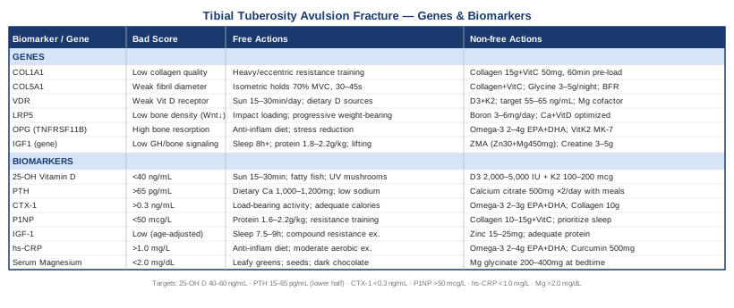
Conclusion
A tibial tuberosity avulsion fracture sits at the intersection of structural vulnerability, biological variability, and mechanical demand. The recovery timeline is not simply a function of how well the surgeon fixed the bone — it is also a reflection of vitamin D status, bone turnover dynamics, collagen gene quality, systemic inflammation, and growth factor availability. Understanding these factors does not replace orthopaedic care; it makes your participation in that care far more informed.
The most practical next step is to request a bone and metabolic panel from your primary care physician or sports medicine doctor that includes 25-OH vitamin D, PTH, hs-CRP, and magnesium at minimum. If bone density or remodeling is a specific concern, add CTX-1 and P1NP. Discuss your genetic testing options if you are interested in identifying structural predispositions. Apply the collagen peptide + vitamin C pre-loading protocol as a low-risk, evidence-supported adjunct to your rehabilitation. And if you have not yet encountered Keith Baar's work on tendon and collagen biology, it is worth your time before your next rehabilitation session. Better biology does not guarantee a faster recovery — but it significantly improves the odds.