This article contains AI generated content.
· UpdatedTransfusional Iron Overload Arthropathy — 5 Genes And 7 Biomarkers To Track
Introduction
If you or someone close to you depends on regular blood transfusions — for thalassemia, sickle cell disease, myelodysplastic syndrome, or aplastic anemia — joint pain probably arrived without a formal warning. Not as a dramatic complication, but as a slow stiffening, an ache that becomes harder to ignore. Transfusional iron overload arthropathy develops precisely this way: the iron accumulating from repeated transfusions eventually reaches synovial membranes and cartilage, where it triggers oxidative damage and inflammation that standard monitoring often catches too late.
Generic clinical advice — keep ferritin below a certain threshold, stay on chelation — is not wrong. But it is incomplete. Iron accumulates differently in different bodies. Some patients develop significant joint damage at ferritin levels that leave others asymptomatic. Some carry genetic variants that accelerate iron loading far beyond what transfusions alone would explain. Some have toxic iron fractions — forms that don't show up on a standard panel — that are driving damage the visible numbers don't capture.
This article is built around that gap. It goes beyond ferritin and looks at the full panel of biomarkers that reveal how iron is actually behaving in joint tissue, how inflammation is responding, and whether cartilage breakdown is already underway. It also examines the specific genes that may explain why iron accumulates faster or damages joints more aggressively in some patients — and what to do about it either way.
Better information doesn't guarantee better outcomes, but it makes more targeted decisions possible. The sections that follow cover seven biomarkers worth tracking, five genes worth knowing about, a book that systematically challenges how medicine currently approaches iron burden, and complementary approaches with genuine clinical evidence for joint protection. Together, they offer a more complete framework than any single specialist appointment usually provides.
Summary
This article covers seven biomarkers — from serum ferritin and transferrin saturation to the more advanced labile plasma iron, hepcidin, and cartilage degradation markers like COMP and CTX-II — that together reveal not just how much iron you carry, but where it is going and how much damage it is already doing to your joints. For each marker, you will find what it costs to measure, what an abnormal result means, and exactly what to do about it — with and without supplements.
The genetics section examines five genes — HFE, HAMP, SLC40A1, BMP6, and ADAMTS5 — that may explain why iron loads faster and attacks joints more aggressively in some patients on identical transfusion protocols. Each gene comes with a practical compensatory plan, including supplement protocols with dosing, cycling, and side effect notes.
Beyond testing and genetics, the article summarizes the key arguments of a book that reframes iron overload management in ways that challenge several standard clinical defaults. Four complementary modalities with meaningful human evidence for joint protection round out the picture.
The goal is not a shortcut around medical care. It is a sharper map for navigating it.
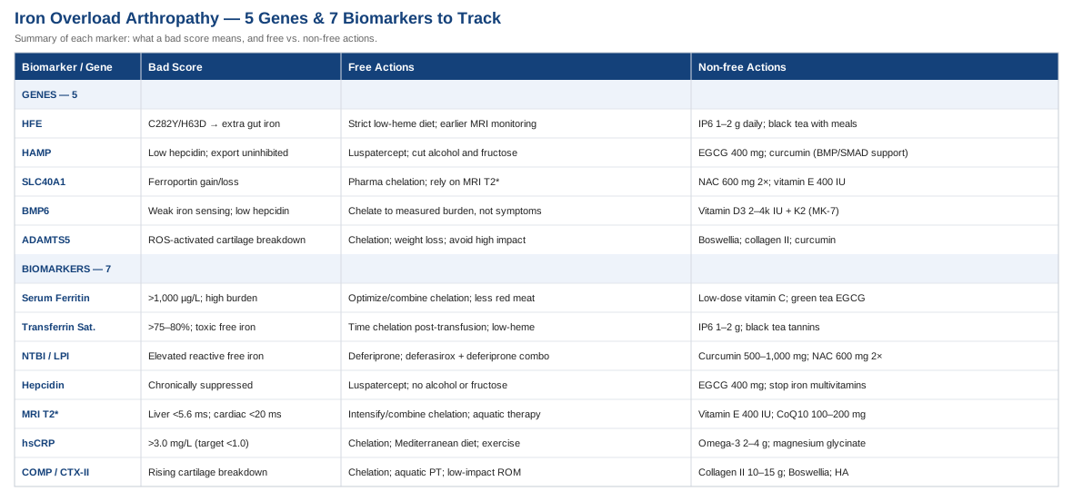
7 Biomarkers to Track
Tracking transfusional iron overload through a single number misses most of what matters for joints. The seven markers below, interpreted together and over time, reveal the full picture: total iron burden, the most toxic free iron fractions, hepcidin regulation, tissue-level iron deposition, systemic inflammation, and cartilage matrix integrity. Several of these are referenced in the clinical literature on iron overload management and in discussions of metabolic biomarker strategy by practitioners like Peter Attia and Thomas Dayspring.
Biomarker 1: Serum Ferritin
Why it matters
Ferritin is the cellular iron storage protein, and its serum concentration reflects cumulative iron burden. In patients receiving regular transfusions, ferritin rises steadily unless chelation keeps pace. For arthropathy, elevated ferritin reflects the systemic iron excess that eventually deposits in synovial tissue and chondrocytes, where it catalyzes hydroxyl radical production and degrades extracellular matrix proteins. The correlation between ferritin trajectory and organ damage — including joint damage — is well established in thalassemia and related conditions.
How to measure it
Standard blood test, ordered as part of any iron panel. Cost: typically $15–$40 in the US, often covered by insurance for transfusion-dependent patients. Results within 24 hours. For thalassemia major, most centers target ferritin below 1,000 µg/L with chelation. For myelodysplastic syndrome, below 2,500 µg/L is a common threshold, though emerging evidence suggests lower targets benefit joint health. Measure every 4–12 weeks depending on transfusion frequency and chelation stability.
If the score is bad — the plan without supplements
The primary intervention is chelation optimization. Three chelators are available: deferoxamine (subcutaneous infusion, typically 5–7 nights per week), deferasirox (oral, once daily), and deferiprone (oral, three times daily). When ferritin remains elevated on monotherapy, combination chelation — particularly deferasirox plus deferiprone — achieves more aggressive reduction. Diet matters too: heme iron from red meat is absorbed at 20–25% efficiency regardless of body iron status. Reducing red meat intake and avoiding cast-iron cookware are accessible, evidence-aligned steps.
If the score is bad — the plan with supplements or equipment
Low-dose vitamin C (100–200 mg daily) enhances deferoxamine by mobilizing stored iron — but this is the ceiling. High-dose vitamin C in iron overload can acutely elevate labile plasma iron and cause cardiac events. Frequency: daily only during deferoxamine infusion, not as standalone. Cycling: ongoing during chelation periods. Green tea extract standardized for EGCG (400–600 mg daily) has mild iron-chelating and anti-inflammatory properties. Evidence in thalassemia is primarily observational. Cycle 8 weeks on, 2 weeks off to limit hepatotoxicity risk at higher doses.
Biomarker 2: Transferrin Saturation
Why it matters
Transferrin is the iron transport protein. When it is fully loaded — typically above 75–80% saturation — iron begins circulating in unbound, toxic forms that reach joint tissue directly. In transfusion-dependent patients, transferrin saturation reaches dangerous levels routinely, particularly in the days following each transfusion. Transferrin saturation gives a real-time gauge of acute iron toxicity risk that ferritin, which reflects accumulated burden more slowly, does not capture.
How to measure it
Ordered as part of an iron panel with serum iron and total iron-binding capacity (TIBC). Cost: $20–$60, often bundled with ferritin testing. Measure fasting in the morning for accuracy. In transfusion-dependent settings, measuring transferrin saturation 24–48 hours before and after transfusion captures the flux and helps calibrate chelation timing.
If the score is bad — the plan without supplements
Chelation timing matters significantly. Starting deferasirox within 24 hours post-transfusion reduces the duration of transferrin saturation at critical levels. Dietary modifications reduce the dietary contribution: avoiding supplemental vitamin C with meals, choosing low-heme protein sources (fish, poultry over red meat), and including calcium-rich foods with higher-iron meals all modestly reduce non-heme iron absorption through the gut.
If the score is bad — the plan with supplements or equipment
Inositol hexaphosphate (IP6), found naturally in whole grains and available as a supplement, chelates iron in the gastrointestinal tract and has shown iron-lowering effects in preliminary studies. Dose: 1–2 g on an empty stomach daily; cycle 3 months on, 1 month off. Monitor zinc and calcium levels — IP6 can reduce mineral absorption broadly. Black tea polyphenols (tannins) inhibit non-heme iron absorption effectively when consumed with or near meals — not a pharmaceutical intervention, but a consistent, evidence-aligned dietary adjunct.
Biomarker 3: Non-Transferrin-Bound Iron and Labile Plasma Iron
Why it matters
When transferrin is saturated, iron circulates as non-transferrin-bound iron (NTBI). Within that pool, labile plasma iron (LPI) is the most chemically reactive fraction: it enters cells without the normal receptor-mediated pathway, generates hydroxyl radicals via the Fenton reaction, and penetrates synovial tissue. LPI is arguably the most proximate driver of iron-mediated joint damage and the primary target of pharmaceutical chelators. Studies in thalassemia have shown that LPI can remain elevated even when serum ferritin appears stable — making it a uniquely sensitive indicator of ongoing joint risk.
How to measure it
NTBI and LPI assays are not standard clinical tests. They are available through specialized reference laboratories at major academic medical centers with thalassemia or hemoglobinopathy programs. Cost: $100–$300, generally not covered by insurance. LPI requires a fluorescence-based assay. If arthropathy is progressing despite apparently controlled ferritin, requesting referral for NTBI or LPI testing is a reasonable clinical escalation.
If the score is bad — the plan without supplements
Chelation timing and drug selection are paramount. Deferiprone has particularly effective LPI-lowering properties because its small molecular size allows it to chelate intracellular as well as plasma iron. Combination therapy — deferasirox plus deferiprone — has shown superior LPI suppression compared to monotherapy in clinical studies. The key question to raise with your hematologist is whether current chelation is actually suppressing LPI, not just reducing ferritin on a quarterly trend.
If the score is bad — the plan with supplements or equipment
Bioavailable curcumin (as phytosome, liposomal, or SLCP formulations for meaningful absorption) chelates iron directly and reduces Fenton reaction-driven oxidative stress. Dose: 500–1,000 mg of a bioavailable curcumin preparation daily; cycle 12 weeks on, 4 weeks off. Side effects: generally well tolerated; may interact with anticoagulants at higher doses. N-acetylcysteine (NAC) replenishes glutathione, the cell's primary antioxidant defense against LPI-generated radicals. Dose: 600 mg twice daily; ongoing during periods of elevated iron burden. Well tolerated at this dose.
Biomarker 4: Hepcidin
Why it matters
Hepcidin is the liver-produced hormone that governs how much iron enters the bloodstream. It works by binding ferroportin — the iron export channel — and triggering its degradation, thereby locking iron inside gut cells and macrophages. In transfusion-dependent conditions with ineffective erythropoiesis (thalassemia intermedia, some myelodysplastic syndromes), hepcidin is chronically suppressed by erythroid signals, meaning the body continues absorbing dietary iron even as transfused iron accumulates. Low hepcidin also means iron-laden macrophages in synovial tissue export more iron into the joint space, feeding local inflammation directly.
How to measure it
Serum hepcidin assays are available through academic medical centers and specialized reference labs. Not yet fully standardized across laboratories. Cost: $75–$200. Urine hepcidin measurement is an emerging alternative. Collect morning samples for consistency; results should be trended within the same laboratory over time rather than compared across labs. A single value is less useful than a direction of change over 6–12 months.
If the score is bad — the plan without supplements
In conditions with ineffective erythropoiesis, luspatercept (a TGF-β ligand trap approved for thalassemia and myelodysplastic syndrome) partially normalizes erythropoiesis and indirectly raises hepcidin toward physiological levels — a significant advance in the management of this specific driver. From a lifestyle standpoint, minimizing alcohol intake (alcohol directly suppresses hepcidin expression), avoiding high-fructose foods (which acutely reduce hepcidin signaling), and maintaining a consistent low-heme diet all reduce the pressure on a dysregulated hepcidin system.
If the score is bad — the plan with supplements or equipment
EGCG from green tea has in vitro evidence for upregulating HAMP gene expression (the gene encoding hepcidin). The clinical translation in humans is preliminary. Dose: 400 mg standardized green tea extract morning; cycle 8 weeks on, 2 weeks off. Iron-containing multivitamins should be eliminated entirely in any patient with confirmed low hepcidin — they represent an avoidable additional iron input into a system that is already failing to regulate absorption.
Biomarker 5: MRI T2-Star — Liver and Heart Iron
Why it matters
MRI T2* is the imaging gold standard for quantifying tissue iron. It measures proton relaxation time in a magnetic field, which shortens as iron concentration rises — allowing non-invasive, quantitative measurement of iron in liver, heart, pancreas, and other organs. Liver T2* correlates closely with total body iron burden (hepatic iron concentration, or HIC). Cardiac T2* detects myocardial iron loading, which is the leading cause of death in transfusion-dependent thalassemia when undetected. For arthropathy, systemic iron burden measured by liver T2* is the most reliable correlate of joint iron deposition, and research protocols are beginning to use MRI directly to image synovial iron in iron overload conditions.
How to measure it
Available at centers with specialized MRI sequences — most major thalassemia treatment centers have this capability. Cost: $500–$2,000 depending on facility, often covered for transfusion-dependent diagnoses. No contrast agent required. Recommended frequency: annually for most patients; every 6 months if cardiac T2* falls below 20 milliseconds. Liver T2* below 5.6 ms indicates severe hepatic iron loading. Both values should be reviewed by a radiologist experienced in iron quantification.
If the score is bad — the plan without supplements
MRI-guided chelation intensification is established best practice when T2* values indicate significant loading. When cardiac T2* falls below 10 ms, combination chelation — typically intravenous or subcutaneous deferoxamine combined with oral deferiprone — is strongly preferred because of its superior cardiac iron extraction. Mechanical joint loading should be moderated during periods of high iron burden; aquatic therapy and cycling reduce joint stress while maintaining circulation that supports tissue recovery.
If the score is bad — the plan with supplements or equipment
No supplement replaces chelation when MRI T2* indicates organ-level loading. Supportive antioxidant coverage is reasonable: mixed tocopherol vitamin E (400 IU daily) and CoQ10 (100–200 mg daily) reduce oxidative stress in iron-loaded tissues and support mitochondrial function. Both are fat-soluble — do not exceed recommended doses. Cycle: ongoing; reassess at 6-month MRI. These are supportive measures only, not primary interventions at this stage of loading.
Biomarker 6: High-Sensitivity C-Reactive Protein
Why it matters
Iron excess generates reactive oxygen species, which trigger systemic inflammation. High-sensitivity CRP (hsCRP) is the most accessible and cost-effective marker of low-grade chronic inflammation — the kind that quietly accelerates cartilage matrix degradation and synovial membrane thickening over months and years. In iron overload arthropathy, elevated hsCRP signals that iron-driven inflammatory processes are active, and it predicts joint symptom progression independently of ferritin levels. Peter Attia has consistently identified hsCRP as one of the most actionable biomarkers in metabolic and inflammatory disease management, precisely because it is sensitive, inexpensive, and responsive to intervention.
How to measure it
Standard blood test, widely available. Cost: $10–$30. Target: below 1.0 mg/L for optimal joint and cardiovascular health; above 3.0 mg/L indicates clinically significant systemic inflammation. Measure during a stable period — acute infections will transiently spike CRP and confound interpretation. Test every 3–6 months alongside the iron panel.
If the score is bad — the plan without supplements
Effective chelation that suppresses NTBI and LPI will, over months, reduce iron-driven oxidative inflammation and lower hsCRP. The Mediterranean dietary pattern — high olive oil, fatty fish, colorful vegetables, minimal ultra-processed food — has among the strongest evidence for reducing hsCRP of any dietary approach. Regular moderate-intensity exercise (30 minutes most days, adapted to joint tolerance) reduces systemic CRP independently of iron status, via multiple anti-inflammatory mechanisms including myokine release.
If the score is bad — the plan with supplements or equipment
Omega-3 fatty acids (EPA plus DHA combined, 2–4 g daily from high-quality fish oil or algal oil) have consistent, well-replicated evidence for reducing hsCRP and IL-6 across diverse populations. Frequency: daily with food; cycle ongoing with quarterly retesting. Side effects: fishy aftertaste — enteric-coated formulations reduce this. High-dose fish oil may modestly affect platelet function; disclose to hematologist. Magnesium glycinate (300–400 mg nightly) reduces NF-κB inflammatory signaling and is commonly deficient in patients on long-term chelation. Ongoing; monitor serum magnesium every 6 months.
Biomarker 7: COMP and CTX-II — Cartilage Degradation Markers
Why it matters
Cartilage Oligomeric Matrix Protein (COMP) is a structural glycoprotein released into serum when cartilage matrix is damaged. CTX-II (the C-terminal telopeptide of type II collagen, measured in urine) is a direct breakdown product of the collagen scaffold that gives articular cartilage its load-bearing properties. In iron overload arthropathy, iron deposits in chondrocytes and synovial tissue generate the reactive oxygen species that degrade these matrix proteins — and COMP and CTX-II capture that degradation in real time. Elevated levels can precede radiographic joint damage by years, offering a critical window for intervention. The logic of tissue-specific structural markers as early warning signals parallels the approach to cardiovascular risk advocated by Allan Sniderman and others who argue that downstream structural markers outperform upstream metabolic markers for predicting irreversible damage.
How to measure it
COMP is a serum test available through rheumatology reference laboratories. Cost: $60–$150. CTX-II is a urine test (second morning void, normalized to creatinine), available at specialty labs. Cost: $80–$200. Neither is routinely ordered, but both are obtainable on request through rheumatology or academic iron overload centers. These markers are most meaningful serially — a single value is less informative than a trend tracked every 6–12 months.
If the score is bad — the plan without supplements
Optimized chelation to reduce synovial iron is the most direct structural intervention. Beyond chelation, joint-protective physical therapy — particularly aquatic therapy and low-impact range-of-motion exercise — reduces mechanical stress on damaged cartilage without adding inflammatory load. Chronic NSAID use should be discussed carefully with a rheumatologist familiar with iron overload: NSAIDs carry renal implications in patients on deferasirox and may affect GI iron absorption patterns when used long-term.
If the score is bad — the plan with supplements or equipment
Hydrolyzed type II collagen peptides (10–15 g daily) have emerging randomized trial evidence for supporting cartilage matrix synthesis by supplying substrates for chondrocyte collagen production. Cycle: 12 weeks on, 4 weeks off; retest CTX-II at cycle end. Minimal side effects. Boswellia serrata standardized to 65% boswellic acids (400–600 mg twice daily) inhibits the 5-LOX enzyme, which is activated by iron-generated oxidative stress and drives prostaglandin-mediated cartilage breakdown. A randomized trial in osteoarthritis patients found significant reductions in pain and functional limitation versus placebo. Cycle: 8–12 weeks; monitor liver enzymes given that iron overload itself carries hepatic risk. Oral hyaluronic acid (80–200 mg daily) supports synovial fluid viscosity and may reduce joint friction in deteriorating cartilage.
Building on the biomarker framework above, the next section examines the genetic architecture that may explain why some patients accumulate iron faster and sustain joint damage more readily — a layer of information that makes biomarker trends more interpretable and interventions more targeted.
5 Genes That May Shape Your Risk
Genetic variation doesn't fix outcomes. But it sets the baseline against which interventions work, and it explains discrepancies that otherwise look like unexplained clinical variability. The five genes below are among the best-studied in iron metabolism and joint tissue biology. Understanding them helps clarify why ferritin trajectories differ between patients on identical transfusion protocols and why arthropathy develops earlier in some.
Gene 1: HFE — The Iron Sensing Gene
HFE is the gene most associated with hereditary hemochromatosis. Its two key variants — C282Y and H63D — impair the body's ability to sense iron adequacy and properly signal the liver to upregulate hepcidin. In transfusion-dependent patients who also carry HFE variants, iron loading accelerates beyond what transfusions alone would cause: dietary iron absorption continues at elevated rates even as transfused iron accumulates. C282Y carriers (even one copy) in a transfusion-dependent setting warrant particular monitoring vigilance, as the combined iron load from transfusion plus unregulated gut absorption can be substantially greater than standard protocols anticipate.
If the gene is problematic — the plan without supplements
More aggressive chelation targets and earlier MRI monitoring intervals are justified. A strict low-heme dietary approach (replacing red meat with fish and poultry, avoiding iron-fortified foods) should be maintained consistently — not just during chelation intensification. Inform your hematologist explicitly that an HFE variant is present; many standard chelation protocols are not calibrated for combined transfusion plus HFE-driven loading.
If the gene is problematic — the plan with supplements or equipment
IP6 (1–2 g daily on an empty stomach) reduces intestinal iron absorption and is a reasonable adjunct for HFE variant carriers. Cycle: 3 months on, 1 month off. Monitor zinc and calcium levels regularly — IP6 binds these minerals non-selectively. Black tea consumed with meals inhibits non-heme iron absorption via tannin binding — a simple, consistent, evidence-supported daily strategy without meaningful side effects at normal dietary intake.
Gene 2: HAMP — The Hepcidin Gene
HAMP encodes hepcidin itself. Variants that reduce HAMP expression or impair its transcriptional response to iron worsen loading by leaving iron export pathways chronically uninhibited. In ineffective erythropoiesis conditions, HAMP is already suppressed by elevated GDF15 and TWSG1 from stressed erythroid precursors — genetic HAMP variants that further reduce expression compound an already dysregulated system. The result: iron streams continuously into plasma and, eventually, joint tissue rather than being retained in regulated storage.
If the gene is problematic — the plan without supplements
Luspatercept addresses the upstream suppression of HAMP by reducing ineffective erythropoiesis. For patients on this therapy, tracking hepcidin response over time reveals whether HAMP expression is recovering. Minimize known hepcidin suppressors: alcohol in any quantity, high-fructose foods, and significant caloric restriction all acutely reduce hepcidin. Even modest reductions in these exposures allow whatever residual HAMP function exists to work more effectively.
If the gene is problematic — the plan with supplements or equipment
EGCG has preclinical evidence for upregulating HAMP gene expression in hepatocyte models. Human data are limited. Dose: 400 mg standardized green tea extract morning; cycle 8 weeks on, 2 weeks off to avoid hepatic stress at sustained higher doses. Curcumin has been shown to modulate BMP/SMAD signaling — the pathway that activates HAMP transcription — offering a complementary mechanism to EGCG. Combining these at moderate doses is unlikely to match pharmaceutical hepcidin regulation but may offer partial supportive benefit.
Gene 3: SLC40A1 — The Ferroportin Gene
SLC40A1 encodes ferroportin, the only known mammalian iron export channel. It is expressed on gut enterocytes, macrophages, and hepatocytes and is the direct target of hepcidin regulation. Gain-of-function mutations in SLC40A1 make ferroportin resistant to hepcidin's inhibitory binding — iron export continues even when the body signals to stop, flooding plasma with iron. Loss-of-function mutations trap iron inside macrophages, including the synovial macrophages in joint tissue. Both phenotypes produce iron overload but with different tissue distribution; both are relevant to arthropathy because in one case the joint is exposed to excess circulating iron, and in the other, the joint's own immune cells become iron-laden and pro-inflammatory.
If the gene is problematic — the plan without supplements
Gain-of-function SLC40A1 variants respond less predictably to hepcidin-based therapeutic approaches and require pharmaceutical chelation as the primary tool. Loss-of-function variants may show deceptively low serum ferritin despite high tissue iron in macrophages, making MRI T2* disproportionately important compared to serum markers in this subgroup. Raise this distinction explicitly with a specialist if ferroportin disease has been identified.
If the gene is problematic — the plan with supplements or equipment
For loss-of-function ferroportin disease, no supplement reliably corrects ferroportin-mediated macrophage iron retention — MRI-guided chelation is the mainstay. Supporting cellular antioxidant defenses in iron-laden macrophages is reasonable: NAC (600 mg twice daily, ongoing) and mixed tocopherol vitamin E (400 IU daily) reduce hydroxyl radical damage inside iron-loaded cells. Monitor fat-soluble vitamin accumulation with periodic serum levels if using vitamin E beyond 6 months.
Gene 4: BMP6 — The Iron Sensor Upstream of Hepcidin
BMP6 encodes bone morphogenetic protein 6, which acts as an iron sensor in the liver: as iron rises in hepatic sinusoids, BMP6 signals through SMAD1/5/8 to activate HAMP transcription and boost hepcidin. Variants in BMP6 that reduce this signaling create a regulatory blind spot — the liver cannot fully perceive iron loading and does not produce enough hepcidin in response. BMP6-linked hemochromatosis (a recognized non-HFE hemochromatosis subtype) can occur even with apparently normal HFE status, and in transfusion-dependent patients with coincident BMP6 dysfunction, standard clinical monitoring calibrated to average hepcidin responses will underestimate real risk.
If the gene is problematic — the plan without supplements
BMP6 variants are rare and typically identified on genetic panels ordered through hemoglobinopathy or hemochromatosis specialist programs. If confirmed, the management principle is: do not rely on hepcidin to rise automatically in response to rising iron burden. Instead, rely on direct iron quantification — ferritin trend, transferrin saturation, MRI T2* — and calibrate chelation to measured burden rather than to clinical symptoms or expected biological response.
If the gene is problematic — the plan with supplements or equipment
BMP6 signaling intersects with the vitamin D receptor pathway, and there is mechanistic plausibility (though no direct human proof) that maintaining optimal vitamin D status preserves BMP pathway function. Maintain serum 25-OH vitamin D between 40 and 60 ng/mL. Supplementation: 2,000–4,000 IU vitamin D3 combined with 100–200 mcg MK-7 (vitamin K2) daily; reassess serum levels every 3 months to avoid over-supplementation. This is a biologically grounded supportive strategy, not a direct BMP6 fix.
Gene 5: ADAMTS5 — The Cartilage Protease Gene
ADAMTS5 encodes a metalloproteinase that cleaves aggrecan, the primary proteoglycan providing compressive resistance in articular cartilage. In a healthy joint, ADAMTS5 activity is tightly regulated. In iron-overloaded joints, the reactive oxygen species generated by iron-catalyzed Fenton chemistry directly activate ADAMTS5 and related matrix metalloproteinases, accelerating cartilage breakdown. Genetic variants that increase ADAMTS5 expression or reduce its endogenous inhibitors amplify this iron-driven destruction, creating a joint that degrades faster at equivalent iron levels compared to someone without the variant. This gene sits precisely at the intersection of iron biology and joint biology, making it particularly relevant for arthropathy risk stratification.
If the gene is problematic — the plan without supplements
Reducing synovial iron through optimized chelation is the most direct intervention against ADAMTS5 hyperactivation. Weight management — even modest reductions in body mass — significantly reduces mechanical stress on the cartilage matrix that ADAMTS5 is degrading. Avoiding repetitive high-impact loading (running on hard surfaces, prolonged standing with joint pain) protects remaining matrix from compounding mechanical damage on top of enzymatic degradation.
If the gene is problematic — the plan with supplements or equipment
Boswellia serrata (400 mg standardized AKBA fraction twice daily) inhibits metalloproteinases through 5-LOX pathway suppression, directly relevant to ADAMTS5-mediated breakdown. Cycle: 12 weeks; monitor liver enzymes in iron overload context. Hydrolyzed collagen type II (10–15 g daily) supports matrix synthesis to partially offset ongoing ADAMTS5-driven degradation. Curcumin (500 mg bioavailable form daily) inhibits NF-κB activation that drives ADAMTS5 gene expression in inflamed chondrocytes. These three can be combined at the above doses; cycle curcumin 12 weeks on, 4 weeks off.
With both the biomarker picture and the genetic framework established, the next step is a broader strategic perspective — one that questions whether standard thresholds and monitoring assumptions are ambitious enough.
What "Dumping Iron" by P.D. Mangan Reveals
Dumping Iron: How to Ditch This Secret Killer and Reclaim Your Health by P.D. Mangan is not a clinical guideline. It is a rigorous survey of peer-reviewed research on iron's role in aging, oxidative disease, and tissue damage — written for an audience that wants to understand the science, not just follow a protocol. Mangan, a biochemist and independent health researcher, draws on hundreds of studies to argue that iron accumulation is one of the most underappreciated drivers of degenerative disease and that the thresholds medicine uses to define "normal" are not the same as thresholds that protect health. For patients managing transfusional iron overload, many of his arguments are directly applicable — in fact, amplified.
10 Insights Worth Knowing
1. Normal ferritin ranges are not optimal ferritin ranges. Laboratory reference intervals for ferritin extend to 200–300 µg/L or higher in men. Mangan cites multiple epidemiological studies showing that cardiovascular risk, cancer risk, and tissue damage risk rise substantially above 80–100 µg/L. For transfusion-dependent patients, this reframes even the clinical "success" threshold of ferritin below 1,000 µg/L as substantially above the biological optimum for joint protection.
2. Iron is fundamentally a pro-oxidant. The Fenton reaction — ferrous iron reacting with hydrogen peroxide to generate hydroxyl radicals — is among the most destructive chemistry in human biology. Mangan explains this accessibly and traces its relevance to cartilage destruction, cardiovascular disease, liver damage, and neurodegeneration. For arthropathy patients, the Fenton reaction inside synovial tissue is the core mechanism they are fighting.
3. The most damaging iron fraction is the one medicine measures least. Mangan makes the case, from the biochemistry literature, that serum iron and ferritin are relatively inert compared to NTBI and LPI. Clinicians who focus on ferritin while ignoring NTBI are watching the wrong number. This is one of the book's most actionable arguments for patients seeking more sophisticated monitoring.
4. Blood donation is an underused iron-lowering intervention — for those who can use it. For individuals without transfusion-dependent conditions but with iron overload (HFE hemochromatosis, relatives of affected patients), regular blood donation robustly reduces ferritin and associated disease risk. Mangan reviews the evidence thoroughly. For transfusion-dependent patients, this is not applicable — but therapeutic phlebotomy for family members who share HFE or other iron-loading variants is worth raising with a physician.
5. Hepcidin suppression explains why lifestyle choices compound iron loading. Mangan documents how alcohol, fructose, sleep disruption, and certain medications suppress hepcidin — opening the gate to dietary iron even in already overloaded conditions. This gives concrete biological meaning to dietary advice that is often presented without explanation.
6. Exercise acutely raises hepcidin and reduces iron absorption. Beyond cardiovascular and metabolic benefits, exercise induces transient hepcidin elevation in the post-exercise window, reducing iron absorption from dietary sources in the hours that follow. Mangan recommends regular moderate-intensity exercise as a non-pharmaceutical iron management strategy. For arthropathy patients, the challenge is finding joint-safe modalities — the complementary approaches section below addresses this directly.
7. Dietary heme iron cannot be meaningfully regulated by the body. Non-heme iron absorption is reduced when body iron is high. Heme iron (from red meat) is absorbed at 20–25% efficiency regardless of iron status. Mangan argues this makes red meat uniquely problematic in iron-loaded conditions and recommends replacing it with fish and poultry rather than attempting to moderate intake.
8. Anti-inflammatory supplements often work partly through iron chelation. Curcumin, quercetin, EGCG, and resveratrol all have demonstrated iron-chelating capacity in addition to their other mechanisms. Mangan reframes these as iron management tools, not just general-purpose anti-inflammatories. This aligns with and contextualizes the supplementary recommendations throughout this article.
9. Iron's relationship to cancer risk is mechanistically clear and clinically underappreciated. Mangan reviews the evidence linking iron excess to cancer promotion through Fenton chemistry, DNA oxidative damage, and tumor microenvironment effects. While this extends beyond arthropathy, it provides additional motivation for aggressive iron management in transfusion-dependent patients who already carry elevated cancer risk in some underlying conditions.
10. Iron damage is long-latency — by the time it is visible, it has been occurring for years. Mangan's central practical argument is that iron-related pathology is preventable earlier than medicine acts on it, and that acting when biomarkers are sub-clinical rather than waiting for symptoms preserves function that cannot be recovered once lost. This is precisely the argument for using the full biomarker panel in this article, beginning well before joints become symptomatic.
The evidence basis throughout this book favors human epidemiological and mechanistic studies. It challenges practitioners to reconsider not just what they measure, but where they set the bar for intervention. For anyone managing iron overload, it is one of the most useful frameworks available outside of specialist clinical literature.
Having established the biomarker, genetic, and strategic frameworks, the following section looks at specific adjunct therapies with meaningful clinical evidence for protecting joint function in relevant populations.
Complementary Approaches With Clinical Evidence
The modalities below are not alternatives to chelation or specialist care. They are adjuncts supported by human clinical evidence for reducing joint symptoms, inflammatory burden, or oxidative stress in conditions mechanistically relevant to iron overload arthropathy. Select based on individual fit, not as a complete protocol.
Low-Level Laser Therapy and Photobiomodulation
Photobiomodulation (PBM) uses red and near-infrared wavelengths (typically 810–904 nm) to stimulate cytochrome c oxidase in the mitochondrial respiratory chain, improving cellular energy production and reducing inflammatory signaling. In iron overload arthropathy, where mitochondrial dysfunction driven by reactive oxygen species is a central mechanism, PBM's ability to partially restore mitochondrial function in joint tissue gives it a mechanistically credible role. PBM also reduces prostaglandin E2 and inflammatory cytokine production in synovial tissue.
A 2021 systematic review and meta-analysis of PBM in inflammatory arthritis conditions found statistically significant reductions in pain intensity and morning stiffness compared to sham treatment, with a favorable safety profile across trials. While this body of evidence is primarily in rheumatoid arthritis rather than iron overload arthropathy specifically, the anti-inflammatory and mitochondrial mechanisms are shared. Evidence in iron overload arthropathy specifically is currently absent from the peer-reviewed literature; PBM should be positioned as a pain and function adjunct.
For practical use: seek physiotherapy or sports medicine clinics offering clinical-grade PBM units (not consumer LED panels, which deliver substantially lower irradiance). A typical protocol for inflammatory joint conditions is 3 sessions per week for 4–6 weeks, with monthly maintenance thereafter. Home-use FDA-cleared near-infrared panels exist but deliver lower therapeutic doses than clinical equipment. Avoid direct application over areas with active infection or malignancy.
Tai Chi
Tai chi is a slow, controlled movement practice combining joint mobility, balance training, postural awareness, and parasympathetic nervous system activation. For patients with iron overload arthropathy, its particular value lies in maintaining joint range of motion, reducing pain-associated muscle guarding, and supporting circulation in periarticular tissues — all without the high mechanical loading that conventional exercise can impose on damaged cartilage. The practice is inherently joint-protective in its execution.
A 2016 randomized controlled trial published in Annals of Internal Medicine by Chenchen Wang and colleagues found that tai chi practice produced improvements in pain, physical function, and depression in knee osteoarthritis equivalent to conventional physical therapy over 12 weeks, with superior results at 24 and 52 weeks in several outcomes. While conducted in osteoarthritis rather than iron overload arthropathy, the joint condition is mechanistically related — cartilage degradation and synovial inflammation are shared pathological features.
Practical protocol: 3–5 sessions per week, 30–45 minutes each. Yang-style tai chi is the most extensively studied for musculoskeletal conditions. Many community centers, YMCAs, and online platforms offer beginner programs. Look for instructors familiar with joint-protective modifications, particularly for avoiding deep knee bends and sudden directional changes during active flares. Seated tai chi modifications are available if standing is limited by pain.
Mindfulness-Based Stress Reduction
Chronic joint pain from arthropathy activates central sensitization — the nervous system progressively amplifies pain signals independently of peripheral joint status. MBSR (Mindfulness-Based Stress Reduction), an 8-week structured program developed by Jon Kabat-Zinn at the University of Massachusetts, directly addresses central pain processing through sustained attention practice and body-awareness exercises. For patients managing transfusional iron overload — with its demanding treatment schedule, frequent monitoring, and chelation side effects — the psychological dimension of the disease also warrants direct, evidence-based intervention.
A 2020 meta-analysis of MBSR across chronic pain conditions found significant reductions in pain intensity, pain-related functional interference, and depression, with effect sizes comparable to pharmacological analgesics used for chronic pain. Several trials within this analysis found that MBSR also reduced inflammatory markers including CRP, consistent with the established relationship between stress physiology and systemic inflammation.
MBSR programs are available through academic medical centers, online platforms (Palouse MBSR offers a freely accessible, evidence-aligned digital version), and apps calibrated to the validated 8-week curriculum. Commit to the full 8 weeks for meaningful benefit — the dose-response relationship in MBSR is strongly tied to program completion. The primary barrier is time: the standard program involves approximately 2.5 hours weekly of guided sessions and 30 minutes daily of home practice. Treat it as a medical adjunct requiring the same consistency as physical therapy.
Microbiome-Directed Therapies
An emerging body of research has identified a gut-iron axis: intestinal iron availability directly shapes microbial community composition, and the resulting dysbiosis amplifies systemic inflammation and impairs iron regulation. In transfusion-dependent patients with high dietary iron exposure, iron-loving pathobionts — particularly certain Enterobacteriaceae — proliferate in the gut, produce endotoxins that enter systemic circulation, and contribute to the low-grade inflammatory state that drives arthropathy. The microbiome represents a modifiable variable that connects iron biology to joint inflammation.
Studies in iron-supplemented populations (including Kenyan children in Jaeggi et al., 2015, and subsequent work in adult iron-overloaded cohorts) have documented that iron availability selectively enriches pro-inflammatory gut bacteria and depletes protective species, while prebiotic and probiotic interventions partially restore microbial balance. This evidence is not yet fully translated to transfusion-dependent iron overload arthropathy specifically, and it should be treated as an evolving research area with practical implications rather than established clinical protocol.
Practical approach: a high-diversity, high-fiber dietary pattern (Mediterranean or whole-foods plant-rich approach) preemptively supports microbial diversity and reduces the relative abundance of iron-dependent pathogens. A multi-strain probiotic including Lactobacillus plantarum and Bifidobacterium longum — both of which have shown iron-binding capacity in controlled studies — may offer supportive benefit. Dose: 10–20 billion CFU daily; cycle 8 weeks, then assess GI symptoms and inflammatory markers. Avoid iron-fortified foods and high-dose inulin supplements that preferentially feed iron-dependent pathobionts. Treat this as a supportive layer within a broader management strategy.
Conclusion
Transfusional iron overload arthropathy is a complication that develops quietly, over years, in bodies already managing significant medical demands. The biomarker framework in this article — seven markers spanning iron burden, toxic iron fractions, hepcidin regulation, tissue iron quantification, systemic inflammation, and cartilage matrix integrity — offers a substantially more complete picture than standard monitoring provides. The genetic context — HFE, HAMP, SLC40A1, BMP6, and ADAMTS5 — explains individual variation in iron loading and joint vulnerability, and points toward personalized strategies that go beyond average-population protocols.
None of this replaces specialist care. But it makes those appointments more productive. Arriving with serial biomarker data, genetic context, and a clear picture of which markers are trending in the wrong direction places you in a far stronger position to ask targeted questions and advocate for protocol adjustments that routine monitoring might otherwise miss.
The next concrete step: identify which of the biomarkers above you are not currently tracking, request them at your next appointment or through a specialty reference laboratory, and begin building the longitudinal picture that makes iron overload arthropathy genuinely manageable rather than reactive. The evidence base exists. The measurement tools are accessible. The difference lies in using them systematically, early, and with clinical support.