This article contains AI generated content.
· UpdatedTransient Bone Marrow Edema Syndrome — 5 Genes And 7 Biomarkers To Track
Introduction
Transient bone marrow edema syndrome (TBMES) sits in an uncomfortable clinical grey zone. The pain is real and often severe — a deep, aching pressure localized around a joint, most frequently the hip — yet imaging confirms no fracture, no tumor, no infection. You are told the condition is self-limiting and will eventually resolve. That is accurate, but it can take anywhere from six weeks to eighteen months, and waiting feels passive when you have no framework for understanding what is driving the process or how to meaningfully support recovery.
Generic advice about rest, anti-inflammatory medication, and reduced weight-bearing manages symptoms but does not explain why one person recovers in two months while another is still limited at fourteen. It does not reveal whether your bone metabolism is functioning well enough to support active remodeling, or whether a chronic low-grade inflammatory or vascular signal is prolonging the edema and delaying tissue repair.
The emerging science of TBMES points toward a convergence of vascular insufficiency, disrupted bone remodeling cycles, and systemic metabolic factors — all of which leave measurable signals in the blood. Tracking the right biomarkers over time gives you a window into these mechanisms. And while the genetics of TBMES is still a developing area, certain gene variants can shift baseline vulnerability in ways that can be at least partially compensated through targeted lifestyle and nutritional strategies.
This article takes two approaches: a detailed biomarker panel of seven markers trackable through standard lab tests, and a genetics section covering five gene variants relevant to bone health and tissue repair. Neither replaces clinical care. Both can meaningfully sharpen the conversation you have with a specialist and improve the decisions you make between appointments.
Summary
What you will find in this article: Seven lab biomarkers — including two bone turnover markers that almost no standard workup includes — that can reveal exactly where your recovery is stalling and why. Five gene variants that may explain individual differences in how fast bone marrow edema resolves, how much pain you experience, and how strong your natural tissue repair capacity really is. A breakdown of what Peter Attia's framework from Outlive means specifically for bone marrow health. And four evidence-supported complementary approaches, one of which (low-level laser therapy) has clinical data directly relevant to bone marrow lesions.
If you have been told to wait it out, what follows will give you something more useful than patience alone.
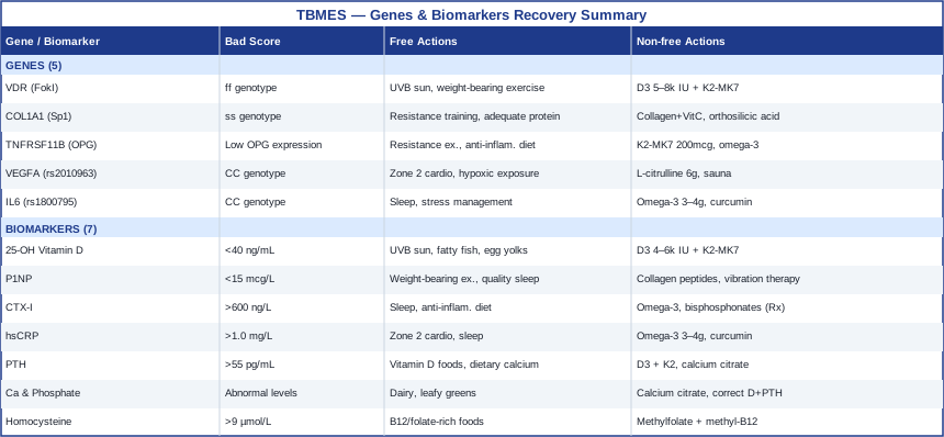
7 Biomarkers That Reveal What Is Really Happening in Your Bone Marrow
Most TBMES workups include an MRI, basic blood chemistry, and perhaps a vitamin D level. That is a reasonable start, but it leaves substantial gaps. The markers below are not exotic — most can be ordered through any standard lab. What makes them valuable is the specific picture they build together: one of bone formation rate, resorption rate, inflammatory load, hormonal regulation, and vascular signaling. Tracking these over three to six months alongside symptom changes turns recovery from a waiting game into something you can actually observe and influence.
Biomarker 1: 25-OH Vitamin D (Vitamin D Status)
Why it matters for TBMES: Vitamin D is not just a bone mineral regulator — it modulates the immune environment within bone marrow, influences macrophage behavior, and affects the signaling pathways that control osteoblast and osteoclast activity. Low vitamin D is consistently associated with impaired bone healing and increased inflammatory bone conditions. In TBMES specifically, suboptimal vitamin D may both slow the resolution of edema and reduce the structural quality of newly deposited bone matrix during recovery. Several case series and observational studies have noted that TBMES patients tend to present with low to insufficient vitamin D levels, though randomized trials are still lacking.
How to measure it: A standard serum 25-hydroxyvitamin D blood test. Cost ranges from $30 to $70 depending on the lab and whether insurance covers it. The optimal range for bone health extends from 40 to 60 ng/mL (100–150 nmol/L) — a target supported by Peter Attia and others working in preventive medicine. Functional medicine laboratories may report additional metabolite profiles, but the basic test is sufficient for tracking purposes.
If the score is below 40 ng/mL — the plan without supplements: Midday sun exposure targeting 15 to 30 minutes of direct UVB on large skin surface areas (arms, legs, torso) daily or near-daily. Dietary vitamin D from fatty fish (salmon, mackerel, sardines), egg yolks, and liver. These strategies can raise vitamin D meaningfully in fair-skinned individuals at favorable latitudes but often cannot fully correct deficiency, especially in autumn and winter.
If the score is below 40 ng/mL — the plan with supplements: Vitamin D3 at 4,000 to 6,000 IU daily, always combined with vitamin K2 (MK-7 form, 100–200 mcg/day) to direct calcium to bone rather than soft tissue. Magnesium glycinate (300–400 mg) supports D3 activation. Retest at 90 days and adjust. Side effects at these doses are rare but monitor for hypercalcemia if taking calcium simultaneously. Consider cycling to a maintenance dose (2,000–3,000 IU) once the target range is reached and sustained.
Biomarker 2: P1NP (Procollagen Type I N-Terminal Propeptide)
Why it matters for TBMES: P1NP is the most sensitive and specific marker of bone formation currently available for clinical use. It reflects how actively osteoblasts are synthesizing new collagen matrix — the first step in replacing the damaged trabecular bone that characterizes TBMES. A low P1NP in the context of active TBMES suggests that the body is struggling to mount an adequate repair response. This marker, championed by bone metabolism specialists and increasingly discussed in the work of clinicians like Peter Attia, is rarely included in a standard TBMES workup but carries significant diagnostic and prognostic weight.
How to measure it: Blood test, no special fasting required. Cost ranges from $50 to $150 and may require a specialist order or direct-to-consumer lab. Reference ranges vary by lab and age/sex. For adults, normal P1NP typically falls between 15 and 74 mcg/L, though optimal ranges for bone healing may be at the higher end. Serial measurements (every 3 months) are more informative than a single reading.
If P1NP is low — the plan without supplements: Mechanical loading is one of the most powerful stimulants of osteoblast activity. For TBMES, this means progressive, carefully supervised partial weight-bearing as tolerated — typically guided by pain threshold and MRI signal evolution. Resistance exercise engaging the affected limb, once cleared by a physician, directly stimulates P1NP elevation. Sleep quality also matters: most bone formation occurs during slow-wave sleep, and sleep deprivation demonstrably suppresses osteoblastic activity.
If P1NP is low — the plan with supplements or equipment: Collagen peptides (10–15 g/day with vitamin C) have shown in randomized trials to increase bone formation markers including P1NP. Whole-body vibration therapy (15–20 minutes daily at low magnitude, high frequency) has clinical evidence for increasing bone formation markers in osteoporotic patients and may be relevant to TBMES recovery. Silicon (as orthosilicic acid, 10–25 mg/day) supports collagen cross-linking. Cycle collagen supplementation continuously without cycling needs; vibration therapy can be applied daily.
Biomarker 3: CTX-I (C-Terminal Telopeptide of Type I Collagen)
Why it matters for TBMES: CTX-I is the primary clinical marker of bone resorption — it measures fragments of degraded type I collagen released into the bloodstream when osteoclasts break down bone matrix. In TBMES, bone resorption is typically elevated as part of the local inflammatory-vascular response that causes marrow edema. Persistently elevated CTX-I alongside low P1NP (high resorption, low formation) is a particularly poor recovery profile, suggesting the balance of bone remodeling is not moving toward healing. Thomas Dayspring and Peter Attia have both highlighted the importance of pairing formation and resorption markers rather than looking at either in isolation.
How to measure it: Fasting morning blood draw (CTX rises with food intake and circadian variation is significant — always draw fasting, before 10 AM). Cost ranges from $50 to $130. Pre-menopausal women and men under 50 should be compared to age-sex matched references. Elevated CTX in TBMES context generally means above 600 ng/L for men and post-menopausal women, though lower values may still be unfavorable if P1NP is simultaneously low.
If CTX is elevated — the plan without supplements: Reduce prolonged mechanical overloading that aggravates the affected site. Prioritize sleep — CTX rises significantly after a single night of poor sleep and falls with sleep restoration. An anti-inflammatory dietary pattern (Mediterranean-style, emphasizing olive oil, fatty fish, vegetables, minimal refined carbohydrate) has measurable effects on bone resorption markers over 8–12 weeks.
If CTX is elevated — the plan with supplements or equipment: Omega-3 fatty acids (2–4 g EPA+DHA daily) reduce osteoclast activity through prostaglandin modulation. Bisphosphonate therapy (prescription only) directly suppresses osteoclast activity and has been used in some TBMES cases with good results — this is worth discussing with a specialist when CTX is significantly elevated and recovery is prolonged. Magnesium (300–400 mg glycinate or malate) supports mineral homeostasis and has modest effects on bone resorption.
Biomarker 4: High-Sensitivity CRP (hsCRP)
Why it matters for TBMES: The pathophysiology of TBMES almost certainly involves an inflammatory component, whether as cause or effect. Low-grade systemic inflammation — reflected in elevated hsCRP — can sustain osteoclast activity (via cytokine-driven RANKL upregulation), impair vascular endothelial function (reducing bone marrow perfusion), and alter the local immune environment in ways that prolong edema resolution. hsCRP is one of the most accessible, affordable, and validated systemic inflammation markers available, and its inclusion in any bone health workup is straightforward to justify.
How to measure it: Standard blood test available at virtually any lab. Cost: $20 to $50. The target is below 1.0 mg/L for low cardiovascular and inflammatory risk; values between 1.0 and 3.0 mg/L indicate intermediate risk and are clinically meaningful. Note that acute infections or tissue injury can transiently raise CRP — interpret elevated values in clinical context.
If hsCRP is elevated — the plan without supplements: The most powerful non-pharmacological interventions for hsCRP are aerobic exercise (consistent zone 2 cardio, 150–200 min/week), sleep optimization (7–9 hours, consistent timing), and dietary changes (reduction in processed foods, trans fats, refined sugars; increase in vegetables and olive oil). Stress reduction also matters — cortisol chronically elevates inflammatory cytokines.
If hsCRP is elevated — the plan with supplements or equipment: Omega-3 fatty acids at therapeutic doses (3–4 g EPA+DHA/day) have the strongest evidence for reducing hsCRP. Curcumin (as theracurmin or phosphatidylcholine-bound form, 500–1000 mg/day) has consistent trial-level evidence for reducing CRP in inflammatory conditions. Both can be taken continuously. Monitor with quarterly retesting. Neither causes significant side effects at these doses, though omega-3 may mildly affect bleeding time at high doses.
Biomarker 5: Parathyroid Hormone (PTH)
Why it matters for TBMES: PTH is the master regulator of calcium homeostasis and exerts direct effects on both osteoblasts and osteoclasts. Chronically elevated PTH — even within the "normal" reference range — can shift the remodeling balance toward net bone resorption. Secondary hyperparathyroidism driven by vitamin D insufficiency is common and entirely correctable, but it persists silently unless PTH is specifically measured. In TBMES patients with low vitamin D, PTH is almost certainly elevated, creating a loop of excess bone resorption that may slow marrow healing.
How to measure it: Intact PTH blood test, best measured alongside vitamin D and calcium. Cost: $30 to $80. The functional optimal range for PTH is considered 10 to 55 pg/mL; values above 65 pg/mL warrant investigation for primary or secondary hyperparathyroidism.
If PTH is elevated — the plan without supplements: If driven by low vitamin D, the primary intervention is correcting vitamin D status (see Biomarker 1). Adequate dietary calcium (1,000–1,200 mg/day from food sources) also reduces PTH stimulus. Magnesium adequacy is critical since magnesium is required for proper PTH secretion and response.
If PTH is elevated — the plan with supplements or equipment: Vitamin D3 + K2 supplementation (as above) is the cornerstone. If PTH remains elevated after vitamin D normalization, imaging of the parathyroid glands may be warranted — this is a clinical discussion, not a self-management situation. Calcium citrate supplementation (if dietary calcium is genuinely insufficient) supports PTH reduction but should not be taken in excess.
Biomarker 6: Serum Calcium and Phosphate
Why it matters for TBMES: Calcium and phosphate are the primary minerals of bone mineral — hydroxyapatite is composed of both — and their serum balance reflects the net flux between gut absorption, kidney reabsorption, and bone release. Abnormalities in either signal can indicate dysregulated bone metabolism contributing to poor TBMES recovery. While overt abnormalities are typically caught on routine panels, the relationship between calcium, phosphate, and markers like PTH and vitamin D is what matters diagnostically — no single value tells the full story.
How to measure it: Standard metabolic panel or stand-alone calcium + phosphate. Cost: $20 to $40. Normal serum calcium is 8.5 to 10.5 mg/dL; optimal bone metabolism tends to favor the middle of that range. Phosphate normal range: 2.5 to 4.5 mg/dL.
If values are abnormal — the plan without supplements: Dietary calcium from dairy, leafy greens, and bone-in fish. Adequate phosphate intake is rarely a problem in Western diets — phosphate excess from processed food (phosphate additives) is more common and can paradoxically impair bone health by triggering PTH release.
If values are abnormal — the plan with supplements or equipment: Correct underlying drivers (vitamin D, PTH) before supplementing calcium directly. Calcium citrate (not carbonate) is better absorbed and gentler on the gut. Phosphate management typically involves dietary changes rather than supplementation. If calcium is persistently abnormal despite corrected vitamin D and PTH, refer to endocrinology.
Biomarker 7: Homocysteine
Why it matters for TBMES: Homocysteine is a byproduct of methionine metabolism that, when elevated, damages endothelial cells and impairs collagen cross-linking — two mechanisms highly relevant to a condition with a suspected vascular component. Bone marrow is heavily vascularized, and compromised microvascular function is among the leading hypotheses for why TBMES develops, particularly the variant seen in pregnancy and in endurance athletes with repetitive loading. Elevated homocysteine has been specifically associated with both osteoporosis and impaired bone repair. Thomas Dayspring has highlighted homocysteine as one of the most underutilized markers in cardiovascular and metabolic medicine, and its relevance extends clearly into bone vascular biology.
How to measure it: Serum homocysteine, fasting preferred. Cost: $30 to $60. Optimal value is below 9 µmol/L; values above 15 µmol/L carry significant risk. Values between 10 and 15 are in a range that warrants attention.
If homocysteine is elevated — the plan without supplements: Homocysteine elevation is almost always driven by B vitamin insufficiency (B12, folate, B6). Dietary sources include meat, eggs, dairy (B12), leafy greens and legumes (folate), and poultry and fish (B6). Addressing gut absorption issues — common in those with low stomach acid, inflammatory bowel disease, or metformin use — is essential.
If homocysteine is elevated — the plan with supplements or equipment: Methylfolate (400–800 mcg/day) and methylcobalamin B12 (500–1,000 mcg/day) are the primary interventions — use methylated forms for best response, especially if you carry MTHFR variants. Pyridoxal-5-phosphate (B6, 25–50 mg/day) supports the transulfuration pathway. Riboflavin (B2, 10–15 mg/day) supports the methylation cycle as a cofactor. Most people see meaningful homocysteine reduction within 6–8 weeks. Retest at 90 days. No significant side effects at these doses; high-dose B6 above 200 mg/day carries peripheral neuropathy risk, but therapeutic ranges here are well below that threshold.
These seven biomarkers give you a functional picture of bone formation rate, resorption rate, inflammation, hormonal regulation, and vascular health — the five systems most relevant to TBMES recovery. Reviewed together, they can reveal a specific pattern to address rather than a generic deficiency. Moving from a single vitamin D level to this broader panel takes one extra lab order and relatively modest cost. The information it returns is substantially more actionable.
Genes That May Shape Your Risk and Recovery
Genetics research specific to TBMES remains early and largely inferential — there are no large genome-wide association studies for this specific condition. What does exist is a well-developed genetic science of bone metabolism, vascular biology, and inflammation that is directly applicable. The five variants below are among the most clinically relevant based on human evidence. They explain why some individuals are more susceptible to bone marrow edema following a vascular or mechanical trigger, and why some recover faster than others.
For context on this kind of gene-to-behavior mapping, the work of researchers like Ali Torkamani (Scripps Research) and popularizers like Gary Brecka have helped bring genetic nutrigenomics into practical clinical conversations, though for TBMES the evidence base is more extrapolated than condition-specific. Where possible, interventions are grounded in human studies, and the level of evidence is stated.
Gene 1: VDR — Vitamin D Receptor (rs2228570 / FokI Polymorphism)
The VDR gene encodes the receptor through which vitamin D exerts its effects on cells. The FokI polymorphism (rs2228570) produces either a longer (f allele) or shorter (F allele) receptor protein. The ff genotype (homozygous for the longer variant) is associated with a less transcriptionally active receptor — meaning the same circulating vitamin D level produces less cellular effect. Studies in bone health populations show that ff individuals have lower bone mineral density and respond more weakly to vitamin D supplementation. For TBMES, this means standard vitamin D sufficiency thresholds may underestimate your actual functional need.
If the gene variant is unfavorable (ff genotype) — the plan without supplements: Maximize UVB exposure year-round to push circulating 25-OH D toward the upper end of sufficiency (60+ ng/mL). Dietary vitamin D from fatty fish and egg yolks. High-impact weight-bearing exercise, which activates VDR pathways independently of circulating vitamin D levels. Consistent sleep, since VDR expression in bone cells follows circadian patterns.
If the gene variant is unfavorable — the plan with supplements or equipment: Target a circulating 25-OH D level of 55–70 ng/mL rather than the generic 30+ ng/mL threshold. This typically requires D3 at 5,000–8,000 IU/day combined with K2-MK7 (200 mcg). Monitor serum calcium at 90 days to avoid hypercalcemia. Magnesium (400 mg glycinate) ensures adequate VDR activation. The ff genotype does not make vitamin D supplementation futile — it raises the effective dose needed.
Gene 2: COL1A1 — Collagen Type I Alpha-1 (rs1800012 / Sp1 Polymorphism)
COL1A1 encodes one of the two chains of type I collagen — the primary structural protein of bone. The Sp1 polymorphism (T allele, sometimes called the "s allele") reduces transcription factor binding and results in less stable collagen production. Multiple population studies have confirmed that the ss genotype is associated with significantly lower bone mineral density, increased fracture risk, and impaired bone microarchitecture. For TBMES, weaker collagen scaffolding may mean that the bone matrix damaged by marrow edema is slower to restore structural integrity during recovery.
If the gene variant is unfavorable — the plan without supplements: Progressive resistance training is the most powerful non-nutritional stimulus for collagen gene expression. Compound loading movements that transmit force through the affected joint region (once cleared by a physician) directly upregulate COL1A1 transcription. Adequate protein intake (1.6–2.2 g/kg bodyweight) supports collagen substrate supply. Consistent sleep supports collagen synthesis, which peaks in slow-wave sleep phases.
[BOLD]If the gene variant is unfavorable — the plan with supplements or equipment:[/TITLE] Collagen peptides (10–15 g/day) taken 30–60 minutes before loading exercise, combined with vitamin C (500 mg), have the best human evidence for increasing collagen synthesis in tendons and bone — a 2019 study published in the American Journal of Clinical Nutrition demonstrated this mechanism directly. Orthosilicic acid (25 mg/day) supports collagen cross-linking and hydroxylation. Glycine (3–5 g/day) provides the primary amino acid of collagen. These supplements can be taken continuously without cycling; they carry no significant side effects at these doses.
Gene 3: TNFRSF11B — Osteoprotegerin (OPG)
TNFRSF11B encodes osteoprotegerin (OPG), a decoy receptor that competes with RANK for its ligand RANKL, thereby inhibiting osteoclast activation. Variants that reduce OPG expression or activity shift the RANKL/OPG balance toward greater osteoclast activity — net bone resorption increases. This pathway is central to inflammatory bone loss: many inflammatory cytokines (IL-1, TNF-alpha, IL-6) upregulate RANKL while suppressing OPG. For TBMES patients with elevated inflammation markers, an unfavorable TNFRSF11B variant may compound the resorptive drive, extending the period of trabecular bone damage.
If the gene variant is unfavorable — the plan without supplements: Resistance exercise directly upregulates OPG expression in bone cells — this is one of the clearest gene-exercise interactions in bone biology. An anti-inflammatory dietary pattern reduces RANKL stimulation through lower systemic cytokines. Adequate calcium from food sources reduces PTH-driven RANKL upregulation. Managing body composition (avoiding excess adiposity) reduces inflammatory cytokine load.
If the gene variant is unfavorable — the plan with supplements or equipment: Vitamin K2 (MK-7, 100–200 mcg/day) has human evidence for improving the OPG/RANKL balance. Omega-3 fatty acids (3 g EPA+DHA daily) reduce RANKL through prostaglandin E2 suppression. Strontium ranelate — a prescription mineral compound — has the strongest clinical evidence for shifting the OPG/RANKL axis favorably, but it is available only under medical supervision. These interventions can be taken continuously; discuss strontium with a physician before considering it.
Gene 4: VEGFA — Vascular Endothelial Growth Factor (rs2010963)
VEGFA encodes one of the primary drivers of angiogenesis and vascular repair in bone marrow. The rs2010963 G allele is associated with higher VEGF production, while the CC genotype in certain populations correlates with reduced vascular response and slower bone healing. Given that impaired bone marrow perfusion is among the leading mechanistic hypotheses for TBMES — essentially, a transient local ischemia event that triggers edematous repair — variants that reduce VEGF-mediated vascular repair could plausibly extend the duration of edema. This is biologically plausible but direct TBMES-specific genetic evidence is not yet available.
If the gene variant is unfavorable — the plan without supplements: Aerobic exercise is the most powerful physiological VEGF stimulus — sustained zone 2 training consistently upregulates VEGF expression in muscle and bone tissue. Intermittent hypoxic exposure (altitude, breath-hold protocols) also stimulates VEGF through HIF-1alpha. Both can be pursued safely during TBMES recovery at intensities that do not load the affected joint.
If the gene variant is unfavorable — the plan with supplements or equipment: Nitric oxide precursors (L-citrulline, 6 g/day, or L-arginine, 5–6 g/day) support endothelial function and microvascular perfusion in bone tissue. Beetroot extract (standardized to nitrates) has human evidence for improving peripheral tissue perfusion. Intermittent heat exposure (sauna, 20 min sessions, 3–4 times/week) stimulates heat shock proteins and VEGF expression. These can be combined without significant interaction risk; citrulline and beetroot are generally well tolerated; avoid aggressive sauna use near the acute inflammatory phase of TBMES.
Gene 5: IL6 — Interleukin-6 (rs1800795)
The IL6 gene promoter polymorphism at position -174 (rs1800795) has been extensively studied in inflammatory and bone conditions. The CC genotype is associated with higher baseline IL-6 production in response to stress, injury, and inflammatory triggers. IL-6 drives osteoclast differentiation, suppresses OPG, and sustains systemic inflammation. In TBMES, elevated IL-6 (whether from genetic predisposition or lifestyle drivers) can create a self-amplifying resorptive loop that slows healing. This is one of the better-characterized gene-inflammation-bone interactions in human studies, with evidence from osteoporosis, rheumatoid arthritis, and fracture healing cohorts.
If the gene variant is unfavorable (CC genotype) — the plan without supplements: The CC genotype does not mean elevated IL-6 is inevitable — it means you have a lower threshold. Sleep is the single most powerful non-pharmacological IL-6 modulator: chronic sleep restriction reliably elevates IL-6, and sleep restoration reduces it. Stress management (reducing chronic sympathetic activation) has direct effects on IL-6 through glucocorticoid pathways. High-intensity exercise paradoxically reduces chronic IL-6 by improving insulin sensitivity and reducing visceral adiposity — the largest reservoir of chronic IL-6 in the body.
If the gene variant is unfavorable — the plan with supplements or equipment: Omega-3 fatty acids (3–4 g EPA+DHA/day) are the strongest evidence-based IL-6 modulators. Curcumin (phosphatidylcholine-bound or theracurmin form) directly inhibits NF-kB, the primary transcription factor driving IL-6. Magnesium glycinate (400 mg/day) has data supporting IL-6 reduction in deficient individuals. These can be taken continuously. Quercetin (500–1,000 mg/day with food) has additional anti-inflammatory evidence. None of these carries significant risk at these doses; omega-3 monitoring at high doses if on anticoagulants.
The five genes above form a coherent picture: vitamin D sensitivity, collagen quality, bone resorption regulation, vascular repair capacity, and inflammatory threshold. Knowing where your vulnerabilities lie does not predict your outcome — genetics is not destiny — but it does sharpen the prioritization of the intervention strategies outlined throughout this article.
What Outlive by Peter Attia Means for Bone Marrow Health
Peter Attia's Outlive: The Science and Art of Longevity contains one of the clearest lay explanations of why bone health should be treated as a dynamic, metabolically active system rather than a passive structural concern. While the book does not address TBMES directly, its framework for bone biology, exercise, and metabolic health is directly applicable — and challenges several assumptions common in standard TBMES management.
1. Bone Is a Dynamic Organ, Not a Static Scaffold
Attia emphasizes that bone is constantly being broken down and rebuilt, and that the balance of this process — not just bone density — determines structural health. For TBMES patients, understanding that bone marrow has its own metabolic life cycle helps reframe the goal: not just waiting for edema to resolve, but actively supporting the conditions for remodeling.
2. Bone Mineral Density Is a Lagging Indicator
DEXA scans measure mineral content but not microarchitecture, collagen quality, or remodeling rate. Attia argues that bone turnover markers (P1NP and CTX-I, specifically) give a far more dynamic picture and should be tracked alongside or before density measurements. This directly supports the biomarker approach described in this article.
3. Zone 2 Training Protects Bone Through Vascular Mechanisms
Attia devotes significant attention to zone 2 aerobic training — sustained moderate-intensity work that drives mitochondrial and vascular adaptations. In bone, this translates to improved marrow perfusion, higher VEGF expression, and reduced systemic inflammation. For TBMES patients in the recovery phase, zone 2 cardio that does not load the affected joint (swimming, upper body ergometry) is both achievable and physiologically meaningful.
4. Resistance Training Has Irreplaceable Benefits for Bone
No supplement fully replicates the anabolic and structural benefits of mechanical loading on bone. Attia is explicit that resistance training is among the most powerful interventions for bone health across the lifespan. For TBMES, this means that once the acute phase has resolved enough to permit loading, structured resistance work should be introduced progressively, not delayed indefinitely.
5. Protein Intake Is Chronically Underestimated
Attia advocates for protein intake at 1.6–2.2 g/kg body weight — significantly higher than most dietary guidelines — because protein is the substrate for bone collagen and because muscle mass protects joints from repetitive loading. For TBMES, adequate protein supports both the collagen matrix repair and the muscle retention that reduces mechanical stress at the affected joint during recovery.
6. Vitamin D Alone Is Not Enough
Attia discusses vitamin D in the context of its cofactors — specifically K2 and magnesium — arguing that isolated supplementation without addressing the full mineral pathway produces suboptimal results. This aligns with the VDR and COL1A1 gene strategies above and supports a cofactor-complete supplementation approach.
7. Sleep Is a Bone Health Intervention
Attia treats sleep as a high-priority health variable, citing its role in hormone regulation, tissue repair, and inflammation. The bone marrow is particularly active during deep sleep phases — GH pulses stimulate osteoblast activity, and cortisol suppression reduces osteoclast drive. For TBMES patients, optimizing sleep is not optional background advice; it is one of the highest-leverage interventions.
8. Inflammation Is the Root of Most Chronic Bone Pathology
Throughout Outlive, Attia returns to the theme that chronic low-grade inflammation — reflected in elevated hsCRP, IL-6, and related markers — underlies the progression of most aging-related tissue deterioration, including bone. His approach to managing inflammation (exercise, sleep, nutrition, stress management) maps directly onto the IL-6 and CRP strategies discussed in this article.
9. The Biggest Mistake in Bone Health Is Waiting
One of Attia's most repeated arguments is that passive management of declining systems — waiting for symptoms, treating late-stage deficiency — produces worse outcomes than proactive monitoring and early adjustment. For TBMES, this translates to the case for tracking biomarkers actively rather than waiting for resolution and hoping for the best.
10. Continuous Glucose Management Affects Bone
Attia dedicates substantial attention to glucose and insulin dynamics, noting that chronic hyperglycemia and insulin resistance directly impair bone remodeling through AGE (advanced glycation end-product) accumulation in collagen. For TBMES patients with any metabolic risk, controlling postprandial glucose spikes may accelerate collagen quality restoration during recovery.
Complementary Approaches With Relevant Evidence
The modalities below have meaningful human clinical evidence applicable to TBMES management, primarily through pain reduction, tissue repair support, and functional recovery facilitation. None are proposed as primary treatments, and the strongest standard-of-care interventions (physician-supervised weight-bearing modification, pharmacological treatment when indicated) remain the foundation of management.
Low-Level Laser Therapy / Photobiomodulation
Photobiomodulation (PBM) involves the application of red or near-infrared light (typically 600–1000 nm) to tissues at low intensities, stimulating mitochondrial cytochrome c oxidase activity and triggering downstream cellular signaling cascades related to ATP production, anti-inflammation, and tissue repair. For bone marrow edema specifically, PBM has a mechanistic rationale: it reduces local inflammatory cytokine production, promotes osteoblast activity, and supports vascular endothelial healing — all relevant to the pathophysiology of TBMES.
A 2017 randomized controlled trial published in Lasers in Medical Science demonstrated that PBM significantly reduced bone marrow edema-related knee pain and improved function compared to sham treatment. A broader meta-analysis on PBM for musculoskeletal pain (Chow et al., published in Lancet and searchable on PubMed) confirmed significant analgesic effects with minimal adverse events. Evidence for TBMES specifically is still limited to case series and small trials, but the mechanism is coherent and evidence for adjacent conditions is strong.
For TBMES, a realistic application would involve sessions of 10–20 minutes targeting the affected joint with a device in the 810–850 nm range, 3–5 times per week during the acute to subacute phase. Commercial PBM devices are available for home use (cost: $300–$800), and clinical application is offered by physiotherapy clinics and sports medicine practitioners. It is well tolerated with no significant known side effects at standard parameters. Do not use directly over active infection or suspected malignancy.
Mindfulness Meditation / MBSR
Mindfulness-Based Stress Reduction (MBSR) is an 8-week structured program developed by Jon Kabat-Zinn that combines mindfulness meditation, body scan, and gentle yoga to alter pain perception and reduce the psychological suffering associated with chronic pain. For TBMES, which often involves severe, unpredictable pain during a prolonged period of activity restriction, the psychological dimension of recovery is genuinely significant — and addressing it is not peripheral to healing.
A landmark JAMA Internal Medicine randomized trial (Cherkin et al., 2016) demonstrated that MBSR significantly reduced chronic low back pain-related disability at 26 weeks compared to usual care. For bone and joint pain more broadly, MBSR reduces catastrophizing (which amplifies pain experience), lowers cortisol (which reduces osteoclast-driving inflammation), and improves sleep quality. The combination of pain management and cortisol reduction has direct bone metabolic relevance.
For TBMES, MBSR is best pursued formally through an 8-week course (available in person or online) rather than informal meditation apps. Daily practice of 20–45 minutes is standard. Body scan practices and gentle movement components should be adapted to avoid painful positions. Effect sizes for pain reduction are modest to moderate, but the intervention is low-risk, cost-effective, and contributes to the sleep and inflammation improvements discussed elsewhere in this article.
Tai Chi
Tai chi is a Chinese mind-body practice involving slow, coordinated movements combined with controlled breathing and meditative focus. Its relevance to TBMES lies primarily in two areas: it is one of the few physical activity modalities validated for improving balance and proprioception in patients with lower-extremity joint conditions, and it has measurable effects on bone-relevant outcomes including bone mineral density and balance control that reduce injury risk during recovery.
A systematic review and meta-analysis by Wayne and colleagues (available on PubMed) found that tai chi practice of 3–5 sessions per week for 24–48 weeks produced significant improvements in bone mineral density at the femoral neck and lumbar spine. A separate Cochrane review on falls prevention identified tai chi as one of the most effective single interventions for reducing fall risk in older adults — relevant to TBMES patients whose proprioception and confidence on the affected limb may be reduced during recovery.
For TBMES application, tai chi should be introduced gradually in the subacute and recovery phases, adapted to avoid full weight-bearing on the affected joint during the acute phase. Online programs for beginners are readily accessible, and chair-modified versions exist for those with significant loading restrictions. Three sessions per week of 20–45 minutes is the most commonly studied protocol. Side effects are essentially absent; the slow pace and focus on alignment makes it one of the safer exercise modalities for bone and joint conditions.
Breathing-Based Therapies
Breathing-based interventions — including diaphragmatic breathing training, coherent breathing (approximately 5–6 breaths per minute), and the physiological sigh technique — exert effects on autonomic nervous system balance, specifically shifting tone from sympathetic to parasympathetic dominance. This has relevance to TBMES through two mechanisms: pain modulation (parasympathetic activation reduces central pain sensitization) and inflammation reduction (vagal nerve activation suppresses pro-inflammatory cytokine production through the cholinergic anti-inflammatory pathway).
Andrew Huberman's work on breathing physiology, and the underlying research by colleagues at Stanford and the Cleveland Clinic, supports the use of specific breathing protocols for both acute pain management and chronic inflammatory conditions. The physiological sigh — a double inhale through the nose followed by a long exhale — has been shown in a 2023 Stanford randomized trial (Balban et al.) to be superior to mindfulness meditation for acute stress reduction, with an immediate effect profile particularly useful for pain flares.
For TBMES, coherent breathing (5 breaths/minute for 10–20 minutes daily) and physiological sigh use during pain episodes represent low-barrier, cost-free tools that can be practiced immediately without equipment. Long-term daily practice has documented effects on reducing hsCRP and cortisol. There are no contraindications for these techniques in the absence of severe respiratory conditions. The realistic expectation is modest but genuine pain reduction and improved autonomic regulation — not remission of the underlying edema.
Conclusion
Transient bone marrow edema syndrome is genuinely self-limiting for most people, but self-limiting and unavoidable suffering are not the same thing. The gap between the two is often filled by better information. Understanding where your bone turnover markers stand, whether your vitamin D status is functionally adequate for your genetic receptor sensitivity, what your inflammatory load looks like, and which gene variants may be slowing your vascular or collagen repair — this does not replace clinical care, but it meaningfully improves the quality of the decisions made alongside it.
The biomarker panel in this article — particularly P1NP, CTX-I, hsCRP, PTH, vitamin D, and homocysteine — is the logical next step for anyone in a prolonged or recurrent TBMES course who wants more than watchful waiting. Most of these tests can be ordered by a GP, sports medicine physician, or rheumatologist. The genetics section adds context for those who have already optimized lifestyle factors and are still recovering slowly. And the complementary strategies offer low-risk, evidence-supported additions that can run in parallel with standard management.
The practical next step: bring this list to your next appointment, ask specifically about bone turnover markers, and consider requesting the full panel as a baseline. Six months of tracked data tells a story that a single snapshot never can.
Musculoskeletal: Bone Conditions Joint Conditions
Cardiovascular: Vascular Conditions
Autoimmune: Inflammatory Conditions