This article contains AI generated content.
· UpdatedTrevor Disease Genes and Biomarkers — 5 Genes and 7 Biomarkers To Track
Introduction
Trevor's disease — formally known as dysplasia epiphysealis hemimelica (DEH) — is one of the rarest skeletal conditions in existence, with fewer than a few hundred confirmed cases described in the medical literature worldwide. If you or your child has received this diagnosis, you have almost certainly encountered the same frustrating reality: most physicians have never seen it, the published guidance is almost entirely based on case series rather than controlled trials, and the clinical conversation tends to begin and end with surgery. That is not a criticism of orthopedic surgeons — in many cases, surgical excision is genuinely necessary. But the purely mechanical view of the condition leaves important biological questions unanswered.
What drives the asymmetric cartilaginous overgrowth from the epiphysis in the first place? Why do some patients see rapid lesion growth while others plateau? Why does recurrence happen after what appears to be complete excision? These questions do not have definitive answers yet, but the molecular biology of endochondral ossification, growth plate regulation, and cartilage matrix metabolism has advanced considerably in the past two decades. Understanding the signaling pathways involved — and tracking the biological markers that reflect how active those pathways are — offers a more granular picture than imaging alone can provide.
Generic advice to "maintain bone health" or "keep inflammation low" is not wrong, but it is too blunt to be useful for a condition as specific as Trevor's disease. The signaling disruptions that cause abnormal epiphyseal proliferation are not the same as those that cause osteoporosis or rheumatoid arthritis. The biomarkers that matter most, and the interventions most likely to support better outcomes, need to be grounded in the actual biology of epiphyseal cartilage development and bone remodeling.
This article approaches Trevor's disease from two angles that most clinical consultations skip entirely. The first examines seven laboratory biomarkers that reflect the bone-formation, cartilage-turnover, growth-factor, and inflammatory activity most relevant to this condition — with practical guidance on how to measure them, what abnormal values may mean, and what steps can move them in a better direction. The second explores five genes central to the signaling pathways disrupted in Trevor's disease, with plans for compensating when a variant is identified. Neither track promises a cure. Both are designed to give you better data and better questions to bring to your care team.
Summary
Trevor's disease is too rare and too biologically specific to be managed well with generic bone-health advice alone. This article covers 7 key biomarkers — including cartilage-specific markers like COMP, bone-turnover pairs like P1NP and CTX-1, and growth-regulating molecules like IGF-1 — that can tell you far more about what is happening in the bone and cartilage than an X-ray alone. It also examines 5 genes that govern the signaling pathways most implicated in epiphyseal overgrowth: EXT1, IHH, PTHLH, RUNX2, and FGFR3. For each gene and each biomarker, you will find concrete protocols — with and without supplements — covering dosing, cycling, and side effects. Beyond the biology, the article includes insights drawn from the most actionable research on skeletal health optimization, plus four evidence-supported complementary modalities that may improve pain, mobility, and recovery outcomes.
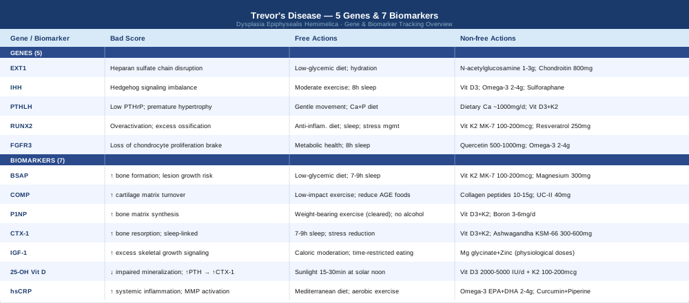
7 Biomarkers Worth Tracking in Trevor's Disease
Most biomarker panels ordered by orthopedic teams for skeletal conditions focus on calcium and phosphate at most. For Trevor's disease, that is not enough. The condition involves active cartilage proliferation, disordered endochondral ossification, and — in many patients — subclinical inflammation that influences how the epiphyseal lesions behave over time. The seven biomarkers below give you a window into each of these domains. None of them replaces imaging. All of them can add clinically meaningful context.
Biomarker 1: Bone-Specific Alkaline Phosphatase (BSAP)
Why it matters: Bone-specific alkaline phosphatase is secreted by osteoblasts and reflects the rate at which new bone matrix is being synthesized. In Trevor's disease, the abnormal epiphyseal masses involve active endochondral ossification — cartilage being converted into bone — and elevated BSAP suggests that this process is biologically active rather than quiescent. Tracking BSAP over time is more informative than a single measurement, since rising values may indicate a period of accelerated lesion growth. Age matters significantly here: BSAP is physiologically elevated during childhood growth spurts, so values must be interpreted against age-adjusted reference ranges, not adult norms.
How to measure it: BSAP is measured via a simple fasting blood draw using an immunoassay or ELISA. Most major reference laboratories offer it. Cost typically ranges from $40 to $90, depending on whether it is bundled with a broader bone-marker panel. Standard total alkaline phosphatase (a common metabolic panel component) is less specific; request bone-specific ALP explicitly. Adult reference range is approximately 11.6 to 30.6 U/L, but again, pediatric ranges differ substantially and must be used for children.
If the score is bad, the plan without supplements: An elevated BSAP in the context of Trevor's disease warrants follow-up imaging to determine whether a known lesion is enlarging or a new site is becoming active. From a lifestyle standpoint, reducing hyperinsulinemia — through lower-glycemic dietary patterns and consistent physical activity — decreases IGF-1 signaling, which in turn reduces the osteoblastic drive. Prioritizing seven to nine hours of sleep per night matters because deep sleep is when cortisol is lowest and bone remodeling is most ordered. Impact-loading exercise, while valuable for bone density in healthy individuals, should be discussed with the treating orthopedic surgeon before starting, given the mechanical stress it places on affected epiphyses.
If the score is bad, the plan with supplements or equipment: Vitamin K2 (MK-7 form) at 100 to 200 mcg per day has the best evidence for regulating osteocalcin carboxylation and modulating osteoblast activity. It is typically cycled continuously rather than in short bursts, and its main safety consideration is interaction with vitamin K antagonist anticoagulants (e.g., warfarin). Magnesium glycinate at 300 to 400 mg per day supports alkaline phosphatase enzyme function and bone matrix quality. Take it in the evening to avoid gastrointestinal effects and separate from calcium supplementation. If zinc is low, 15 to 25 mg of zinc bisglycinate daily supports osteoblast function as well.
Biomarker 2: Cartilage Oligomeric Matrix Protein (COMP)
Why it matters: Cartilage oligomeric matrix protein is a pentameric glycoprotein released into the bloodstream when cartilage matrix is being turned over — either through normal chondrocyte metabolism or as a result of pathological degradation and remodeling. It is arguably the single most specific circulating biomarker of cartilage activity currently available in clinical practice. Given that Trevor's disease is fundamentally a disease of cartilage — specifically, a disorder of epiphyseal cartilage that undergoes dysregulated proliferation and endochondral transformation — COMP levels give a real-time signal of how metabolically active that cartilage is. Elevated serum COMP is associated with progressive cartilage conditions including hip dysplasia, osteoarthritis, and other epiphyseal disorders. Published research supports its use as a prognostic biomarker in skeletal cartilage diseases.
How to measure it: COMP is measured from serum via ELISA, typically at specialty reference labs (e.g., Eurofins, Mayo Clinic Laboratories). It is not a standard panel item, so you may need to request it specifically. Cost ranges from $100 to $220. Normal adult serum COMP is generally below 12 U/L, but reference ranges vary by laboratory. Pediatric norms are less standardized and should be discussed with the ordering physician.
If the score is bad, the plan without supplements: Elevated COMP calls for reducing mechanical stress on affected joints and discussing with your orthopedic team whether the timing of surgical intervention needs reassessment. Sleep quality is disproportionately important here: chondrocyte repair and proteoglycan synthesis occur predominantly during restorative sleep. Minimizing dietary advanced glycation end products (AGEs) — which means reducing charred, fried, and ultra-processed foods — reduces RAGE-mediated cartilage inflammation. Low-impact movement (swimming, cycling) maintains synovial fluid circulation to cartilage without compressive loading.
If the score is bad, the plan with supplements or equipment: Hydrolyzed collagen peptides at 10 to 15 grams per day taken with vitamin C have shown, in randomized trials, to increase serum COMP-favorable markers and support cartilage matrix synthesis in cartilaginous joint conditions. Consume 30 to 60 minutes before mechanical activity for optimal tissue delivery. Side effects are minimal. Undenatured type II collagen (UC-II) at 40 mg per day is a distinct mechanism: it works via oral tolerance to downregulate collagen-reactive T cells. No significant cycling required. Some practitioners also use low-level laser therapy (LLLT) devices (Class IIIb/IV) targeting affected joints to support mitochondrial activity in chondrocytes; see the complementary approaches section for more detail.
Biomarker 3: P1NP (Procollagen Type I N-Terminal Propeptide)
Why it matters: P1NP is widely considered the most sensitive and reproducible serum marker of bone formation. It reflects how actively osteoblasts are synthesizing type I collagen — the predominant structural protein of bone matrix. The International Osteoporosis Foundation and ISCD jointly designated P1NP and CTX-1 as the two reference bone turnover markers for clinical use and research, precisely because of their strong correlation with bone formation rates and their responsiveness to interventions. In Trevor's disease, tracking P1NP over time — particularly alongside CTX-1 — gives you the bone-remodeling ratio: how much bone is being built relative to how much is being broken down. Disproportionate formation activity may signal active lesion development.
How to measure it: P1NP is measured from a morning fasting blood sample (bone turnover markers are subject to diurnal variation, with levels being highest in the early morning). Most major labs offer it; cost is approximately $50 to $100. It is important to test under standardized conditions — same time of day, same fasting state — to make longitudinal comparisons meaningful.
If the score is bad, the plan without supplements: Elevated P1NP without a corresponding rise in CTX-1 suggests an uncoupled bone formation response — new bone being added faster than old bone is being resorbed, which may reflect active lesion-associated ossification. From a lifestyle standpoint, weight-bearing exercise of appropriate intensity (cleared by your orthopedic team) normalizes bone remodeling coupling. Reducing alcohol intake, which disrupts osteoblast function, and eliminating smoking are straightforward steps that have dose-response relationships with bone formation markers.
If the score is bad, the plan with supplements or equipment: The key nutrient driving appropriate P1NP modulation is vitamin D3 (discussed in more detail in Biomarker 6). Specifically, combined D3 + K2 supplementation supports orderly mineralization of newly formed collagen matrix. Boron at 3 to 6 mg per day modulates sex steroid metabolism and has been shown in small human studies to influence bone formation markers. Cycle every 8 weeks on, 2 weeks off to avoid accumulation. Strontium ranelate — once used clinically — is now restricted in most countries due to cardiovascular concerns and should not be self-administered.
Biomarker 4: CTX-1 (C-Terminal Telopeptide of Type I Collagen)
Why it matters: CTX-1 — sometimes called beta-CrossLaps — is the partner marker to P1NP. Where P1NP reflects how fast bone is being built, CTX-1 reflects how fast osteoclasts are breaking it down. The ratio of P1NP to CTX-1 provides a functional picture of whether bone remodeling is balanced, dominated by formation, or dominated by resorption. Elevated CTX-1 in isolation suggests increased bone breakdown, which can weaken the bone adjacent to Trevor's disease lesions and contribute to structural instability. Chronic elevation of CTX-1 is also closely linked to elevated cortisol and sleep deprivation — making lifestyle inputs directly legible through this biomarker.
How to measure it: CTX-1 is measured from a morning fasting serum sample (it is highly sensitive to recent food intake; even coffee can suppress it acutely). Some laboratories offer a urine CTX-1/creatinine ratio as an alternative, though serum is more standardized. Cost is $40 to $80. Morning fasting is non-negotiable for valid comparisons over time.
If the score is bad, the plan without supplements: The most powerful lifestyle intervention for elevated CTX-1 is sleep quality improvement. A study published in the Journal of Clinical Endocrinology and Metabolism demonstrated that CTX-1 rises significantly after sleep restriction. Targeting seven to nine hours of sleep per night, reducing ambient light after 9 PM, and maintaining a cool sleeping environment (18 to 20°C) are all practical steps. Reducing perceived stress — through whatever mechanism is realistic for the individual — lowers cortisol, the primary hormone that elevates osteoclast activity. Vigorous resistance exercise (again, cleared by the orthopedic team) acutely elevates CTX-1 but normalizes the remodeling cycle long-term.
If the score is bad, the plan with supplements or equipment: Calcium from whole foods (dairy, leafy greens, fortified foods) suppresses parathyroid hormone-driven CTX-1 elevation more safely than high-dose supplement calcium, which has raised cardiovascular concerns in older individuals. Vitamin D3 in combination with K2 regulates osteoclast differentiation and is the most important pairing here. If cortisol-driven CTX-1 elevation is suspected, ashwagandha (KSM-66) at 300 to 600 mg per day has demonstrated cortisol-lowering effects in double-blind RCTs; cycle 8 weeks on, 2 weeks off; avoid during pregnancy.
Biomarker 5: IGF-1 (Insulin-Like Growth Factor 1)
Why it matters: Insulin-like growth factor 1 is the primary mediator of growth hormone's anabolic effects on the skeleton. It stimulates chondrocyte proliferation, bone matrix synthesis, and osteoblast differentiation. In children and adolescents — the population most commonly affected by Trevor's disease — IGF-1 is physiologically elevated and plays a central role in the endochondral ossification process that governs normal long-bone growth. When IGF-1 signaling is excessive or poorly regulated, it may amplify abnormal proliferative signaling in a dysplastic epiphysis. Tracking IGF-1 over time provides insight into the hormonal environment driving skeletal growth, and helps distinguish age-appropriate levels from pathological elevation.
How to measure it: IGF-1 is measured from a standard blood draw; no specific fasting is required, though morning sampling is conventional. Cost ranges from $50 to $120. Reference ranges are highly age- and sex-dependent — a level that is normal for a 12-year-old would be high for a 40-year-old. Always compare against pediatric reference charts, not adult ranges.
If the score is bad, the plan without supplements: Chronically elevated IGF-1 (above the 95th percentile for age) warrants endocrinological evaluation to rule out growth hormone excess. In the absence of a pituitary or endocrine disorder, the most effective lifestyle modifiers are caloric moderation (IGF-1 rises with energy surplus), protein intake normalization (very high protein intake chronically elevates IGF-1), and improving sleep architecture (IGF-1 is partly regulated by nocturnal growth hormone pulses). Time-restricted eating patterns (e.g., a 10 to 12-hour eating window) have demonstrated modest IGF-1-lowering effects in intervention studies.
If the score is bad, the plan with supplements or equipment: Direct IGF-1 suppression via supplementation is not advisable without medical supervision — this is a domain for endocrinologists, not self-directed supplementation. However, magnesium glycinate and zinc bisglycinate at physiological doses support appropriate, rather than excessive, IGF-1 signaling. Both are cofactors in growth hormone receptor activation. Avoid high-dose isolated amino acid supplements (arginine, glutamine in gram doses), which are sometimes marketed for growth hormone release and may inappropriately elevate IGF-1.
Biomarker 6: 25-Hydroxyvitamin D
Why it matters: Vitamin D is not optional background noise in bone health — it is a prerequisite for almost every aspect of calcium and phosphate metabolism, osteoblast function, and immune regulation that affects skeletal tissue. Deficiency disrupts the normal mineralization of newly formed bone matrix, weakens growth plate function, and elevates PTH, which in turn elevates CTX-1 and drives excessive bone resorption. In a condition like Trevor's disease, where maintaining bone quality in the non-affected portions of the skeleton matters for compensating around structural lesions, vitamin D sufficiency is foundational. Clinicians like Peter Attia recommend a target of 40 to 60 ng/mL (100 to 150 nmol/L), which is substantially higher than the deficiency threshold of 20 ng/mL used in conventional medicine. This NIH Office of Dietary Supplements factsheet summarizes the evidence base.
How to measure it: The standard test is serum 25-hydroxyvitamin D, available from any laboratory. Cost is $30 to $60 (often covered by insurance with a documented deficiency or at-risk status). Retest every 90 days after starting supplementation until the target range is achieved, then twice annually.
If the score is bad, the plan without supplements: Direct sunlight exposure to the face, arms, and legs for 15 to 30 minutes at solar noon (when UV index is at least 3) is the most physiological way to raise 25-OH vitamin D. Skin tone, geographic latitude, and season heavily influence conversion efficiency. Dietary sources — wild salmon, mackerel, egg yolks, vitamin D-fortified dairy or plant milks — contribute but rarely normalize deficiency alone.
If the score is bad, the plan with supplements or equipment: Vitamin D3 (cholecalciferol) at 2,000 to 5,000 IU per day is appropriate for most deficient adults; pediatric dosing should be guided by a physician and body weight. Always pair with vitamin K2 (MK-7) at 100 to 200 mcg daily to direct calcium to bone rather than soft tissue. Take both with the fattiest meal of the day for optimal absorption. Continuous use is standard; there is no need to cycle unless serum levels rise above 80 ng/mL, at which point dose reduction should be considered. Recheck serum levels at 90 days. Side effects at standard doses are rare; toxicity requires very high doses (typically above 10,000 IU/day for extended periods).
Biomarker 7: hsCRP (High-Sensitivity C-Reactive Protein)
Why it matters: High-sensitivity CRP is the most widely accessible measure of systemic low-grade inflammation. While Trevor's disease is not primarily an inflammatory condition in the way that rheumatoid arthritis or lupus are, subclinical chronic inflammation interacts with the cartilage signaling pathways involved in epiphyseal development. Elevated interleukin-1β and TNF-α — the upstream mediators reflected by elevated hsCRP — directly inhibit chondrocyte anabolism and promote matrix metalloproteinase activity, accelerating cartilage degradation. Peter Attia advocates for an hsCRP target below 0.5 to 1.0 mg/L for optimal metabolic and cardiovascular health, well below the conventional "normal" cutoff of 3.0 mg/L. For someone with an active skeletal condition, keeping the inflammatory environment as quiet as possible makes biological sense.
How to measure it: hsCRP is a blood test available from virtually any laboratory. Cost is $20 to $50. It is standard on many comprehensive metabolic or cardiovascular panels. Because CRP rises with any acute infection or injury, avoid testing within two weeks of illness, vaccination, or surgery. A resting baseline value is most useful.
If the score is bad, the plan without supplements: The most effective non-pharmacological strategies for lowering hsCRP are weight normalization (adipose tissue produces inflammatory cytokines), dietary elimination of ultra-processed foods and refined carbohydrates, consistent aerobic exercise (even moderate walking reduces CRP significantly over 8 to 12 weeks), improved sleep quality, and stress reduction. A Mediterranean-style dietary pattern has more RCT support for CRP reduction than any specific supplement.
If the score is bad, the plan with supplements or equipment: Omega-3 fatty acids (EPA + DHA) at 2 to 4 grams per day from high-quality fish oil or algal oil have robust evidence for reducing hsCRP and inflammatory cytokines. The NIH ODS review of omega-3 fatty acids summarizes the evidence. Take daily with food; no cycling required. Common side effects are fishy aftertaste and loose stools at higher doses; enteric-coated versions minimize both. Curcumin with piperine (500 to 1,000 mg curcuminoids with 5 to 10 mg piperine per day) has demonstrated anti-inflammatory effects in several RCTs; take with food. Avoid high-dose curcumin if on blood thinners. Berberine at 500 mg twice daily also reduces inflammatory markers through AMPK activation; cycle 8 weeks on, 4 weeks off, and monitor liver enzymes with extended use.
Using These 7 Biomarkers Together
The value of tracking these markers lies not in any single number but in the pattern across them. A useful starting framework: run a full panel at baseline, then recheck every 90 to 120 days after making targeted interventions. Look for the P1NP-to-CTX-1 ratio to narrow (indicating better-coupled remodeling), COMP to trend downward (indicating reduced cartilage turnover), and hsCRP to fall below 1 mg/L. If BSAP and P1NP remain elevated despite lifestyle and nutritional optimization, bring the data to your orthopedic or metabolic specialist as a reason to re-examine imaging frequency or timing.
The goal is not to self-treat Trevor's disease with supplements. The goal is to give the clinicians responsible for surgical decision-making a richer biological picture — and to ensure that the tissue environment is as supportive as possible before and after any intervention.
The Genes Behind Epiphyseal Overgrowth in Trevor's Disease
Trevor's disease does not yet have a definitively identified monogenic cause. Most cases are sporadic, and genetic testing does not currently include a "Trevor's disease gene panel." What research into skeletal dysplasias, growth plate biology, and related conditions does provide is a clear picture of the molecular pathways that govern epiphyseal cartilage development — and therefore the pathways most likely disrupted when those processes go wrong. The five genes below are central to those pathways. Understanding their roles and checking for relevant variants through clinical-grade genetic testing or direct-to-consumer platforms like 23andMe or Nebula Genomics (interpreted by a clinical geneticist) can be informative, though the evidence linking specific variants to Trevor's disease remains incomplete.
Gene 1: EXT1 — The Heparan Sulfate Gate
What it does: Exostosin Glycosyltransferase 1 (EXT1, NCBI Gene ID 2131) encodes a glycosyltransferase enzyme critical for heparan sulfate chain elongation. Heparan sulfate proteoglycans are essential co-receptors for multiple growth factor signaling pathways — including FGF, BMP, and Hedgehog signaling — that regulate chondrocyte proliferation and differentiation in the growth plate. Loss-of-function mutations in EXT1 cause hereditary multiple exostoses (HME), a condition characterized by multiple osteochondromas (benign bone-capped cartilaginous tumors) that share histological features with Trevor's disease lesions. This mechanistic overlap suggests that EXT1-related heparan sulfate disruption may be relevant to the pathogenesis of some Trevor's disease cases, though this connection has not been formally established in the literature via published genetic studies.
Evidence level: mechanistic/analogical (HME research); direct Trevor's disease genetic data is early and limited.
If the gene is bad, the plan without supplements: Heparan sulfate proteoglycan function is sensitive to metabolic health. Hyperglycemia and elevated insulin directly disrupt proteoglycan synthesis through oxidative stress mechanisms. A low-glycemic, whole-food dietary pattern reduces this burden. Avoiding chronic low-level dehydration (proteoglycans require water for their space-filling function in cartilage matrix) and reducing pro-inflammatory dietary patterns are the primary non-supplemental levers. Physical activity that promotes synovial fluid circulation — walking, cycling, swimming — supports cartilage proteoglycan hydration without adding excessive mechanical load.
If the gene is bad, the plan with supplements or equipment: N-acetylglucosamine (NAG) at 1 to 3 grams per day provides a substrate for heparan sulfate biosynthesis via the hexosamine pathway. It is distinct from glucosamine sulfate. Take daily with food; no specific cycling required. Gastrointestinal tolerance is generally good. Chondroitin sulfate at 800 to 1,200 mg per day provides a sulfated glycosaminoglycan substrate relevant to cartilage matrix support. Combined collagen peptide and chondroitin formulations (as studied in osteoarthritis RCTs) provide the broadest substrate coverage.
Gene 2: IHH — The Hedgehog Signal
What it does: Indian Hedgehog Signaling Molecule (IHH, NCBI Gene ID 3549) is a secreted signaling protein produced by prehypertrophic chondrocytes in the growth plate. It acts as one half of the crucial IHH–PTHrP negative feedback loop that controls the pace of chondrocyte maturation during endochondral ossification. When IHH signaling is disrupted — either through gain-of-function or loss-of-function variants — the timing and location of chondrocyte hypertrophy become disordered, potentially contributing to ectopic or asymmetric epiphyseal cartilage proliferation consistent with Trevor's disease anatomy. This is one of the most mechanistically compelling pathways to examine in epiphyseal dysplasias.
Evidence level: strong mechanistic basis from growth plate biology; clinical genetic confirmation in Trevor's disease is pending.
If the gene is bad, the plan without supplements: The IHH pathway is modulated by mechanical forces on growth plate cartilage — which means the type and intensity of physical activity during growth matters. Low-to-moderate compressive loading has a normalizing effect on IHH expression in healthy chondrocytes. Extreme loading during the growth years may disrupt this balance. Work with a pediatric physical therapist to identify appropriate activity types and intensities. Sleep sufficiency is also critical: growth hormone pulses during deep sleep drive orderly IHH-PTHrP cycling.
If the gene is bad, the plan with supplements or equipment: There is no direct dietary or supplement-based intervention proven to normalize IHH pathway function. The indirect approaches are those that support overall Hedgehog pathway health: vitamin D3 (IHH downstream targets are partly regulated by VDR signaling) and omega-3 fatty acids (reduce inflammatory suppression of hedgehog co-receptors). Sulforaphane from broccoli sprouts at 50 to 100 mcg glucoraphanin daily has demonstrated modulation of hedgehog pathway activity in cancer research (early evidence; relevance to Trevor's disease is theoretical). No cycling required at vegetable-dose levels.
Gene 3: PTHLH — The Brake Pedal
What it does: Parathyroid Hormone Like Hormone (PTHLH, NCBI Gene ID 5744) — more commonly known as PTHrP — is the counter-signal to IHH in the growth plate feedback loop. Produced by the periarticular perichondrium, PTHrP keeps chondrocytes proliferating and delays their hypertrophy, preventing premature ossification. When the IHH-PTHrP balance is disrupted — either through a variant that reduces PTHrP activity or through a receptor defect that diminishes PTHrP responsiveness — chondrocytes progress to hypertrophy too quickly and in dysregulated spatial patterns. This can produce exactly the kind of asymmetric epiphyseal proliferation and early ossification seen in Trevor's disease.
Evidence level: mechanistic, based on growth plate signaling research; direct Trevor's disease correlation is theoretical.
If the gene is bad, the plan without supplements: PTHrP is responsive to mechanical strain — moderate joint loading helps maintain appropriate PTHrP expression in periarticular tissues. Avoiding prolonged immobilization is important; even children with Trevor's disease should maintain as much gentle movement as their surgical history allows. Adequate dietary calcium and phosphate intake is essential, as mineral imbalances alter PTHrP secretion through secondary hyperparathyroidism mechanisms.
If the gene is bad, the plan with supplements or equipment: Calcium from whole foods (dairy, fortified plant milks, leafy greens) at approximately 1,000 mg per day for children (1,300 mg for adolescents) normalizes PTH-related signaling without the risks associated with high-dose isolated calcium supplements. Vitamin D3 + K2 remains the cornerstone: vitamin D ensures calcium absorption and PTHrP receptor sensitivity. There are no direct PTHrP-modulating supplements available outside clinical research settings.
Gene 4: RUNX2 — The Bone Program Switch
What it does: RUNX Family Transcription Factor 2 (RUNX2, NCBI Gene ID 860) is the master transcription factor that drives osteoblast differentiation and activates the genes responsible for bone matrix synthesis. It also promotes chondrocyte hypertrophy — the penultimate step before cartilage converts to bone in endochondral ossification. Overactivation of RUNX2, or loss of the normal regulatory brakes on RUNX2 (including the Twist family proteins and HDAC4), leads to premature and excessive endochondral ossification. In the context of a dysplastic epiphysis, abnormal RUNX2 activity levels could explain why cartilaginous epiphyseal masses in Trevor's disease progressively ossify in a disordered fashion.
Evidence level: mechanistic, based on skeletal development research; specific RUNX2 variants have not been systematically studied in Trevor's disease.
If the gene is bad, the plan without supplements: RUNX2 activity is regulated by mechanical loading, oxidative stress, and inflammatory cytokines. The same foundational habits apply: anti-inflammatory diet, sleep optimization, stress management. Reducing chronic low-grade inflammation (and therefore NF-κB activation) helps prevent the inflammatory upregulation of RUNX2 that can accelerate pathological ossification.
If the gene is bad, the plan with supplements or equipment: Vitamin K2 (MK-7) is the most directly relevant supplement here. Research has shown that K2 influences RUNX2-driven osteocalcin transcription and modulates osteoblast differentiation — the K2/carboxylation axis helps direct bone formation away from soft tissue and toward appropriate skeletal sites. At 100 to 200 mcg MK-7 per day, it is safe, well-tolerated, and compatible with long-term use in the absence of anticoagulant therapy. Resveratrol at 250 to 500 mg per day activates SIRT1, which deacetylates and inactivates excessive RUNX2; early evidence from cell and animal studies is promising, though human data in skeletal conditions is limited. Cycle 8 weeks on, 2 weeks off.
Gene 5: FGFR3 — The Chondrocyte Proliferation Brake
What it does: Fibroblast Growth Factor Receptor 3 (FGFR3, NCBI Gene ID 2261) is uniquely important among growth factor receptors in the skeleton because it acts as a negative regulator of chondrocyte proliferation. While most growth factor receptors promote growth when activated, FGFR3 signaling through the STAT1 and MAPK pathways inhibits chondrocyte proliferation and bone elongation. This is why gain-of-function FGFR3 mutations cause achondroplasia and thanatophoric dysplasia — conditions of severe growth suppression. Conversely, when FGFR3 signaling is reduced or dysregulated in a specific epiphyseal region, the braking mechanism on chondrocyte proliferation is lost, potentially allowing the asymmetric, region-specific overgrowth characteristic of Trevor's disease to occur. This is a biologically compelling hypothesis, though again: direct genetic studies specifically in DEH are limited.
Evidence level: strong mechanistic basis from achondroplasia/FGFR3 research; application to Trevor's disease is inferential.
If the gene is bad, the plan without supplements: There is no direct lifestyle intervention to correct FGFR3 signaling. The indirect approach is to reduce FGF ligand oversupply: since FGF pathway activity is amplified by obesity, hyperglycemia, and chronic inflammation, maintaining metabolic health minimizes the likelihood of aberrant FGF signaling. Ensuring adequate sleep is relevant because growth hormone and FGF signaling interact during nocturnal bone growth cycles.
If the gene is bad, the plan with supplements or equipment: Mead Johnson (inositol hexaphosphate / IP6) and quercetin at 500 to 1,000 mg per day have shown FGFR pathway-modulating activity in cell studies, though human evidence in skeletal conditions is absent. These are low-risk additions at standard doses, but should be framed as experimental rather than evidence-based for this specific application. The more evidence-backed approach is omega-3 supplementation (2 to 4 g EPA+DHA), which modulates receptor tyrosine kinase sensitivity broadly and reduces inflammatory amplification of FGF signaling.
What Peter Attia's Approach to Skeletal Health Reveals About Trevor's Disease
Peter Attia's Outlive: The Science and Art of Longevity (2023) is not written about Trevor's disease — but its framework for musculoskeletal health and metabolic optimization contains insights that translate directly to the biology underlying this condition. Attia's core argument is that medicine as currently practiced is reactive, intervening only after disease has become overtly symptomatic, and that the most powerful moves happen much earlier. For Trevor's disease, where the orthopedic team typically acts only when a lesion is large enough to cause pain or deformity, this early-action philosophy offers a genuinely different frame.
1. Bone mineral density is a lagging indicator
Attia argues that by the time bone density shows up as abnormal on a DXA scan, years of metabolic mismanagement have already occurred. For Trevor's disease, the same logic applies to imaging: by the time a lesion is visible enough to require surgical planning, its biological activity has been running for months or years. Earlier biomarker surveillance — as outlined in the previous section — is the equivalent of what Attia calls "early warning signal monitoring."
2. The musculoskeletal system is the most under-appreciated longevity organ
Attia dedicates significant space in Outlive to the argument that muscle and bone health are the single strongest predictors of healthy aging — not cardiovascular fitness, not metabolic panels. For Trevor's disease patients, this means that preserving muscle strength around affected joints is not cosmetic or supplementary: it is structurally necessary. Stronger surrounding musculature reduces joint stress on an epiphysis that may have already compromised structural integrity.
3. Protein intake is systematically underestimated
Outlive recommends 1.6 to 2.2 grams of protein per kilogram of body weight per day — far above most dietary guidelines. For bone and cartilage repair, collagen synthesis requires both adequate total protein and specific amino acids: glycine, proline, and hydroxyproline. This is not adequately met by standard dietary advice to "eat enough protein."
4. Zone 2 aerobic training changes bone metabolism
Attia's emphasis on Zone 2 training (low-intensity aerobic exercise at approximately 60 to 70% of max heart rate for extended periods) is supported by evidence showing that it reduces systemic inflammation, improves mitochondrial density in osteocytes, and normalizes bone remodeling markers over time. For Trevor's disease patients with joint restrictions, Zone 2 exercise in non-weight-bearing formats (cycling, swimming) provides these benefits without compressive risk to affected epiphyses.
5. Sleep is the most powerful bone-repair tool available
Attia frames sleep as the period during which the body does its most intensive repair work — including bone remodeling and growth hormone secretion. CTX-1 rises acutely with sleep deprivation. Growth hormone — the primary upstream driver of IGF-1 and skeletal growth — is secreted almost entirely during slow-wave sleep. This is not a soft claim: it has significant implications for how seriously sleep quality should be prioritized in pediatric Trevor's disease patients.
6. Insulin sensitivity is a master regulator of skeletal health
Elevated insulin and hyperglycemia increase the production of advanced glycation end products, which cross-link collagen in bone and cartilage matrix and make it stiffer and more brittle. Attia's emphasis on maintaining tight fasting glucose (below 90 mg/dL) and insulin sensitivity directly supports cartilage matrix quality maintenance.
7. Omega-3 dosing matters more than most people realize
Attia recommends at least 2 grams of combined EPA + DHA per day, and often more in high-inflammatory states. The dose commonly found in retail "fish oil" capsules (300 to 500 mg per capsule) falls far short of what the research demonstrating meaningful anti-inflammatory effects actually used. High-quality, triglyceride-form fish oil at 2 to 4 grams per day is the target.
8. Diagnostic tools should be used proactively, not reactively
Outlive's "Medicine 3.0" framework calls for using laboratory and imaging tools before symptoms demand them, in order to detect trajectory changes early. For Trevor's disease, this means establishing a baseline biomarker panel at diagnosis — not waiting for pain or functional decline to prompt testing.
9. Pharmacological interventions exist but carry underappreciated trade-offs
Bisphosphonates and other bone-modifying agents occasionally appear in discussions of skeletal dysplasias. Attia's framework appropriately highlights that these drugs have long skeletal half-lives and downstream effects on bone remodeling that are not fully benign. They should not be considered without specialist guidance and should never be self-initiated.
10. The relationship between metabolic health and skeletal outcomes is bidirectional
Attia's synthesis of the research makes clear that skeletal disease worsens metabolic health (through reduced mobility, inflammation, and hormonal disruption), and poor metabolic health accelerates skeletal disease. For Trevor's disease, this means that optimizing the metabolic environment is not peripheral to orthopedic management — it is directly relevant to how the condition progresses and how well patients recover from surgical intervention.
Complementary Approaches With Relevant Evidence
No complementary modality will excise an osteocartilaginous lesion. But for the pain, mobility limitation, and recovery challenges that accompany Trevor's disease — especially in the post-surgical period — several evidence-supported approaches offer meaningful adjunctive benefit.
Low-Level Laser Therapy (LLLT) / Photobiomodulation
Photobiomodulation uses low-intensity red and near-infrared light (typically 630 to 1064 nm wavelengths) to stimulate mitochondrial activity in target tissues via cytochrome c oxidase. In musculoskeletal applications, this translates to accelerated tissue repair, reduced inflammatory cytokine production, and improved local microcirculation. For Trevor's disease specifically, its relevance spans two domains: post-surgical wound and bone healing, and chronic joint pain management in areas where lesion-related deformity generates ongoing discomfort. A growing body of clinical trials supports LLLT for bone and cartilage healing applications.
A systematic review published in Lasers in Medical Science (2017) and subsequent meta-analyses have demonstrated reduced pain and improved function in knee osteoarthritis using LLLT at 5 to 50 mW/cm² with doses of 2 to 8 J/cm² per session. While Trevor's disease is not osteoarthritis, the tissue targets — cartilage and periarticular bone — are the same. Sessions typically last 5 to 15 minutes per site, three times per week for 4 to 8 weeks, then maintenance as needed.
For practical application, a Class IIIb or Class IV LLLT device (808 nm or 904 nm) used by a trained physiotherapist or physical medicine specialist is the appropriate setting. Home devices exist at lower power levels but have less evidence. Do not apply directly over active surgical wounds. The treating orthopedic surgeon should be informed before starting photobiomodulation, particularly in the post-surgical recovery period.
Mindfulness Meditation and MBSR
Mindfulness-Based Stress Reduction (MBSR), developed by Jon Kabat-Zinn, is an 8-week structured program combining mindfulness meditation, body scan, and gentle movement. Its relevance to Trevor's disease is primarily in the domain of chronic pain management and the psychological burden of living with a rare, chronic skeletal condition — particularly significant for parents of affected children, and for adult patients navigating ongoing pain and repeat surgical interventions. MBSR has perhaps the most robust evidence base of any mind-body intervention for chronic pain, having been validated in multiple systematic reviews and RCTs.
A landmark 2011 RCT in JAMA Internal Medicine demonstrated that MBSR produced clinically significant reductions in pain intensity and pain interference in chronic low back pain, with effects sustained at 26 weeks. For chronic musculoskeletal pain conditions more broadly, a 2021 Cochrane-adjacent meta-analysis found consistent moderate-quality evidence for pain reduction. The pain reduction mechanism involves downregulation of the brain's default mode network, reduced catastrophizing, and changes in cortisol regulation.
For practical application: the standard 8-week MBSR program is available in-person or via validated online platforms (including those based on the original UMass Medical School curriculum). For children with Trevor's disease, developmentally adapted mindfulness programs (MindUP, Mindful Schools) are appropriate. Even daily practice of 10 to 20 minutes shows measurable pain and anxiety reduction within 4 to 8 weeks. There are no meaningful contraindications.
Yoga
Yoga — particularly gentle and restorative styles — is relevant to Trevor's disease for its effects on joint mobility preservation, proprioceptive training around affected limbs, and pain modulation. Many Trevor's disease patients develop compensatory movement patterns in response to limb deformity or post-surgical restrictions, and yoga provides a structured, low-impact framework for working through range of motion limitations systematically. A substantial and growing RCT literature supports yoga for musculoskeletal pain and mobility outcomes.
A multicenter RCT published in Spine (2011) comparing yoga to conventional exercise for chronic musculoskeletal pain found yoga superior for pain reduction, function, and mood. For lower-extremity joint conditions specifically, studies using Iyengar yoga — a props-supported, alignment-focused style — have demonstrated improved balance, proprioception, and pain in hip and knee pathologies.
For Trevor's disease patients, the most practical approach is to work initially with a yoga therapist (C-IAYT certified) rather than in a general group class. This ensures that postures are modified for the specific limb affected and that compression on dysplastic epiphyses is avoided. Yin yoga (long-hold, low-load passive stretching) and restorative yoga (fully supported poses with props) are appropriate starting points. Avoid dynamic inversions and deep knee-loaded postures until surgical clearance is confirmed.
Massage Therapy
Manual massage therapy for Trevor's disease serves a specific, practical role: managing the soft tissue tension, lymphatic congestion, and compensatory muscle hypertonicity that develop around affected joints, particularly post-surgically. Patients with lower-extremity Trevor's disease often develop muscle imbalances in the hip abductors, quadriceps, and calf muscles as they load-compensate around a deformed ankle or knee. Structural massage that addresses these secondary soft tissue changes can improve gait quality, reduce referred pain, and support post-surgical rehabilitation. Evidence for massage in musculoskeletal conditions is reviewed in multiple systematic analyses.
A systematic review in the Journal of Pain (2015) found massage therapy to provide short-to-medium-term benefit for musculoskeletal pain conditions with effect sizes comparable to exercise therapy for pain and function outcomes. For post-surgical orthopedic recovery, manual lymphatic drainage in the first 4 to 8 weeks reduces edema, accelerates tissue remodeling, and improves scar mobility — particularly relevant after lower-extremity surgeries.
For practical application: sessions of 45 to 60 minutes every one to two weeks by a registered massage therapist (RMT) with experience in orthopedic or post-surgical massage are the realistic target. Manual lymphatic drainage (MLD) requires a specifically trained practitioner. In the post-surgical period, massage should not begin until wound integrity is confirmed. Avoid direct massage over an unresected lesion without physician guidance, as the tissue response is unpredictable.
Conclusion
Trevor's disease demands more from its management than a schedule of imaging appointments and surgical decisions. The biology underlying epiphyseal overgrowth — involving cartilage matrix metabolism, growth plate signaling, and the inflammatory environment — is measurable, and meaningful parts of it are modifiable. Seven biomarkers give you a practical window into what is happening in bone and cartilage between clinical visits. Five genes point to the signaling pathways most likely disrupted and suggest mechanistic directions for compensation. The lifestyle, nutritional, and complementary frameworks reviewed here will not replace the orthopedic care that Trevor's disease requires — but they can make the biological terrain more favorable and the recovery from intervention more complete.
The next smart step is to bring a specific request to your clinician: run a baseline biomarker panel including BSAP, COMP, P1NP, CTX-1, IGF-1, 25-OH vitamin D, and hsCRP. From there, track the trends over time and use the data to have more targeted conversations. Better information does not guarantee a better outcome, but it meaningfully improves the conditions under which good outcomes become possible.
Musculoskeletal Endocrine & Metabolic
Musculoskeletal: Bone Conditions Joint Conditions
Autoimmune: Inflammatory Conditions