This article contains AI generated content.
Tripartite Patella: 4 Genes And 7 Biomarkers To Track
If you've been told you have a tripartite patella, chances are it showed up almost by accident. A knee X-ray after a fall, an MRI ordered for something else, or a persistent ache along the front of the knee that finally got imaged properly. What you got back was a name for something most people have never heard of, followed by generic reassurance: it's usually harmless, rest if it hurts, come back if it doesn't improve.
That advice isn't wrong, but it isn't especially useful either. It doesn't explain why your kneecap developed this way, why some tripartite patellae stay silent for a lifetime while others flare up after a specific twist or fall, or what you can actually do — beyond waiting — to support the tissue around it. Generic knee-pain advice is built for the average patient, not for someone with a specific, congenital variation in how their patella ossified.
This article takes a narrower and more useful angle. Instead of repeating "rest and ice," it looks at the biological terrain around a tripartite patella: the blood-measurable indicators of bone formation, mineralization, and inflammation that plausibly influence whether the fibrous junctions between the patellar fragments stay stable or become a source of pain, and the small set of genes involved in how ossification centers form and fuse in the first place.
None of this rewrites the anatomy you were born with. But better information about bone metabolism, inflammation, and the developmental biology behind joint formation can support better decisions — about training load, nutrition, and when it's worth investigating further. This article covers seven biomarkers worth tracking, four genes with early but genuine relevance, a bone-health podcast episode worth your time, and a short set of complementary approaches with real supporting evidence for knee pain.
Summary
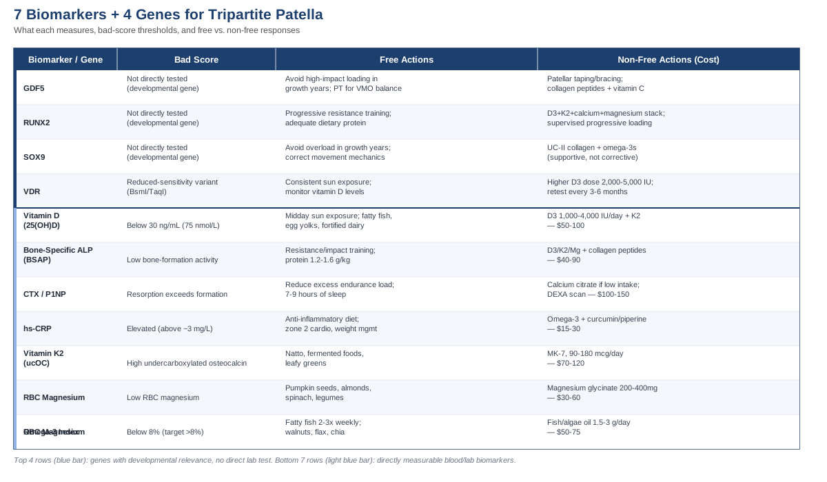
Most tripartite patellae never cause a single symptom — they're discovered incidentally and never need active management. But a meaningful minority become painful, usually after trauma, overuse, or a growth spurt that stresses the fibrous junctions holding the fragments together, and generic knee advice rarely explains what's actually happening at those junctions or what a person can do about it. This article walks through seven blood- and lab-based biomarkers — vitamin D, bone-specific alkaline phosphatase, the CTX/P1NP bone turnover pair, hs-CRP, vitamin K2 status, magnesium, and the omega-3 index — that reflect the bone-formation, mineralization, and inflammatory processes most relevant to how well those junctions are supported, plus exactly how to test each one, what a poor result plans out to (with and without supplements), and realistic cost ranges. It then covers four genes — GDF5, RUNX2, SOX9, and VDR — that shape how ossification centers form and fuse in the skeleton generally, with honest caveats about how much of that evidence is patella-specific versus extrapolated. A breakdown of a highly relevant bone-health podcast episode, and a short review of complementary approaches with real clinical evidence for knee pain, round out a genuinely practical way to think about a diagnosis that usually comes with far too little context.
What a Tripartite Patella Actually Is
The patella normally forms from a single ossification center that appears around age three to five and consolidates into one solid bone. In a small percentage of people, additional accessory ossification centers form — usually at the superolateral corner of the kneecap — and fail to fuse fully with the main body of the patella. When there are two separate fragments, it's called a bipartite patella; when there are three, it's tripartite. The fragments are usually held together by fibrocartilage or fibrous tissue rather than solid bone, which is the structural detail that matters most for anyone with symptoms.
The Oohashi classification, still the most widely used framework for describing these variants, groups them by the location of the accessory fragment and its relationship to the quadriceps and vastus lateralis attachments, which correlates with how likely a given pattern is to become symptomatic under load (Oohashi et al., Knee Surg Sports Traumatol Arthrosc, 2010). Multipartite patellae are estimated to occur in roughly 0.2 to 1.7 percent of the population, are more common in males, and are bilateral in a meaningful minority of cases — findings consistent across the case-series literature on symptomatic presentations (Symptomatic bipartite patella: three subtypes, three representative cases).
Most people never know they have one. When symptoms do appear, they typically follow direct trauma, a fall, repetitive loading in sport, or a rapid increase in training volume — the fibrous junction between fragments gets irritated or, rarely, traumatically separated, as described in a recent case report of a fall-related tripartite patella injury managed conservatively with rest, anti-inflammatories, and physical therapy (Tripartite Patella: A Rare Case Report, J Orthop Case Rep, 2025). That last detail is the throughline for everything below: this isn't a condition you reverse, but the biological environment around bone and connective tissue — how well it mineralizes, how it responds to load, how much background inflammation it's dealing with — is something you can actually measure and influence.
Seven Biomarkers Worth Tracking If You Have a Tripartite Patella
None of these biomarkers are specific tests "for" tripartite patella — no such test exists. What they reflect instead is the state of the bone-formation, mineralization, and inflammatory systems that plausibly determine how well the fibrous junctions between your patellar fragments tolerate load, and how quickly they recover from irritation. This is the same logic Peter Attia and Thomas Dayspring apply to cardiometabolic risk: track the mechanistic drivers, not just the symptom.
1. 25-Hydroxyvitamin D
Vitamin D governs intestinal calcium absorption and is required for normal bone mineralization; serum 25(OH)D below roughly 30 ng/mL (75 nmol/L) is associated with impaired mineralization of the skeleton (Effectiveness and safety of vitamin D in relation to bone health). For anyone managing a fibrous junction between bone fragments, adequate vitamin D is a baseline requirement, not an optional extra.
How to measure it
A standard serum 25(OH)D blood draw, available through a physician or direct-to-consumer lab. Cost typically runs $50 to $100 without insurance, often less if bundled into a routine panel.If the score is bad: the plan without supplements
Ten to thirty minutes of midday sun exposure on bare skin, several times a week, adjusted for skin tone and latitude; regular intake of fatty fish, egg yolks, and fortified dairy; outdoor weight-bearing activity, which supports both vitamin D synthesis and mechanical bone loading at once.If the score is bad: the plan with supplements or equipment
Vitamin D3, 1,000 to 4,000 IU daily depending on how deficient the baseline result is, taken with a fat-containing meal. Retest after eight to twelve weeks, then move to a maintenance dose and retest every six to twelve months. Pairing with vitamin K2 (see below) is common practice to direct absorbed calcium toward bone. Side effects are rare at these doses but sustained intake above 10,000 IU/day can cause hypercalcemia, nausea, and kidney stone risk — this is a supplement worth retesting rather than guessing on long-term.2. Bone-Specific Alkaline Phosphatase (BSAP)
BSAP is produced by active osteoblasts and reflects how actively new bone is being formed. It's a useful proxy for whether the bone-forming machinery around a fibrous patellar junction is working at a normal rate, particularly in adolescents and young adults whose accessory ossification centers may still have some capacity to consolidate.
How to measure it
A specialty blood test, not part of routine metabolic panels — typically ordered through a physician, endocrinologist, or direct-access lab. Cost is roughly $40 to $90.If the score is bad: the plan without supplements
Progressive resistance and impact training, which mechanically stimulates osteoblast activity; adequate dietary protein (roughly 1.2 to 1.6 g/kg body weight daily); avoiding smoking and excess alcohol, both of which suppress osteoblast function.If the score is bad: the plan with supplements or equipment
A vitamin D3, K2, and magnesium stack to support mineralization once formation is stimulated; collagen peptides (5 to 15 g/day, ideally with a source of vitamin C) as a building-block supplement. For equipment, supervised progressive loading — a leg press, controlled squat pattern, or a vibration plate for those who can't yet tolerate axial load — two to three times weekly. Build load gradually around a sensitive patella; the main side effect risk here is local irritation from progressing too quickly, not the supplements themselves.3. CTX and P1NP (Bone Turnover Pair)
CTX (C-terminal telopeptide) reflects bone resorption, while P1NP (procollagen type 1 N-terminal propeptide) reflects new bone formation; together they're the reference markers recommended by international bone-health standardization bodies for tracking whether remodeling is balanced or skewed toward breakdown (National Bone Health Alliance recommendations on CTX and P1NP standardization).
How to measure it
A fasting morning blood draw is recommended, since both markers show diurnal variation. Cost for the pair is typically $100 to $150; often ordered through rheumatology or endocrinology rather than primary care.If the score is bad: the plan without supplements
Reduce chronic high-volume endurance training without adequate recovery, which can skew turnover toward resorption; prioritize seven to nine hours of sleep, since most bone remodeling activity is concentrated overnight; manage chronic stress, which elevates cortisol and suppresses formation relative to resorption.If the score is bad: the plan with supplements or equipment
Calcium citrate or malate, 500 to 1,000 mg daily in split doses, only if dietary calcium intake is genuinely low; vitamin D3 and K2 as above. A DEXA scan is a reasonable complementary measurement if turnover markers are persistently abnormal. Prescription options such as bisphosphonates are only appropriate under physician supervision for diagnosed pathology — not a self-directed step. Excess supplemental calcium (as opposed to dietary) has been debated for cardiovascular effects, so food sources are the first-line approach.4. High-Sensitivity CRP (hs-CRP)
hs-CRP is a general marker of low-grade systemic inflammation. In knee osteoarthritis research, elevated hs-CRP correlates modestly but significantly with pain and functional limitation, even when it doesn't track closely with structural radiographic changes (Circulating C-reactive protein in osteoarthritis: a systematic review and meta-analysis). For anterior knee pain associated with a symptomatic tripartite patella, this same inflammatory pathway is a plausible contributor.
How to measure it
A standard hs-CRP blood test, widely available and often included in cardiometabolic panels. Cost is typically $15 to $30.If the score is bad: the plan without supplements
An anti-inflammatory whole-food diet with reduced ultra-processed food and refined sugar; regular moderate (zone 2) cardiovascular activity; consistent sleep; weight management to reduce mechanical and metabolic load on the knee.If the score is bad: the plan with supplements or equipment
Omega-3 fatty acids (see below) and curcumin with piperine, 500 to 1,000 mg standardized curcuminoids daily, cycled for eight to twelve weeks before reassessing hs-CRP. Side effects can include mild GI upset, and both omega-3s and curcumin have mild blood-thinning effects worth discussing with a physician if you're on anticoagulants.5. Vitamin K2 Status (via Undercarboxylated Osteocalcin)
Vitamin K2 is required to carboxylate osteocalcin so it can bind calcium into the hydroxyapatite matrix of bone. High levels of undercarboxylated osteocalcin (ucOC) indicate that circulating calcium isn't being efficiently directed into bone, and vitamin K2 supplementation has been shown to reduce ucOC and support bone mineral density in postmenopausal populations (Effect of vitamin K2 treatment on carboxylation of osteocalcin).
How to measure it
A ucOC blood test through a specialty lab, roughly $70 to $120. Not a routine test — you'll likely need to request it specifically or infer status from dietary intake if it isn't available.If the score is bad: the plan without supplements
Fermented foods, especially natto (highest natural MK-7 content), grass-fed dairy, egg yolks, and leafy greens for vitamin K1.If the score is bad: the plan with supplements or equipment
Vitamin K2 as MK-7, 90 to 180 mcg daily, taken long-term — a three-year randomized controlled trial found sustained MK-7 supplementation improved bone mineral density and microarchitecture in postmenopausal women with osteopenia (MK-7 bone mineral density and microarchitecture, 3-year RCT). The one meaningful caution: vitamin K interacts directly with warfarin and other vitamin K antagonists, so this supplement requires physician sign-off if you're on anticoagulation therapy.6. Red Blood Cell (RBC) Magnesium
Magnesium is a required cofactor for the liver and kidney enzymes that activate vitamin D, and low magnesium status is independently associated with poorer bone health outcomes (Impact of magnesium on bone health in older adults: a systematic review and meta-analysis). It's also relevant to quadriceps function around the knee — muscle tightness and cramping can alter patellar tracking and load distribution across the fibrous junctions.
How to measure it
RBC magnesium, not standard serum magnesium (which is a poor indicator of tissue status since only about 1 percent of body magnesium is extracellular). Cost is roughly $30 to $60.If the score is bad: the plan without supplements
Pumpkin seeds, almonds, spinach, dark chocolate, and legumes as regular dietary staples.If the score is bad: the plan with supplements or equipment
Magnesium glycinate, 200 to 400 mg nightly — better tolerated than magnesium oxide, which frequently causes loose stools at effective doses. No cycling is required; reassess RBC magnesium after roughly three months.7. Omega-3 Index
The omega-3 index reflects long-term EPA and DHA incorporation into red blood cell membranes and is a better indicator of anti-inflammatory status than a one-off dietary recall. A meta-analysis found omega-3 supplementation meaningfully reduces pain and improves joint function in osteoarthritis patients (Effect of omega-3 polyunsaturated fatty acids supplementation for patients with osteoarthritis: a meta-analysis), and separately reduces circulating CRP (Efficacy of omega-3 fatty acids supplementation on inflammatory biomarkers: an umbrella meta-analysis).
How to measure it
A finger-prick mail-in test (commercial options such as OmegaQuant), roughly $50 to $75. Target an index above 8 percent.If the score is bad: the plan without supplements
Fatty fish (salmon, sardines, mackerel) two to three times weekly; walnuts and flax or chia seeds, though plant-based ALA converts to EPA/DHA inefficiently in most people.If the score is bad: the plan with supplements or equipment
Fish oil or algae-based EPA/DHA, 1.5 to 3 g combined daily, retested after three to four months. Mild GI upset and fishy aftertaste are the most common side effects; at higher doses there's a mild blood-thinning effect worth flagging before any planned surgery or if you're on anticoagulants.Tracking this panel isn't about chasing perfect numbers — it's about identifying which specific lever, if any, is actually out of range before spending money or effort correcting something that was never the problem.
The Genetic Side: What Early Research Suggests About Ossification and Fusion
No gene has been definitively proven to cause tripartite patella specifically — this is an important caveat to state upfront, in the spirit of researchers like Ali Torkamani, who has spent much of his career cautioning against overreading polygenic and single-gene associations, and consumer-genomics educators like Gary Brecka, who popularized looking at genes such as VDR as a starting point for personalized supplementation rather than a diagnosis. What follows is drawn from the broader developmental biology of how ossification centers and joints form in the human skeleton, extrapolated — carefully — to what's known about accessory ossification centers in the patella. Family clustering of bipartite and tripartite patella has been observed clinically, suggesting some heritable component exists, even though the specific genes involved haven't been mapped in humans.
GDF5
GDF5 is one of the earliest markers of joint formation, expressed precisely at the boundary where a joint cavity should form to separate one skeletal element from another; mouse studies show that disrupting Gdf5 causes multiple joint and skeletal patterning defects, and later work has shown Gdf5 expression is controlled by joint-specific enhancers across the skeleton (Modular Gdf5 enhancers control different joints in the vertebrate skeleton; GDF5 coordinates bone and joint formation during digit development). The theoretical link to tripartite patella: if the signaling that specifies where cartilage stops and a joint boundary starts is even slightly mistimed within the developing patella, it's plausible — though unproven — that this contributes to an accessory ossification center forming as a distinct segment rather than fusing.
If the gene is bad: the plan without supplements
Since this is a developmental patterning gene, there's nothing to "fix" post-formation. The practical response is protecting whatever fusion pattern already exists: avoiding high-impact loading during the growth years when accessory fragments are still maturing, and working with a physical therapist on quadriceps and vastus medialis balance to reduce shear stress across the fibrous junction.If the score is bad: the plan with supplements or equipment
No supplement targets GDF5 expression directly. The realistic equipment-based approach is patellar taping or bracing during high-load activity to offload the accessory fragment, alongside general connective-tissue support (collagen peptides, adequate vitamin C) — supportive, not corrective.RUNX2
RUNX2 is the master transcription factor for osteoblast differentiation, and its dosage directly affects whether and when ossification centers fuse elsewhere in the skeleton. Haploinsufficiency in RUNX2 causes cleidocranial dysplasia, a human disorder marked by delayed closure of cranial sutures and persistently open ossification gaps — a direct, well-documented mechanism for exactly the kind of "failure to fuse" seen at a much smaller scale in a tripartite patella (Mutations in the RUNX2 gene in patients with cleidocranial dysplasia). This is a stronger mechanistic analogy than most, though it has not been specifically studied in patella development.
If the gene is bad: the plan without supplements
Mechanical loading upregulates RUNX2 activity through mechanotransduction pathways, so progressive resistance training is the main lifestyle lever here — not a supplement, but a signal. Adequate dietary protein supports the downstream osteoblast output RUNX2 drives.If the score is bad: the plan with supplements or equipment
The vitamin D3, K2, calcium, and magnesium stack described above supports the mineralization RUNX2-driven osteoblasts perform. Equipment-wise, supervised progressive loading (leg press, controlled squat patterns, or a physio-guided program) two to three times weekly is more useful than passive rest.SOX9
Before the patella ossifies, it exists as a cartilage template within the quadriceps tendon, and SOX9 is the master regulator that establishes and maintains that cartilage template, directly activating genes like type II collagen and aggrecan. Mutations in SOX9 cause campomelic dysplasia, a severe human skeletal disorder, which confirms its importance to skeletal patterning broadly — but its specific role in how the patellar cartilage template patterns into one, two, or three ossification centers is mechanistic inference, not a tested hypothesis (Incoherent feedforward regulation via Sox9 and Erk underpins cartilage development).
If the gene is bad: the plan without supplements
There's no proven lifestyle lever that modifies SOX9 expression in humans for this purpose. The realistic response is protecting the cartilage environment broadly: avoiding high-impact overload during growth years, and correcting movement mechanics that place uneven stress on the patellofemoral joint.If the score is bad: the plan with supplements or equipment
Type II collagen (UC-II) and omega-3s may support the general cartilage environment, though neither has been shown to alter SOX9 activity itself — this is supportive nutrition, not a targeted fix, and should be framed that way.VDR (Vitamin D Receptor)
Unlike the three developmental genes above, VDR polymorphisms (such as BsmI and TaqI) have been studied directly in human bone mineral density and osteoporotic fracture risk, with meta-analyses showing modest but real associations, particularly in specific populations (Evaluation of association studies and updated meta-analysis of VDR polymorphisms in osteoporotic fracture risk). This is the most actionable of the four genes precisely because it doesn't require guessing — it changes how you'd interpret the vitamin D biomarker above.
If the gene is bad: the plan without supplements
More consistent, regular sun exposure and dietary vitamin D sources, with closer monitoring of actual blood levels rather than assuming a standard dose is adequate.If the score is bad: the plan with supplements or equipment
If a reduced-sensitivity VDR variant is identified through a consumer genetic panel, a higher maintenance vitamin D3 dose (2,000 to 5,000 IU daily) combined with more frequent retesting — every three to six months rather than annually — is a reasonable, personalized adjustment, paired with magnesium and K2 as described earlier. The same hypercalcemia caution applies at the higher end of that dosing range.A Bone-Health Podcast Worth Your Attention
The Peter Attia Drive episode featuring Dr. Belinda Beck, a Griffith University exercise physiologist and director of The Bone Clinic, is one of the more useful deep dives available on how bone actually adapts — and it challenges a lot of conventional "be gentle with your bones" advice that also gets applied, often unnecessarily, to anyone with an unusual bone structure like a tripartite patella (Bone health for life: building strong bones, preventing age-related loss, and reversing osteoporosis with evidence-based exercise). Here are the ten most useful points from that conversation.
1. Bone Is "Use It or Lose It" Tissue Governed by Wolff's Law
Bone remodels in direct response to the mechanical stress placed on it. Under-load it and it weakens; load it progressively and appropriately, and it strengthens — a principle that applies just as much to the region around a fibrous patellar junction as anywhere else in the skeleton.2. Childhood and Adolescence Set the Ceiling for Lifelong Bone Strength
Peak bone mass is largely established by early adulthood. For anyone whose tripartite patella was identified young, this is the highest-leverage window for supporting overall skeletal density, even if the patella itself doesn't fully fuse.3. Heavy, Progressive Loading Outperforms Walking for Bone Density
Low-impact activity like walking is good for general health but provides insufficient mechanical stimulus to meaningfully build bone density. Resistance training with real, progressive load is what drives adaptation.4. The LIFTMOR Trial Rewrote the Rules on "Safe" Exercise for Fragile Bone
Beck's own randomized controlled trial found that high-intensity resistance and impact training significantly improved bone mineral density and physical function in postmenopausal women with osteopenia and osteoporosis — populations previously told to avoid heavy loading entirely (High-Intensity Resistance and Impact Training Improves Bone Mineral Density: The LIFTMOR Randomized Controlled Trial).5. Menopause Changes the Remodeling Equation, Not Just the Fracture Rate
Estrogen decline accelerates bone resorption relative to formation. While this is most relevant to older patients, it's a useful reminder that the CTX/P1NP balance discussed earlier shifts with hormonal status over a lifetime, not just with training.6. DEXA T-Scores and Z-Scores Tell Different Stories
T-scores compare you to a young healthy adult; Z-scores compare you to your own age group. Misreading which one applies leads to unnecessary alarm or false reassurance — worth clarifying with whoever orders your scan.7. Supervision Matters More Than Raw Intensity
High-load exercise for anyone with reduced bone density or an atypical joint structure carries real fracture or injury risk without proper form and progression — this is squarely why a physical therapist familiar with patellofemoral anatomy is worth involving before ramping up load around a symptomatic tripartite patella.8. Calcium and Vitamin D Are Necessary, Not Sufficient
Beck is direct that nutrient supplementation alone, without mechanical loading, does very little for bone density. This reframes the biomarker section above: nutrients set the ceiling, but loading is what actually builds toward it.9. Bone Adapts Locally, Not Globally
Loading the hip doesn't meaningfully strengthen the wrist. This is directly relevant here — general fitness doesn't automatically translate to better support for the knee; targeted quadriceps and lower-limb loading does.10. Reversal Is a Realistic Goal, Not Just Maintenance
Contrary to older clinical assumptions that bone loss can only be slowed, Beck's data shows meaningful density gains are achievable with the right protocol — a genuinely hopeful, evidence-backed reframe for anyone assuming their skeletal situation is fixed.Beyond structured exercise and biomarker tracking, a handful of complementary approaches have real, condition-relevant clinical evidence behind them for the anterior knee pain pattern that can accompany a symptomatic tripartite patella.
Complementary Approaches Worth Considering
Biofeedback
Electromyographic (EMG) biofeedback trains a person to consciously activate specific muscles — most relevantly, the vastus medialis oblique — using real-time visual or auditory feedback from surface electrodes. Because uneven quadriceps activation is a known contributor to abnormal patellar tracking and anterior knee pain, this is directly relevant to symptomatic tripartite patella, where reducing shear across the fibrous junction is a primary goal.
A recent systematic review of biofeedback in patellofemoral pain found consistent improvements in quadriceps activation ratios and pain outcomes when biofeedback was added to a standard exercise program (The Role of Biofeedback in Patellofemoral Pain Conservative Treatment: A Systematic Review), building on earlier controlled trials showing EMG biofeedback improved vastus medialis activity beyond exercise alone.
Realistically, this means working with a physical therapist who has surface EMG equipment, typically for two to three sessions weekly over four to eight weeks, transitioning to home exercises once the correct muscle firing pattern is established. Evidence is encouraging but still based on relatively small trials, so treat it as a solid adjunct rather than a standalone fix.
Tai Chi
Tai chi combines slow, controlled weight-shifting movements with balance training and mindful breathing, and it's one of the better-studied movement therapies for knee pain generally. Its relevance here is indirect but real: it builds lower-limb strength and proprioception under low, controlled load — a reasonable entry point for someone whose knee can't yet tolerate heavier resistance training.
A large randomized trial found tai chi produced pain and functional improvements equivalent to standard physical therapy in patients with symptomatic knee osteoarthritis over 52 weeks (Comparative Effectiveness of Tai Chi Versus Physical Therapy for Knee Osteoarthritis: A Randomized Trial). While this trial studied osteoarthritis rather than tripartite patella specifically, the shared mechanism — improved neuromuscular control and reduced pain sensitization around the knee — makes it a reasonable extrapolation.
In practice, twice-weekly instructor-led classes for eight to twelve weeks is the typical dose used in research; it's low-risk and can run alongside more targeted strength work rather than replacing it.
Low-Level Laser Therapy / Photobiomodulation
Low-level laser therapy (LLLT) applies specific wavelengths of light to soft tissue, theorized to reduce local inflammation and modulate pain signaling at the cellular level. It's been most rigorously tested in knee osteoarthritis, which shares the anterior knee pain and localized inflammation pattern relevant to a symptomatic tripartite patella.
A systematic review and meta-analysis of randomized, placebo-controlled trials found LLLT produced statistically significant reductions in pain and disability in knee osteoarthritis patients (Efficacy of low-level laser therapy on pain and disability in knee osteoarthritis: systematic review and meta-analysis), with the World Association for Photobiomodulation Therapy recommending wavelengths in the 780–860 nm and 904 nm ranges for musculoskeletal conditions.
A typical protocol is two to three sessions weekly for four to six weeks, delivered by a physical therapist or sports medicine clinic with the appropriate device. It's well tolerated with minimal side effects, though evidence is stronger for pain reduction than for functional or structural improvement, so it's best used as one part of a broader plan rather than a substitute for strength work.
Massage Therapy
Therapeutic massage around the knee and quadriceps can reduce muscle tension and guarding patterns that contribute to abnormal patellar loading, and it has one of the more concrete dosing studies available among complementary approaches for knee pain.
A randomized dose-finding trial found that 60-minute massage sessions once weekly produced significantly greater improvement in knee osteoarthritis symptoms than usual care, with benefits plateauing around a cumulative 480 minutes of treatment (Massage therapy for osteoarthritis of the knee: a randomized dose-finding trial).
Applied to a symptomatic tripartite patella, this suggests a realistic starting point of once-weekly, hour-long sessions for around eight weeks, focused on the quadriceps and IT band rather than direct pressure over the tender fibrous junction itself — a detail worth communicating clearly to the therapist.
Yoga
Modified yoga postures, particularly in the Iyengar tradition, use props and careful alignment cues to build strength and flexibility around a joint without the ballistic loading of many other forms of exercise — a reasonable fit for someone easing back into activity after a symptomatic flare.
A pilot study of modified Iyengar yoga in yoga-naive adults over 50 with knee osteoarthritis found reductions in pain and disability after eight weeks of once-weekly 90-minute classes (Iyengar yoga for treating symptoms of osteoarthritis of the knees: a pilot study). It's worth noting this was a small pilot study, not a large randomized trial, so the evidence base is real but limited.
The practical version: a single weekly class with an instructor experienced in modifying poses for knee issues, avoiding deep unsupported knee flexion (like full lotus or unsupported pigeon pose) until symptoms are stable.
Bringing It Together
A tripartite patella is a structural fact, not something biomarkers or genes will reverse. What actually changes with better information is the quality of the decisions around it: whether training load is progressed sensibly, whether vitamin D, K2, magnesium, and omega-3 status are actually supporting bone and connective tissue rather than working against them, and whether persistent anterior knee pain gets investigated as inflammation and mechanics rather than dismissed as "just the extra bone piece." The genetics covered here are early and partly extrapolated — worth knowing, not worth overweighting — while the biomarker panel and the complementary approaches above are grounded in real, if not condition-specific, human evidence.
If you're dealing with a symptomatic tripartite patella, the most useful next step is a concrete one: get the core biomarker panel done, especially vitamin D and hs-CRP, track how your knee responds to a specific, progressive loading plan over eight to twelve weeks, and bring both sets of data to a physical therapist or sports medicine physician who can tailor recommendations to your particular Oohashi classification and activity level — rather than treating this as a single, generic diagnosis.
Musculoskeletal: Bone Conditions Joint Conditions Sports Injuries