This article contains AI generated content.
· UpdatedTrochlear Dysplasia — 5 Genes And 6 Biomarkers To Track
Introduction
Trochlear dysplasia is one of those diagnoses that tends to arrive with an imaging report and not much else. You learn that the groove at the base of your femur — the trochlea — is shallower or flatter than it should be, and that this is why your kneecap does not track properly. What you rarely get is a clear picture of what is happening inside the joint right now: how inflamed it is, how fast cartilage is turning over, and whether the tissue environment is actively working against you or holding up reasonably well.
That gap matters more than it might seem. Two people with the same Dejour classification on MRI can follow very different trajectories over ten years. One develops early patellofemoral osteoarthritis; the other maintains reasonable joint health with conservative management. The structural abnormality is identical on the scan. What differs is the joint environment — the inflammatory load, the cartilage matrix quality, the rate of collagen turnover — and that is something anatomy alone cannot explain.
This is where a more targeted approach becomes genuinely useful. Specific biomarkers can tell you whether cartilage is being broken down faster than it is being repaired, whether systemic or local inflammation is amplifying the mechanical damage, and whether your vitamin and collagen levels support the kind of tissue maintenance that protects a dysplastic joint over time. Genetics adds a second layer: understanding which molecular pathways are inherently more vulnerable in your case can help you prioritize interventions and understand why certain approaches may matter more for you than for someone else.
This article covers both. The main focus is six biomarkers — practical, measurable, and directly informative for anyone managing trochlear dysplasia — with specific guidance on what to do if any of them is out of range. The second section covers five genetic variants with meaningful evidence in joint morphology, cartilage metabolism, and connective tissue resilience. Neither section promises reversal of the structural condition itself. What both offer is better information — and better information leads to better decisions about how to protect this joint over the long term.
6 Biomarkers to Track If You Have Trochlear Dysplasia
Trochlear dysplasia creates a mechanical environment where specific areas of the patellofemoral cartilage absorb disproportionate stress. That stress pattern does not show up on a blood test, but its downstream consequences do — in the form of elevated degradation markers, inflammatory cytokines, and disrupted collagen synthesis signals. The following six biomarkers are the most informative for monitoring the joint environment and identifying where intervention can have the most impact.
Biomarker 1: CTX-II — Cartilage Breakdown in Real Time
Why it matters
CTX-II (C-terminal crosslinked telopeptide of type II collagen) is released into the urine when type II collagen — the dominant structural protein in articular cartilage — is degraded by proteolytic enzymes. It is one of the most specific markers of cartilage catabolism currently measurable outside a research laboratory, and it can rise meaningfully before any change appears on standard MRI.
In a dysplastic trochlea, the abnormal patellar tracking creates focal pressure zones that accelerate local cartilage wear. CTX-II gives you a systemic signal of whether that wear is happening at a rate your repair machinery can handle. Consistently elevated CTX-II over months indicates a net catabolic state — cartilage is being lost faster than it is being rebuilt — and is associated in cohort studies with faster progression to radiographic osteoarthritis in patellofemoral joints.
How to measure it
Measured from a second morning void urine sample (fasting preferred for consistency). Available through specialty labs and some integrative medicine providers. Cost range: $50–$120 USD, usually out of pocket. Retest every 6–12 months to track trends; single values matter less than trajectory.
If the score is high — plan without supplements
The most effective non-supplemental interventions target both load reduction and repair quality. Replace high-impact activities — running on pavement, court sports with hard deceleration — with low-impact cyclical loading such as cycling, swimming, or elliptical training at 150–200 minutes per week. This provides the mechanical stimulus cartilage needs for metabolic activity without the impact spikes that worsen degradation.
Engage a physiotherapist experienced in patellofemoral rehabilitation. Targeted VMO (vastus medialis oblique) activation, hip abductor strengthening, and foot mechanics correction all reduce medial-lateral patellar stress and off-load focal cartilage zones. Three sessions per week for 8–12 weeks makes a meaningful difference in patellar tracking mechanics. Sleep is the third lever: cartilage matrix repair is concentrated during slow-wave sleep, and even moderate sleep disruption is associated with elevated CTX-II. Seven to nine hours is not optional when joint tissue is under sustained mechanical and biochemical stress.
If the score is high — plan with supplements or equipment
Type II undenatured collagen (UC-II) at 40 mg per day taken before breakfast has shown reduction in joint-degradation markers in randomized trials, likely through oral tolerance mechanisms that modulate immune activity against cartilage collagen. Cycle 3 months on, 1 month off to monitor baseline response. Well-tolerated; no significant side effects at standard dose.
Curcumin with piperine or phospholipid delivery system at 500–1000 mg per day inhibits NF-κB and matrix metalloproteinase expression, reducing the upstream signaling that drives collagen degradation. Take with meals. Cycle 8 weeks on, 2–3 weeks off. Side effect: loose stools at higher doses. Start low.
Pulsed electromagnetic field (PEMF) therapy applied 20–30 minutes daily directly over the knee has shown cartilage-protective and anabolic effects in several controlled clinical trials for knee osteoarthritis. Home devices range from $200–$800. Use daily for 3 months and retest CTX-II.
Omega-3 fatty acids at 2–3 g EPA+DHA per day reduce systemic protease activity and are among the most evidence-backed anti-catabolic interventions. Take continuously with the fattiest meal of the day.
Biomarker 2: Serum COMP — A Sensitive Early Warning Sign
Why it matters
COMP (Cartilage Oligomeric Matrix Protein) is a structural protein that stabilizes collagen fiber organization within the cartilage extracellular matrix. When cartilage is under mechanical stress or undergoing degradation, COMP fragments are released into the bloodstream. Serum COMP is particularly sensitive to early chondrocyte damage — it can be elevated before symptoms worsen and before structural changes appear on imaging.
For trochlear dysplasia, where abnormal patellar tracking creates areas of concentrated focal pressure, COMP provides a signal that those mechanical stresses are translating into actual cellular damage. Studies in patellofemoral syndrome and early knee OA populations show that elevated serum COMP in younger adults is associated with faster cartilage volume loss on follow-up MRI. This makes it one of the more useful early-stage markers for anyone managing this condition conservatively.
How to measure it
Serum COMP is measured through a standard blood draw using ELISA analysis. It requires a lab with this capability — not universally available at standard panels but accessible through functional medicine, sports medicine, and some academic medical centers. Cost range: $80–$200 USD. Reference ranges vary by laboratory; use the same lab for longitudinal comparisons.
If the score is high — plan without supplements
Load management with structured periodization is the foundation: alternate higher-load and lower-load training days and avoid consecutive days of significant patellofemoral stress. For knee alignment, a podiatric or biomechanical assessment of foot pronation and arch mechanics is worth pursuing — malalignment at the foot is often an unaddressed driver of excessive patellofemoral contact forces in trochlear dysplasia.
Targeted VMO work with terminal knee extensions and shallow single-leg presses (0–30° range) at 3–4 sessions per week reduces medial patellar tilt and redistributes cartilage loading toward more anatomically appropriate zones. Reduce prolonged sitting with deep knee flexion (beyond 90°) — this position maximizes patellofemoral contact pressure and should be interrupted every 30–45 minutes.
If the score is high — plan with supplements or equipment
Hydrolyzed collagen peptides at 10–15 g per day, taken with 50 mg vitamin C 30–60 minutes before exercise, provide the amino acid substrate for cartilage matrix synthesis at the moment when mechanical loading is stimulating chondrocyte activity. Clinical data shows measurable improvement in cartilage density and composition markers with this protocol over 12–24 weeks. Continuous use; no cycling required; no known side effects at standard doses.
Glucosamine sulfate at 1500 mg per day (split into two or three doses) has modest evidence for reducing serum COMP in early knee OA populations over 8–16 weeks. Avoid if shellfish allergy (most formulations are shellfish-derived). Continuous.
A patellar stabilizing brace or sleeve worn during physical activity directly reduces lateral patellar displacement and lowers patellofemoral contact stress — a mechanical protection for cartilage under COMP-elevated conditions. Low cost ($20–$80); should fit snugly without compressing the popliteal space.
Biomarker 3: hs-CRP — Measuring the Inflammatory Multiplier
Why it matters
High-sensitivity C-reactive protein (hs-CRP) is the most widely used systemic inflammation marker, and it matters in trochlear dysplasia for a specific reason: inflammation acts as a multiplier on mechanical damage. Elevated hs-CRP reflects an environment in which synoviocytes are more active, matrix metalloproteinases are more abundantly expressed, and the repair balance tips toward catabolism. Two people with the same degree of mechanical abnormality in their knee have very different outcomes depending on whether their systemic inflammatory load is well-managed or chronically elevated.
Inflammatory drivers are often systemic — poor diet, excess body fat, disrupted sleep, chronic stress — but they directly affect the joint microenvironment. Managing hs-CRP is not separate from managing trochlear dysplasia; it is central to it.
How to measure it
hs-CRP is a standard blood test included in many preventive health panels and often covered by insurance. Cost range: $10–$30 USD. Optimal target is below 0.5 mg/L for cardiovascular and joint health purposes (a threshold regularly referenced by Peter Attia in his work on longevity markers); levels above 1.0 mg/L warrant investigation and intervention.
If the score is high — plan without supplements
Dietary pattern has the strongest evidence for non-pharmacological hs-CRP reduction. An anti-inflammatory dietary structure — fatty fish three times per week, colorful vegetables daily, olive oil as primary fat, nuts and seeds regularly, elimination of high-glycemic ultra-processed foods and refined seed oils — reduces hs-CRP measurably over 8–12 weeks. This is not about any single food; the pattern matters.
Consistent moderate aerobic exercise at 150 or more minutes per week is independently associated with hs-CRP reduction over the same timeframe. Body composition is a third lever: adipose tissue actively secretes IL-6 and TNF-α, so even a 5–10% reduction in excess body fat produces measurable reductions in systemic CRP. Sleep quality — consistently 7–9 hours with stable timing — is the fourth, and it is often the most overlooked. Chronic partial sleep deprivation reliably elevates hs-CRP within days.
If the score is high — plan with supplements or equipment
Omega-3 fatty acids (EPA+DHA) at 2–4 g per day is among the most replicated supplement interventions for hs-CRP reduction. Take with meals continuously. Side effect: fishy aftertaste in some formulations; enteric-coated versions mitigate this.
Magnesium glycinate at 300–400 mg taken at night reduces NF-κB activation, improves sleep quality, and has modest evidence for hs-CRP reduction. Continuous; side effect of loose stools at doses above 500 mg.
Infrared sauna at 80–90°C for 20–30 minutes, three to four times per week, has consistent evidence for hs-CRP reduction over 8–12 weeks, as well as benefits for joint flexibility and subjective pain. Home units range from $1,500–$4,000; commercial sauna facilities offer the same benefit at $20–$40 per session.
Biomarker 4: IL-6 — The Upstream Driver of Joint Inflammation
Why it matters
Interleukin-6 (IL-6) is a pro-inflammatory cytokine that operates upstream of hs-CRP — hs-CRP is a downstream acute-phase reactant that the liver produces in response to IL-6. In the joint context, IL-6 stimulates synoviocytes to produce metalloproteinases, promotes osteoclast activity in subchondral bone, and directly inhibits chondrocyte anabolic signaling. Elevated IL-6 in the patellofemoral joint environment is associated with accelerated cartilage degradation and greater symptom severity in patellofemoral syndrome populations.
Measuring IL-6 alongside hs-CRP gives complementary information. hs-CRP tells you the net inflammatory state; IL-6 suggests the degree to which the joint or systemic environment is actively driving it. In trochlear dysplasia, where repetitive mechanical irritation of the synovium sustains a low-grade inflammatory cycle, IL-6 is often the more informative early marker.
How to measure it
Serum IL-6 is measured via ELISA blood test. Less routinely ordered than hs-CRP but available through functional medicine, sports medicine, and academic laboratory services. Cost range: $30–$90 USD. Optimal level is below 2 pg/mL for joint health; levels above 7 pg/mL warrant active intervention.
If the score is high — plan without supplements
Cold water immersion — 10–15 minutes in water at 10–15°C, three times per week — reduces synovial IL-6 acutely through vasoconstriction and cytokine diffusion suppression, without blocking downstream anabolic repair signals the way NSAIDs do. A cold bath or cold shower achieves the same effect.
Moderate-intensity resistance training consistently reduces resting IL-6 over 8–12 weeks through muscle-derived anti-inflammatory myokines. Time-restricted eating within an 8-hour feeding window reduces inflammatory cytokine cycling that occurs during prolonged postprandial states. Managing chronic psychological stress through structured breathing or mindfulness practice has measurable effects on IL-6 via HPA axis modulation; stress is a direct driver that is rarely addressed in patellofemoral management plans.
If the score is high — plan with supplements or equipment
Quercetin at 500 mg per day with meals directly inhibits IL-6 production in synoviocytes and has anti-inflammatory evidence across several human studies. Cycle 8 weeks on, 4 weeks off. Well-tolerated; no significant side effects at standard doses.
Vitamin D3 with K2 at doses sufficient to maintain serum 25-OH D at 50–80 ng/mL (see below): vitamin D deficiency is independently associated with elevated IL-6, and correction of deficiency produces measurable cytokine reduction.
Boswellia serrata extract (standardized for AKBA) at 100–200 mg AKBA per day is a well-documented inhibitor of the 5-lipoxygenase pathway and IL-6 production in joint tissue, with multiple clinical trials supporting its use in knee inflammatory conditions. Cycle 3 months on, 1 month off. Occasional GI discomfort at higher doses; take with food.
Biomarker 5: 25-OH Vitamin D — The Under-Appreciated Joint Regulator
Why it matters
Vitamin D receptors are present on chondrocytes, synoviocytes, and periarticular muscle fibers. Adequate vitamin D supports cartilage matrix homeostasis, reduces synovial inflammation, enhances calcium regulation in subchondral bone, and promotes the neuromuscular function — VMO activation timing, hip abductor recruitment — that is central to patellar tracking quality in trochlear dysplasia.
Deficiency below 30 ng/mL is extremely common in indoor-predominant populations and is independently associated with accelerated cartilage degradation, elevated IL-6, and increased patellofemoral pain severity in multiple observational studies. This is also one of the cheapest and most actionable interventions available — deficiency correction typically costs less than $20 per month in supplement form.
How to measure it
25-OH Vitamin D is measured from a standard blood draw. Cost range: $30–$60 USD, frequently covered by insurance. Optimal target is 50–80 ng/mL — the range recommended by Peter Attia and Thomas Dayspring for metabolic and musculoskeletal health, as opposed to the often-cited "sufficient" cutoff of 30 ng/mL which represents deficiency avoidance, not optimization.
If the score is low — plan without supplements
Direct midday sun exposure on arms and legs for 20–30 minutes, five times per week, produces meaningful vitamin D synthesis via UVB in summer months at most latitudes. This is insufficient for correction and maintenance during winter months above 35°N latitude, or for those who work indoors consistently. Dietary sources — fatty fish, egg yolks, fortified dairy — provide modest contributions but rarely enough to correct established deficiency on their own.
If the score is low — plan with supplements or equipment
Vitamin D3 at 4,000–6,000 IU per day is appropriate for restoration of deficiency; take with the fattiest meal of the day for optimal absorption. Always co-administer vitamin K2 (MK-7 form, 90–200 mcg/day) to direct calcium toward bone and away from soft tissue — this is not optional when supplementing D3 at these doses. Retest serum 25-OH D after 8–12 weeks to calibrate the maintenance dose. Continuous. Side effects at therapeutic doses are rare; at doses above 10,000 IU per day, periodic calcium and parathyroid hormone monitoring is prudent.
At-home vitamin D testing kits (dried blood spot) are available for $50–$80 through services such as Everlywell or ZRT Laboratory, offering quarterly monitoring without a clinic visit.
Biomarker 6: PIIANP — Are You Actually Building Cartilage?
Why it matters
PIIANP (procollagen type IIA N-terminal propeptide) is released during the active synthesis of new type IIA collagen — the form produced during cartilage repair and maintenance. It is a synthesis marker, not a degradation marker. Paired with CTX-II, it completes the picture: you can now see both sides of the cartilage turnover equation — how much is being broken down and how much is being rebuilt.
For anyone actively working to support joint tissue through training, nutrition, and supplementation, PIIANP is the marker that tells you whether those interventions are producing actual cartilage anabolism — not just whether pain has changed, but whether the biology is moving in the right direction. Low PIIANP in the context of elevated CTX-II is a clear signal that the catabolic process is not being matched by repair.
How to measure it
Measured from serum or urine through specialty ELISA testing. Less universally available than hs-CRP or vitamin D, but increasingly accessible through functional medicine and sports medicine providers. Cost range: $90–$180 USD. Trend over time is more informative than any single measurement.
If the score is low — plan without supplements
Moderate cyclic mechanical loading — walking, cycling, elliptical at 150–200 minutes per week — is a direct stimulus for chondrocyte anabolic activity. Both immobilization and excessive impact loading suppress PIIANP; the therapeutic window is moderate cyclical load. Adequate dietary protein at 1.6–2.2 g per kilogram of body weight per day provides the amino acid substrate for matrix synthesis; leucine-rich protein sources are particularly important for chondrocyte anabolic signaling. Cold-to-warm temperature cycling (2–3 minutes cold followed by 5 minutes warm) after exercise may stimulate localized tissue repair signaling through enhanced circulation.
If the score is low — plan with supplements or equipment
The hydrolyzed collagen plus vitamin C pre-exercise protocol (10–15 g collagen with 50 mg vitamin C, 30–60 minutes before loading) is the most evidence-supported intervention for increasing PIIANP. A controlled study by Clark and colleagues (2017, American Journal of Clinical Nutrition) demonstrated increased collagen synthesis markers in connective tissue following this protocol, with dose-dependent effects. Continuous use; no cycling needed.
Glycine supplementation at 3–5 g before sleep provides the primary limiting amino acid for collagen synthesis during the overnight repair window. No cycling required; no known side effects at this dose; also improves sleep quality in some individuals.
Red light therapy (photobiomodulation) at 660–850 nm applied 10–15 minutes over the knee 4–5 times per week stimulates mitochondrial activity in chondrocytes and has been shown in several controlled trials to increase collagen synthesis markers and reduce cartilage degradation markers in knee OA populations. Home devices range from $150–$600.
The Genetic Angle: 5 Variants That Shape Your Risk Profile
Genetics cannot explain all of trochlear dysplasia — environmental and developmental factors matter — but certain genetic variants meaningfully influence the quality of joint tissue, the rate of cartilage metabolism, and the likelihood of progressive damage under abnormal mechanical conditions. Understanding these variants helps prioritize interventions that matter most for your specific biological profile.
GDF5 — The Joint Architecture Gene
GDF5 (Growth Differentiation Factor 5) encodes a signaling protein critical for joint formation and cartilage differentiation during skeletal development. The rs143383 single nucleotide polymorphism in the GDF5 promoter region reduces gene expression by approximately 27%, altering the growth factor signaling that shapes joint geometry during embryogenesis — including trochlear groove depth and morphology. This variant is one of the most consistently replicated genetic associations in knee and hip joint morphology research.
Importantly, the evidence for GDF5 rs143383 extends beyond osteoarthritis risk to joint shape phenotypes that overlap with features of trochlear dysplasia. A large meta-analysis confirmed the knee OA association across multiple ethnicities, and developmental biology studies support GDF5 as a determinant of groove geometry during fetal joint formation. The connection to trochlear dysplasia specifically is mechanistically plausible; direct causal evidence is still emerging.
Evidence strength: strong for OA risk; moderate-emerging for joint morphology effects.
If the gene is unfavorable — plan without supplements
Start joint surveillance earlier — annual assessment from age 25–30 rather than waiting for symptom escalation. Prioritize cartilage-stimulating loading (walking, cycling, swimming) consistently throughout life; the GDF5 T allele means baseline matrix quality is lower and requires more consistent mechanical stimulus to maintain. Build periarticular muscle strength from adolescence: VMO, hip abductors, and hip external rotators are the architectural compensators for reduced bony groove integrity.
If the gene is unfavorable — plan with supplements or equipment
Hydrolyzed collagen (10–15 g/day) with vitamin C continuously supports extracellular matrix maintenance in a joint with inherently lower GDF5-driven matrix quality. Curcumin plus boswellia combination (6–8 weeks on, 2 off) addresses the downstream inflammatory amplification of matrix vulnerability. PEMF therapy at 20–30 minutes daily has shown upregulation of chondrogenic differentiation signaling in controlled studies, partially compensating for reduced GDF5 anabolic signaling downstream; use 3 months on, 1 month off.
COL2A1 — The Cartilage Collagen Blueprint
COL2A1 encodes the alpha-1 chain of type II collagen — the primary structural scaffold of articular cartilage. Variants in COL2A1 affect the quality, cross-linking density, and mechanical resilience of the cartilage matrix. Rare pathogenic mutations produce chondrodysplasias with severe joint involvement; more common mild variants influence cartilage quality along a gradient in the general population and are associated with osteoarthritis susceptibility in genome-wide association studies.
In trochlear dysplasia, COL2A1 variants matter because the patellar and trochlear cartilage in a dysplastic groove is already absorbing abnormal stress patterns. A collagen scaffold that is intrinsically less resilient due to COL2A1 variants will deteriorate faster under the same mechanical conditions. The combination of abnormal geometry and suboptimal collagen quality creates a compounding vulnerability.
Evidence strength: strong for rare pathogenic mutations; moderate for common variants in OA susceptibility.
If the gene is unfavorable — plan without supplements
Prioritize movement quality over movement quantity: correct valgus knee collapse during gait and exercise, address foot pronation with appropriate footwear or orthotics, and avoid high-shear loading patterns on already-compromised cartilage. Patellar stabilizing bracing during high-demand activity reduces lateral stress on trochlear cartilage. Periodic MRI with cartilage-sensitive sequences (T2 mapping or dGEMRIC) every 2–3 years allows monitoring of cartilage thickness and composition before symptoms escalate.
If the gene is unfavorable — plan with supplements or equipment
Type II undenatured collagen (UC-II, 40 mg/day) supports joint immune tolerance and reduces collagen-directed inflammatory signaling. MSM (methylsulfonylmethane) at 1500–3000 mg per day provides sulfur as a substrate for collagen cross-linking; well-tolerated continuously, takes 4–6 weeks for effect. A patellar stabilizing brace is both a mechanical and a biological tool here — by reducing abnormal forces on a fragile collagen matrix, it reduces the activation of degradative enzymes that would otherwise be triggered by excessive tissue strain.
ACAN — The Aggrecan Gene and Cartilage Hydration
ACAN encodes aggrecan, the large proteoglycan responsible for cartilage's capacity to attract and retain water and absorb compressive loads. Aggrecan variants affect cartilage hydration, thickness, and shock-absorbing properties. Pathogenic ACAN mutations cause short stature and early-onset joint degeneration; common variants influence cartilage quality and OA risk in the general population and are included in multi-locus joint disease risk models.
In a dysplastic patellofemoral joint, aggrecan integrity is central to load distribution across uneven contact areas. A compromised aggrecan network dehydrates more rapidly under cyclical loading, increases subchondral bone stress, and accelerates cartilage thinning at the exact focal zones that trochlear dysplasia already creates.
Evidence strength: strong for severe mutations; moderate for common variants in OA risk.
If the gene is unfavorable — plan without supplements
Aquatic exercise three times per week (swimming, water aerobics) provides cartilage metabolic stimulation with joint unloading — the optimal mechanical environment for aggrecan-compromised cartilage. Maintain systemic hydration at 2–3 L of water per day; synovial fluid volume and quality are partially dependent on systemic hydration status, and cartilage has no direct blood supply. Interrupt prolonged static loading (extended standing, heavy squatting) with regular position changes; cyclical loading and unloading is more cartilage-protective than sustained compression.
If the gene is unfavorable — plan with supplements or equipment
Chondroitin sulfate at 800 mg per day directly supports aggrecan structure and reduces aggrecanase activity; takes 2–3 months to assess effect; often paired with glucosamine; continuous. Oral high-molecular-weight hyaluronic acid at 200 mg per day has emerging evidence for improving synovial fluid viscosity and cartilage hydration markers in knee OA populations. Continuous; well-tolerated.
COMP Gene — When the Structural Protein Itself Is Affected
The COMP gene encodes Cartilage Oligomeric Matrix Protein, which acts as an adapter that bridges collagen fibers and other matrix components within the cartilage extracellular network. Pathogenic COMP mutations cause pseudoachondroplasia and multiple epiphyseal dysplasia; milder variants affect the structural organization and resilience of cartilage matrix in less dramatic but clinically relevant ways.
For trochlear dysplasia, COMP gene variants affect the ability of patellar and trochlear cartilage to maintain structural organization under the mechanical stress patterns created by the abnormal groove geometry. A lower-quality COMP network means faster matrix disorganization at exactly the focal contact zones where dysplastic trochleae create the most stress. Serum COMP (discussed above as a biomarker) can serve as a functional readout of COMP gene variant impact in practice.
Evidence strength: strong for pathogenic mutations; early-moderate for common variants affecting cartilage matrix quality.
If the gene is unfavorable — plan without supplements
Track serum COMP longitudinally as the primary monitoring tool. Eccentric quadriceps loading — slow eccentric squats, step-downs, reverse sled pulling — loads cartilage through optimal ranges and has been associated with improved patellar cartilage quality on imaging in patellofemoral populations. Maintain healthy body weight: each additional kilogram of body weight adds approximately 3–5 kg of patellofemoral contact force, and a COMP-compromised matrix handles excess load poorly.
If the gene is unfavorable — plan with supplements or equipment
The collagen plus vitamin C pre-exercise protocol (10–15 g hydrolyzed collagen, 50 mg vitamin C, 30 min before loading) compensates for lower intrinsic COMP network quality by stimulating matrix synthesis during the loading window. PEMF therapy has shown upregulation of chondrocyte COMP production in laboratory models; 20 minutes daily for 3 months on, 1 month off.
MMP3 — The Matrix-Degrading Enzyme Gene
MMP3 (Matrix Metalloproteinase 3, stromelysin-1) is a zinc-dependent enzyme that degrades multiple extracellular matrix components including aggrecan, and collagen types II, IV, and IX. The rs679620 and rs591058 variants in MMP3 are associated with increased enzyme expression and elevated protease activity, which accelerates cartilage matrix breakdown in a load-dependent joint environment.
For someone with trochlear dysplasia, a high-activity MMP3 genotype creates a biochemically hostile joint environment precisely where the mechanical condition is already creating tissue stress. The gene does not cause trochlear dysplasia, but it significantly worsens the trajectory for those who have it.
Evidence strength: moderate-strong. MMP3 variants are associated with OA risk and progression in multiple human studies, particularly for knee and hip joints.
If the gene is unfavorable — plan without supplements
Reducing dietary inflammatory load is the primary non-supplemental MMP3 lever — the enzyme is upregulated by NF-κB, which is directly activated by excess omega-6 fatty acids, trans fats, and high glycemic foods. Maintain healthy body composition: adipose tissue directly upregulates MMP3 in synoviocytes through adipokine signaling. Moderate aerobic exercise consistently reduces circulating MMP3 and systemic protease activity over 8–12 weeks. Avoid chronic NSAID use: while NSAIDs reduce pain, they may impair downstream repair processes alongside inflammation.
If the gene is unfavorable — plan with supplements or equipment
Curcumin with piperine at 500–1000 mg per day directly inhibits MMP3 expression at the transcriptional level through NF-κB suppression. Cycle 8 weeks on, 3 weeks off; use hs-CRP as a proxy marker for MMP3 pathway activity. Green tea extract (EGCG) at 300–500 mg per day is a potent MMP inhibitor with good human evidence; cycle 3 months on, 1 month off; avoid taking near iron-rich meals as EGCG chelates iron. Discuss sub-antimicrobial dose doxycycline (20 mg twice daily) with a rheumatologist if MMP3 activity is clinically suspected as a major driver — this is an FDA-recognized MMP inhibitor at sub-antibiotic doses, not appropriate for self-supplementation.
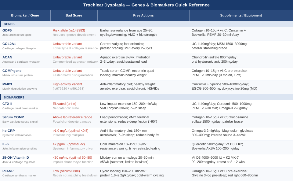
Quick Reference: Genes and Biomarkers at a Glance
10 Things the Huberman Lab Protocols Reveal About Connective Tissue Health
Andrew Huberman's work on connective tissue health, collagen synthesis, and joint recovery — spread across several Huberman Lab podcast episodes — contains practical, science-backed insights that are directly applicable to anyone managing a structural knee condition like trochlear dysplasia. Most of these insights are not part of standard orthopaedic advice, and several challenge common assumptions about how joints are managed and repaired.
1. Pre-Exercise Collagen Timing Is More Effective Than Post-Exercise
Taking hydrolyzed collagen (with 50 mg vitamin C) 30–60 minutes before loading — not after — amplifies collagen synthesis in connective tissue during and after exercise. The amino acids from collagen circulate in the bloodstream precisely when mechanically stimulated tissue is most receptive to matrix building. This is counter-intuitive for people used to thinking about protein as a post-workout intervention. For cartilage, tendons, and ligaments specifically, the pre-loading window is the critical one.
2. Loading Velocity Must Progress Through Three Phases
Huberman distinguishes between slow isometric loading (ideal for pain management in acute phases), eccentric loading (ideal for tendon and cartilage matrix remodeling), and fast dynamic loading (required for full functional tissue quality restoration). People with trochlear dysplasia commonly remain in phase one indefinitely — avoiding dynamic load because it triggers symptoms — without progressing to the eccentric and explosive phases that actually remodel tissue architecture. Staying in pain-avoidance mode indefinitely keeps the joint in a suboptimal repair state.
3. Slow-Wave Sleep Is the Primary Cartilage Repair Window
Most tissue repair occurs during slow-wave sleep, when growth hormone secretion drives collagen synthesis and matrix maintenance across the body. Disrupted sleep architecture — even subclinically — compresses slow-wave sleep duration and meaningfully reduces the biological repair window. Huberman frames sleep optimization not as a wellness bonus but as a primary physiological intervention for anyone dealing with joint tissue damage or instability.
4. Cold Exposure Reduces Joint Inflammation Without Blocking Repair
Unlike chronic NSAID use — which suppresses prostaglandins needed for downstream repair signaling — cold water immersion (10–15 minutes at 10–15°C) reduces localized synovial inflammation acutely without blocking anabolic repair cascades. Huberman recommends cold exposure before exercise or at least four hours after, never immediately post-exercise, to avoid blunting beneficial adaptation signals. For trochlear dysplasia with elevated IL-6, this is a clinically relevant distinction from drug-based anti-inflammatory approaches.
5. Vitamin D Is Foundational, Not Optional
Huberman consistently frames vitamin D as non-negotiable for musculoskeletal health, noting global deficiency prevalence. He recommends morning sunlight for circadian regulation and midday sun for UVB-mediated synthesis. For joints specifically, deficiency impairs chondrocyte function, reduces calcium regulation in subchondral bone, and elevates inflammatory cytokines — all factors that compound the already-unfavorable mechanical environment in trochlear dysplasia.
6. Heat Exposure Independently Improves Synovial Fluid Quality
Regular sauna use — 15–20 minutes at 80–100°C — increases synovial fluid fluidity, reduces joint stiffness, and has been associated with reduced inflammatory cytokines with repeated sessions. For trochlear dysplasia, where stiffness increases patellofemoral contact pressure and exacerbates abnormal tracking, regular heat exposure is a simple structural support tool that requires no supplementation.
7. Omega-3s Act as Membrane Lubricants for Chondrocytes
Beyond anti-inflammatory signaling, EPA and DHA are incorporated into chondrocyte cell membranes, making them more flexible and mechanically responsive. Huberman emphasizes 2–3 g EPA+DHA daily as a baseline for anyone with joint concerns. In a dysplastic joint where chondrocytes are regularly exposed to abnormal loading patterns, membrane integrity directly affects cellular resilience.
8. Breathing Protocols Can Acutely Lower Inflammatory Cytokines
Cyclic hyperventilation protocols (Wim Hof-style breathing) produce a sympathoadrenal response that measurably reduces circulating IL-6 and other pro-inflammatory cytokines in controlled studies. While this is not joint-specific, for individuals with chronically elevated IL-6 markers, structured breathing represents a zero-cost intervention with documented biochemical effects that go beyond stress management.
9. Whole-Body Strength Training Supports Connective Tissue Systemically
Huberman references grip strength as a proxy for systemic connective tissue and musculoskeletal integrity, underscoring that whole-body resistance training — not only isolated knee rehabilitation — supports joint tissue quality systemically. People with trochlear dysplasia often limit their training to knee-focused physiotherapy protocols; the evidence suggests that full-body strength training at 3 sessions per week independently supports connective tissue health across the entire kinetic chain.
10. Interventions Need at Least Two Weeks Before Assessing Effect
Connective tissue markers and joint symptoms typically lag behind actual biological change by 10–21 days. Stopping a new protocol because pain has not resolved within several days leads to a cycle of abandoning strategies that were beginning to work. Huberman frames the two-week minimum as a neurobiological and connective tissue reality, not a patience recommendation. This is particularly relevant for trochlear dysplasia management, where improvement timelines are longer than in soft tissue conditions.
Complementary Approaches with Relevant Clinical Evidence
The following approaches have meaningful human clinical evidence for joint pain, cartilage health, or patellofemoral conditions and may serve as useful adjuncts to the biomarker-informed and genetics-informed strategies above.
Biofeedback for VMO Activation and Patellar Tracking
Biofeedback — using surface electromyography (EMG) to provide real-time visual or auditory feedback about muscle activation — is directly relevant to trochlear dysplasia because patellar tracking quality depends heavily on the timing and magnitude of VMO (vastus medialis oblique) contraction relative to the vastus lateralis. In a dysplastic trochlea, even small improvements in VMO activation timing can meaningfully reduce lateral patellar displacement and patellofemoral contact stress.
A randomized controlled trial by Ng and colleagues examined EMG biofeedback training for patellofemoral pain syndrome and found significantly greater improvements in VMO:VL activation ratios and pain scores in the biofeedback group compared to standard physiotherapy alone. The mechanism is neuromuscular re-education: biofeedback helps patients learn to activate VMO at the correct moment in the gait and loading cycle, which is difficult to achieve through verbal instruction alone.
Practically, 6–8 weeks of biofeedback-assisted physiotherapy (2–3 sessions per week, 20–30 minutes per session) is a realistic protocol for people with trochlear dysplasia. Home EMG biofeedback devices are available in the $100–$300 range. This approach is particularly well-suited to the period following surgical stabilization, when restoring proper neuromuscular control is critical to long-term outcomes, but it is equally relevant for conservative management.
Low-Level Laser Therapy (Photobiomodulation) for Joint Tissue Support
Photobiomodulation (PBM) at wavelengths of 660–850 nm delivers low-energy photons to tissue, stimulating cytochrome c oxidase in mitochondria and triggering a cascade of cellular effects: increased ATP production, reduced oxidative stress, upregulated collagen synthesis, and modulated inflammatory mediator expression. In joint tissue, these effects translate to measurable reductions in cartilage degradation markers and improvements in chondrocyte anabolic activity.
A Cochrane-informed systematic review and multiple RCTs on photobiomodulation for knee osteoarthritis have shown significant reductions in pain, stiffness, and inflammatory markers with LLLT applied to the knee joint. While direct evidence for trochlear dysplasia specifically is not available, the mechanism is well-matched: reducing synovial inflammation, stimulating cartilage matrix synthesis, and supporting subchondral bone quality are all relevant to the challenges this condition creates. Evidence is stronger for symptomatic pain management than for structural reversal.
A realistic protocol is 810 nm at 50–100 mW per cm², applied 10–15 minutes over the patellofemoral joint, 4–5 times per week for 8–12 weeks. Class IV clinical devices are available through physiotherapy and sports medicine clinics. Home devices in the 660–850 nm range (typically 50–100 mW output) are available for $150–$600. Combine with the collagen plus vitamin C pre-exercise protocol for potential synergy on cartilage synthesis.
Tai Chi for Proprioception and Patellofemoral Load Management
Tai chi — a slow, controlled, mind-body movement practice — has a consistent body of evidence for improving proprioception, balance, and lower extremity joint control in populations with knee conditions. In trochlear dysplasia, proprioceptive deficits are common: the abnormal patellar tracking disrupts the normal sensory feedback from mechanoreceptors in the patellofemoral joint, reducing the joint's ability to self-regulate loading and position during movement.
A randomized trial by Lee and colleagues published in Arthritis and Rheumatism found that 12 weeks of tai chi practice significantly improved knee proprioception, pain scores, and functional mobility in knee OA participants compared to a control group. The slow, eccentric-loaded movement patterns in tai chi also provide gentle cyclical cartilage loading without the impact spikes of conventional exercise — an important feature for a joint with abnormal stress distribution.
A practical entry point is a supervised beginner tai chi class (60–90 minutes, 2×/week) for the first 8–12 weeks to ensure correct form, followed by a self-directed daily 20-minute practice. The movements can be adapted with the guidance of a physiotherapist familiar with patellofemoral conditions to minimize deep knee flexion angles during the learning phase. Evidence is limited for trochlear dysplasia specifically, but the mechanism — improved neuromuscular control, enhanced proprioception, and low-impact cartilage loading — is directly relevant.
Conclusion
Trochlear dysplasia is a structural condition, and no amount of biomarker tracking changes the anatomy of the trochlear groove. But the joint environment around that anatomy — the inflammatory state, the cartilage turnover balance, the quality of the collagen matrix — is dynamic and responsive to what you do. The six biomarkers covered here give you a real-time window into that environment; the five genetic variants help explain your individual risk profile and prioritize which interventions matter most in your case.
The most practical next step is to choose one or two of the most accessible markers — hs-CRP and 25-OH Vitamin D are the most affordable and widely available starting points — and run a baseline panel. If either is out of optimal range, follow the specific plan outlined here before adding complexity. If you have access to genetic testing through a sports medicine or functional medicine provider, GDF5 and MMP3 are the variants most worth knowing about in a patellofemoral context.
Work with a physiotherapist experienced in patellofemoral rehabilitation and, where possible, a sports medicine or musculoskeletal specialist who can help interpret your biomarker trends over time. The goal is not to fix the anatomy; it is to give the joint the best possible biochemical and mechanical environment to function in over the years ahead. That is achievable — and the information in this article is a solid starting point for getting there.
Musculoskeletal: Bone Conditions Joint Conditions Muscle Conditions Sports Injuries
Autoimmune: Connective Tissue Conditions