This article contains AI generated content.
Trochlear Groove Bone Marrow Edema: 5 Genes And 7 Biomarkers To Track
Introduction
An MRI report mentioning bone marrow edema in the trochlear groove tends to raise more questions than it answers. The scan shows something real and often painful, yet the advice that follows it is frequently generic: rest, modify activity, maybe an anti-inflammatory, come back in a few months. For someone who wants to understand what is actually happening inside the joint, and why recovery timelines vary so much from one person to another, that is rarely enough.
Generic guidance struggles here because bone marrow edema of the trochlear groove is not one uniform event. It can reflect mechanical overload at the patellofemoral joint, a local bone remodeling imbalance, a systemic vitamin D or inflammatory issue, or some combination of the three. Two people with an identical-looking MRI can have very different underlying biology, and that biology is what actually determines whether the edema resolves in eight weeks or lingers for a year.
This article looks past the generic advice and toward the data that can actually be checked: blood biomarkers that reflect bone formation, bone resorption, inflammation, and vitamin D status, alongside the genetic and epigenetic factors that shape how a person's bone metabolism responds to load and repair in the first place. Neither layer is a substitute for a proper orthopedic or sports-medicine evaluation, but both can sharpen the questions asked in that evaluation and guide day-to-day decisions in the meantime.
None of this promises a fixed timeline or a guaranteed outcome. What better information reliably does is narrow the range of plausible explanations and turn a vague "wait and see" into a more specific, trackable plan. The sections ahead cover the biomarkers worth measuring, what current genetic research suggests, a longevity framework that reframes how joint injuries should be thought about at all, and a set of complementary approaches with real supporting evidence.
Summary
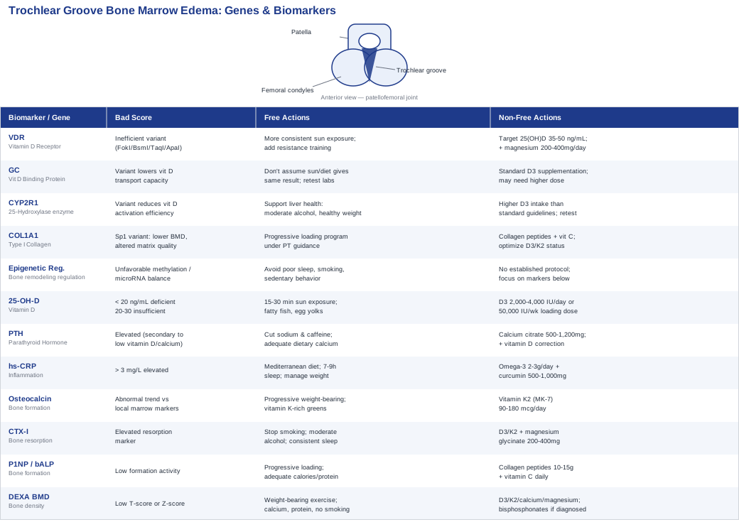
Bone marrow edema in the trochlear groove is rarely just a local mechanical problem. Research on bone marrow edema syndrome consistently points to a handful of measurable blood markers, most notably vitamin D status, parathyroid hormone, and the balance between bone formation and bone resorption, that differ between people who recover quickly and those who do not. This article walks through the seven biomarkers worth asking a physician to check, what each one is likely to reveal, realistic cost ranges, and two-tier plans (with and without supplements) for correcting the ones that come back unfavorable.
Beyond the lab work, there is a genetic layer. Variants in the vitamin D receptor gene and in collagen-related genes appear to influence how efficiently someone processes vitamin D and builds bone matrix, which may partly explain why some people are more prone to marrow edema and bone stress reactions than others. A longevity-focused reframing of exercise and injury recovery, and a short list of complementary therapies with actual clinical trial support in knee conditions, round out the picture. The diagram below maps how these pieces connect.
The Biomarkers Most Worth Tracking
Before ordering another scan, it is worth building a basic lab picture. Bone marrow edema syndrome research has repeatedly found that people with this condition differ from healthy controls on a specific set of blood markers, mostly related to vitamin D metabolism, calcium regulation, inflammation, and the balance between bone formation and breakdown. A retrospective study of a large bone marrow edema syndrome cohort assessed calcium, phosphate, 25-OH-vitamin D, bone-specific alkaline phosphatase, parathyroid hormone, and osteocalcin as part of routine workup, and a scoping review focused specifically on the role of vitamin D and vitamin C in bone marrow edema syndrome found deficiency to be common in affected patients. These are not exotic tests; most can be ordered through a standard blood panel.
The seven markers below are ranked by how directly they connect to bone marrow edema and how actionable they are.
Serum 25-Hydroxyvitamin D (25-OH-D)
This is the single most consistently implicated marker in bone marrow edema syndrome research. Vitamin D deficiency has been identified as a common finding in patients with primary bone marrow edema syndrome across multiple body regions, and correcting it is one of the few interventions with a clear biological rationale: low vitamin D impairs calcium absorption, which forces the body to pull calcium from bone and disrupts the formation-resorption balance right at the site under mechanical stress.
How to measure it: a standard blood draw measuring 25-hydroxyvitamin D, typically $40 to $100 out of pocket if not covered by insurance, reported in ng/mL (below 20 ng/mL is generally considered deficient, 20 to 30 ng/mL insufficient, above 30 ng/mL sufficient by most lab reference ranges).
If the score is bad, the plan without supplements: aim for 15 to 30 minutes of midday sun exposure on bare arms and legs, several times a week, adjusted for skin tone and latitude, since darker skin and higher latitudes both require more exposure time. Add dietary sources such as fatty fish (salmon, mackerel, sardines), egg yolks, and fortified dairy or plant milk. Retest after 8 to 12 weeks, since sun and diet alone often produce only modest gains, especially in winter months or northern climates.
If the score is bad, the plan with supplements or equipment: vitamin D3 (cholecalciferol) at 2,000 to 4,000 IU per day is a reasonable maintenance dose for mild insufficiency. For more significant deficiency (below 20 ng/mL), some clinicians use a supervised loading protocol, such as 50,000 IU once weekly for 6 to 8 weeks, followed by a switch to daily maintenance dosing. A UVB lamp is a legitimate equipment-based alternative for people with limited sun access, used for a few minutes per session per manufacturer guidance. Retest at 3 months rather than adjusting dose blindly. Side effects at appropriate doses are rare, but excessive, unsupervised dosing (sustained intake above 10,000 IU/day for months) can cause hypercalcemia, nausea, and kidney stone risk, so retesting rather than guessing is the safer approach.
Parathyroid Hormone (PTH)
PTH rises when the body senses low blood calcium, often secondary to vitamin D deficiency, and it directly drives bone remodeling by activating osteoclasts to release calcium from bone. Chronically elevated PTH tips the formation-resorption balance toward breakdown, which is exactly the kind of imbalance implicated in marrow edema. PTH was included in the same bone marrow edema syndrome cohort studies that tracked vitamin D, precisely because the two are metabolically linked.
How to measure it: intact PTH blood test, roughly $50 to $100, usually interpreted alongside calcium and vitamin D rather than in isolation.
If the score is bad, the plan without supplements: correcting vitamin D status (above) is the primary lever, since secondary hyperparathyroidism from vitamin D deficiency typically normalizes once vitamin D is repleted. Reducing excess sodium and caffeine intake, both of which increase urinary calcium loss, and ensuring adequate dietary calcium (dairy, leafy greens, canned fish with bones) can help.
If the score is bad, the plan with supplements or equipment: calcium citrate or calcium carbonate, 500 to 1,200 mg per day in divided doses, if dietary intake is genuinely low, combined with the vitamin D correction above (PTH usually falls as a secondary effect rather than needing direct treatment). Retest in 8 to 12 weeks. Side effects include constipation, particularly with calcium carbonate, and a theoretical increased kidney stone risk with high-dose calcium supplementation in people not also getting enough vitamin K2, so pairing the two is preferred over calcium alone.
High-Sensitivity C-Reactive Protein (hs-CRP)
Bone marrow lesions in the knee are not purely mechanical; a cross-sectional and longitudinal study found that serum hs-CRP levels correlate with the presence of knee bone marrow lesions and with knee pain, including pain at night and while sitting. This supports a broader shift in how osteoarthritis and marrow lesions are understood, as a partly inflammatory process rather than pure wear-and-tear degeneration. A persistently elevated hs-CRP suggests systemic low-grade inflammation that may be slowing local healing.
How to measure it: hs-CRP blood test, $20 to $50, often bundled with a standard metabolic or cardiovascular panel. Under 1 mg/L is considered low risk, 1 to 3 mg/L average, above 3 mg/L elevated.
If the score is bad, the plan without supplements: shift toward a Mediterranean-style eating pattern emphasizing fish, olive oil, vegetables, and legumes while cutting ultra-processed food and added sugar, both linked to CRP reduction in multiple trials. Prioritize 7 to 9 hours of sleep, since short sleep duration reliably raises inflammatory markers. Manage body weight, since adipose tissue itself produces inflammatory cytokines, and modify (rather than eliminate) knee-loading activity to avoid the deep flexion and high-impact loading patterns that provoke patellofemoral irritation.
If the score is bad, the plan with supplements or equipment: omega-3 fish oil, 2 to 3 grams combined EPA/DHA daily, retested at 8 to 12 weeks. Curcumin extract with piperine for absorption, 500 to 1,000 mg daily, cycled 8 to 12 weeks on and 4 weeks off rather than taken indefinitely without reassessment. Side effects: fish oil can cause GI upset and has a mild blood-thinning effect, requiring caution alongside anticoagulants; curcumin can cause GI upset and should be avoided or discussed with a physician if there is a history of gallstones.
Osteocalcin
Osteocalcin is produced by osteoblasts and is a standard marker of bone formation activity. Interestingly, in bone marrow edema syndrome, serum osteocalcin can appear entirely normal even as bone turnover markers measured directly from marrow aspirate at the lesion site run high, according to research on biochemical markers of bone turnover and their association with bone marrow lesions. That mismatch matters practically: a normal serum osteocalcin does not rule out active local remodeling, which is why it is most useful as a trend marker over time rather than a single snapshot, and as an indirect check on vitamin K status, since osteocalcin requires vitamin K-dependent carboxylation to function properly.
How to measure it: fasting morning blood draw (osteocalcin has diurnal variation), $40 to $80.
If the score is bad, the plan without supplements: once pain allows, progressive weight-bearing and resistance exercise is the primary non-supplement driver of healthy osteoblast activity, alongside a diet rich in vitamin K from leafy greens (kale, spinach, broccoli).
If the score is bad, the plan with supplements or equipment: vitamin K2 (as MK-7), 90 to 180 mcg per day, paired with the vitamin D3 protocol above since the two work synergistically in calcium handling. Side effects are minimal at these doses, but anyone on warfarin or other vitamin K-antagonist anticoagulants should not add vitamin K2 without direct physician guidance, since it can interfere with anticoagulation control.
CTX-I (C-Terminal Telopeptide)
CTX-I is the standard marker of bone resorption, the breakdown side of the remodeling equation. Read alongside a formation marker like P1NP or osteocalcin, it indicates whether bone at a given moment is in net breakdown or net repair, which is directly relevant to a lesion that is either resolving or being aggravated by ongoing loading.
How to measure it: fasting, early-morning blood draw is important due to strong diurnal variation, $60 to $120.
If the score is bad, the plan without supplements: eliminate smoking, which is one of the more reliably documented factors that raises bone resorption markers, moderate alcohol intake, and prioritize consistent sleep timing, since resorption markers are tied to circadian rhythm disruption. Ensure adequate dietary protein, since insufficient protein intake can worsen bone turnover imbalance.
If the score is bad, the plan with supplements or equipment: the same vitamin D3/K2 correction described above, plus magnesium glycinate, 200 to 400 mg per day, which supports several enzymatic steps in bone metabolism. Reassess in 3 months rather than adjusting more frequently, since bone turnover markers move slowly. Side effects: magnesium in excess can cause loose stools, which is typically self-limiting at lower doses.
P1NP or Bone-Specific Alkaline Phosphatase (Formation Markers)
P1NP and bone-specific alkaline phosphatase (bALP) are the two most common formation-side markers, used to complement CTX-I and confirm whether bone is actively rebuilding. Tracking a formation marker alongside a resorption marker over the course of recovery gives a clearer picture than either alone, since a lesion resolving well should generally show formation activity keeping pace with or exceeding resorption.
How to measure it: blood draw, $70 to $130 depending on which marker is ordered.
If the score is bad, the plan without supplements: progressive, clinician-approved loading (walking progressing to resistance training) is the strongest non-supplement stimulus for bone formation markers, and adequate total caloric and protein intake matters, since athletes and very active people in energy deficit can suppress bone formation markers even while training hard, a pattern seen in relative energy deficiency in sport.
If the score is bad, the plan with supplements or equipment: collagen peptides, 10 to 15 grams per day, combined with vitamin C, which is a required cofactor for collagen synthesis. No significant side effects at typical doses beyond occasional mild GI upset or known allergy.
DEXA Bone Mineral Density (BMD)
A DEXA scan does not diagnose marrow edema itself, but it provides useful structural context, particularly for anyone with recurrent lesions, low body weight, a history of disordered eating, amenorrhea, or other osteoporosis risk factors. It also serves as a meaningful baseline to track over subsequent years.
How to measure it: DEXA scan, $100 to $400 out of pocket, often covered by insurance if risk factors are documented, low radiation exposure. Results reported as T-score (compared to young healthy adults, most relevant post-menopause) or Z-score (compared to age-matched peers, more relevant for younger adults).
If the score is bad, the plan without supplements: weight-bearing and resistance exercise, adequate dietary calcium and protein, smoking cessation, and moderated alcohol intake.
If the score is bad, the plan with supplements or equipment: the vitamin D3, K2, calcium, and magnesium approach outlined above, applied consistently over months rather than weeks, since bone density changes slowly. If a true osteoporosis diagnosis is confirmed, pharmaceutical options such as bisphosphonates exist, but that decision belongs with a physician managing bone health directly rather than as a self-directed supplement choice.
What Genetics and Epigenetics May Add to the Picture
Genetic research on bone marrow edema syndrome specifically is thin, most of what exists comes from the broader field of bone metabolism genetics, the kind of work popularized by researchers like Ali Torkamani and practitioners like Gary Brecka, who both emphasize using genetic data to explain why identical interventions produce different results in different people. Applied here, that means the genes below are best understood as modifiers of vitamin D and collagen metabolism rather than direct causes of trochlear groove edema, and the evidence should be read as suggestive rather than conclusive.
VDR (Vitamin D Receptor)
Common VDR variants, referred to by the restriction enzymes historically used to detect them (FokI, BsmI, TaqI, ApaI), affect how efficiently the vitamin D receptor binds to active vitamin D and switches on downstream genes involved in calcium absorption and bone remodeling. Someone with a less efficient VDR variant may need a higher circulating 25(OH)D level to achieve the same cellular effect as someone with a more efficient variant, which could partly explain why some people with "normal" vitamin D labs still show signs of impaired bone metabolism.
If the gene is bad, the plan without supplements: this is not a gene that can be edited, so the practical response is behavioral, more consistent sun exposure and dietary vitamin D intake than someone without the variant might need, plus resistance training, which independently supports calcium handling in bone regardless of receptor efficiency.
If the score is bad, the plan with supplements or equipment: because a less efficient receptor may need more circulating substrate to compensate, targeting the higher end of the normal 25(OH)D range (35 to 50 ng/mL rather than just above 30) is a reasonable, low-risk adjustment, done under retesting every 3 months rather than by feel. Magnesium (a required cofactor for vitamin D activation) at 200 to 400 mg daily is often paired with this approach. Side effects mirror those of standard vitamin D supplementation: risk of hypercalcemia only at sustained high doses without monitoring.
GC (Vitamin D Binding Protein)
The GC gene encodes the protein that transports vitamin D through the bloodstream. Variants in this gene are associated with different baseline levels of circulating vitamin D independent of sun exposure or intake, meaning two people with identical lifestyles can land in different labs purely due to this gene.
If the gene is bad, the plan without supplements: since this gene affects baseline transport capacity rather than something behavior can directly fix, the main non-supplement lever is simply not assuming that a given amount of sun or diet will produce the same lab result as it does for someone else, and retesting rather than assuming.
If the score is bad, the plan with supplements or equipment: standard vitamin D3 supplementation as described earlier, with attention to the fact that people with less favorable GC variants may need a somewhat higher maintenance dose to sustain the same serum level, again confirmed by retesting rather than guessed.
CYP2R1 (Vitamin D 25-Hydroxylase)
CYP2R1 encodes the liver enzyme responsible for the first activation step of vitamin D, converting it into 25(OH)D, the form measured on standard blood tests. Variants here are among the most consistently replicated genetic contributors to baseline vitamin D levels in large population studies.
If the gene is bad, the plan without supplements: no behavioral fix directly targets enzyme efficiency, but supporting general liver health (moderate alcohol intake, maintaining a healthy weight) keeps the enzyme system functioning as well as it can.
If the score is bad, the plan with supplements or equipment: this variant is one of the stronger reasons some people simply need more vitamin D3 intake than standard guidelines suggest to reach the same blood level, reinforcing the value of retesting after 8 to 12 weeks rather than relying on generic dosing charts.
COL1A1 (Collagen Type I Alpha 1)
COL1A1 encodes the main protein chain of type I collagen, the principal structural protein in bone. A well-studied polymorphism in the gene's Sp1 binding site has been associated with lower bone mineral density and altered bone matrix quality in research on bone complications and polymorphisms affecting bone mineral and matrix formation. This variant does not cause marrow edema on its own, but it may make bone matrix somewhat less resilient under repetitive mechanical load, which is the same load pattern implicated in trochlear groove overuse injuries.
If the gene is bad, the plan without supplements: since matrix quality partly depends on mechanical stimulus, a structured, progressive loading program (not avoidance of load altogether) is what actually signals bone to reinforce itself, done under physical therapy guidance while the lesion is active.
If the score is bad, the plan with supplements or equipment: collagen peptides with vitamin C, as described in the P1NP section above, plus ensuring the vitamin D and K2 status described earlier is optimized, since matrix formation depends on all of these working together rather than any single input. No specific side effects beyond those already listed for each component.
Epigenetic Regulation of Bone Remodeling
Beyond fixed gene sequence, early research is exploring how DNA methylation and microRNA activity regulate the balance between osteoblast and osteoclast gene expression, essentially controlling how strongly the "build" versus "break down" signals are expressed regardless of the underlying gene sequence. This is genuinely early-stage science, mostly cell and animal studies with limited human trial data, and should be treated as a developing area rather than an actionable target today.
If the gene is bad, the plan without supplements: the honest answer is that no established consumer intervention currently modifies these epigenetic marks, so the more grounded approach is simply to remove known negative epigenetic influences: chronic sleep deprivation, smoking, and sedentary behavior all shift methylation patterns unfavorably in bone tissue in animal models, and are worth avoiding independent of this particular mechanism.
If the score is bad, the plan with supplements or equipment: no specific supplement protocol currently has strong human evidence for directly favorable epigenetic modification of bone remodeling; the honest position is to focus supplement effort on the well-established biomarkers above rather than speculative epigenetic products.
The Longevity Framework That Reframes Joint Recovery
Physician Peter Attia's book Outlive is not written about bone marrow edema, but its central argument, that most people wait far too long to train the physical capacities that prevent injury and preserve function, applies directly to a joint injury like this one. Attia frames exercise around what he calls the "Centenarian Decathlon," working backward from the physical tasks someone wants to still perform decades from now to figure out what needs training today. For a knee-region injury specifically, several of his arguments are worth pulling out individually.
Muscle Is the Organ of Longevity
Attia argues that skeletal muscle mass and strength predict long-term function and mortality risk more strongly than most people assume. For a trochlear groove issue, adequate quadriceps and hip strength directly changes how much load the patellofemoral joint absorbs versus how much is redirected through surrounding musculature, making strength training a direct, not incidental, part of recovery once cleared for loading.
Zone 2 Training Supports the Repair Machinery
Zone 2 cardio, sustained aerobic effort at a conversational pace, improves mitochondrial density and metabolic efficiency throughout the body, including in tissues undergoing repair. Attia recommends roughly 3 to 4 hours per week as a baseline, and low-impact versions (cycling, swimming, incline walking) are usually well tolerated even with an irritated knee.
VO2 Max Is the Single Strongest Mortality Predictor
Attia cites data showing that moving from the bottom quartile to the top quartile of VO2 max is associated with a roughly fivefold reduction in mortality risk, a larger effect than quitting smoking. While not directly about bone healing, cardiovascular fitness supports the circulatory delivery of nutrients and repair signals to healing tissue throughout the body.
Stability Is the Most Skipped Pillar
Of his four training pillars (strength, zone 2, VO2 max, stability), Attia considers stability the most commonly neglected, and the one that makes the other three safe to pursue. For patellofemoral and trochlear groove issues specifically, poor hip and core stability is one of the more common mechanical contributors to abnormal joint tracking and repetitive overload.
The "Marginal Decade" Argument for Patience[TITLE]
Attia frequently argues that the payoff for physical training compounds over decades, not weeks. Applied to injury recovery, this reframes an eight-month rehabilitation timeline as a reasonable investment rather than a frustrating delay, particularly if it is done correctly rather than rushed.
Reverse-Engineering the Centenarian Decathlon
Rather than training generically, Attia suggests identifying specific physical tasks someone wants to be able to do at 80 or 90 (carrying groceries, getting up off the floor, hiking) and training backward from there. For a recovering knee, this means rehabilitation should be built around real future movement goals, not just pain-free walking.
Medicine 3.0: Proactive Rather Than Reactive
Attia's broader argument for what he calls Medicine 3.0 is to track relevant biomarkers proactively rather than waiting for a problem to become symptomatic. This is the same logic behind the biomarker panel described earlier in this article: checking vitamin D, inflammatory markers, and bone turnover before or during a minor issue, rather than only after a more serious one develops.
Eccentric Strength and Rate of Force Development
Attia emphasizes eccentric loading and rate of force development as underappreciated components of joint resilience, since most real-world injuries happen during deceleration or unexpected loading rather than during controlled concentric effort. Slow, controlled eccentric quadriceps work is a common and evidence-supported component of patellofemoral rehabilitation programs for exactly this reason.
A Single Injury Can Cascade
Attia repeatedly notes that an injury in one's 40s or 50s can trigger a downward spiral of deconditioning that is disproportionate to the original injury, since reduced activity leads to lost muscle and cardiovascular fitness, which increases the risk of the next injury. This is a direct argument for staying as active as safely possible during recovery, rather than full inactivity, guided by a physical therapist.
Sleep Underpins All Tissue Repair
Attia treats sleep as foundational rather than optional, citing its role in hormonal regulation, inflammation control, and tissue repair. This aligns directly with the hs-CRP and CTX-I discussion earlier in this article, both of which are measurably worsened by poor sleep.
Complementary Approaches Worth Considering
A handful of mind-body and manual therapies have real clinical trial evidence in knee conditions closely related to trochlear groove issues, mainly knee osteoarthritis and patellofemoral pain, even though direct trials in bone marrow edema specifically are limited. These are reasonable additions alongside, not instead of, medical evaluation and the biomarker-driven plan above.
Tai Chi
Tai chi combines slow, controlled weight-shifting movements with balance and proprioceptive training, both of which are directly relevant to patellofemoral joint mechanics, since poor neuromuscular control around the knee is a known contributor to abnormal trochlear loading. Because sessions are low-impact, it is one of the few structured movement practices generally tolerable even during an active flare.
A randomized controlled trial of group and home-based tai chi in elderly subjects with knee osteoarthritis found meaningful improvements in pain and physical function compared to an attention-control group, using a modified Yang-style protocol delivered in supervised group sessions followed by home practice.
Realistically, this means 2 to 3 sessions per week of a beginner-level Yang-style or Sun-style tai chi program, ideally starting with instructor guidance to ensure knee alignment is correct rather than compensatory, and continuing for at least 8 to 12 weeks before judging effect. Evidence in bone marrow edema specifically is absent, so this should be framed as general joint-friendly movement therapy rather than a targeted treatment.
Massage Therapy
Massage therapy addresses the surrounding soft tissue (quadriceps, IT band, hip musculature) that influences how load is distributed across the patellofemoral joint, and may reduce the muscular guarding and stiffness that often accompanies joint pain.
A randomized controlled trial published in the Annals of Internal Medicine found that standard Swedish massage therapy produced significant improvements in pain, stiffness, and physical function in knee osteoarthritis compared to a delayed-treatment control group, using twice-weekly sessions for the first four weeks tapering to once weekly for the following four weeks.
A realistic starting protocol mirrors the study design: two sessions weekly for the first month, then weekly for a second month, focused on the quadriceps, hamstrings, and IT band rather than the joint itself. This is a supportive therapy, not a substitute for addressing the underlying biomarker or mechanical picture.
Mindfulness Meditation and MBSR
Chronic joint pain has a well-documented central sensitization component, meaning the nervous system's pain processing itself can become part of the problem over time, independent of ongoing tissue damage. Mindfulness-based approaches target this pathway directly rather than the joint itself.
A systematic review and meta-analysis on mindfulness meditation for chronic pain, which included osteoarthritis among the conditions studied, found modest but consistent improvements in pain and function compared to usual care, though the authors note effect sizes are generally smaller than for more targeted therapies. Trials specific to knee osteoarthritis using mindfulness-based stress reduction protocols have shown mixed but generally positive results, and evidence in bone marrow edema specifically does not yet exist.
A standard MBSR-style approach involves an 8-week structured program with weekly sessions of roughly 45 minutes plus daily home practice of 10 to 20 minutes. Given the modest but real effect size, this is best used as a pain-management and stress-reduction layer alongside, not in place of, the biomechanical and lab-based interventions above.
Photobiomodulation (Low-Level Laser Therapy)
Photobiomodulation uses red and near-infrared light to stimulate mitochondrial activity in cells, with documented anti-inflammatory effects and, in bone-specific research, enhanced osteoblast activity and alkaline phosphatase expression, both directly relevant to the bone formation side of the remodeling equation discussed earlier.
A randomized clinical trial examining photobiomodulation therapy's impact on swelling reduction and recovery after total knee arthroplasty found measurable improvements in post-surgical swelling and recovery metrics, supporting its anti-inflammatory and tissue-repair effects in a knee-adjacent context, even though this trial was in a post-surgical rather than bone marrow edema population specifically.
In practice, this typically means a series of sessions with a clinical-grade device, 2 to 3 times weekly for 4 to 6 weeks, applied directly over the anterior knee under professional guidance rather than with unsupervised consumer devices of uncertain dosing. Evidence directly in trochlear groove bone marrow edema is still preliminary, so this is best considered a reasonable adjunct where available rather than a primary strategy.
Conclusion
Trochlear groove bone marrow edema is rarely explained by mechanics alone. Vitamin D status, parathyroid hormone, inflammatory tone, and the balance between bone formation and resorption all shape how quickly a lesion resolves, and each of those can be measured rather than guessed at. Genetic variants in vitamin D and collagen metabolism add a layer of "why me" context, even if they cannot yet be directly changed. A longevity-minded approach to the surrounding musculature and cardiovascular system, and a small set of complementary therapies with real trial support in knee conditions, round out a plan that goes well beyond "rest and reassess."
The next practical step is straightforward: ask for the vitamin D, PTH, hs-CRP, and bone turnover panel described above at the next appointment, track the results over time rather than as a single snapshot, and bring both the numbers and this framework into the conversation with an orthopedic specialist or sports medicine physician who can weigh them against the actual imaging and mechanical exam findings.