This article contains AI generated content.
Trochlear Groove Chondropathy: 4 Genes And 7 Biomarkers To Track
If you've been told you have trochlear groove chondropathy, chondromalacia patellae, or "cartilage wear" behind the kneecap, you've probably already heard the standard advice: strengthen your quads, lose a little weight, avoid deep squats, maybe try a brace. None of that is wrong, but it also isn't built around you. It doesn't explain why your cartilage is behaving the way it is, what's actively driving the breakdown, or which of the dozen possible contributing factors actually applies to your knee.
Generic advice treats chondropathy as one condition with one playbook. In reality, the same MRI finding — thinning or fissured cartilage in the groove where the kneecap tracks — can come from mechanical causes (trochlear shape, patellar tracking, muscle imbalance), metabolic causes (glycation, insulin resistance), inflammatory causes (systemic low-grade inflammation), or a genetic tendency toward slower cartilage repair. Treating all four the same way is why so many people follow "the protocol" for months and see their imaging stay flat.
This article takes a more specific approach. Instead of one generic list, it looks at the actual biological signals — blood and urine biomarkers, and a handful of well-studied genes — that can tell you which of those drivers is most active in your case, and what a targeted response looks like with and without supplements or equipment.
None of this replaces an orthopedic or sports medicine evaluation, and no biomarker or gene rewrites your anatomy. But better information about what's happening at the tissue level tends to produce better decisions than a one-size-fits-all handout — and that's the practical hope this article is built around.
Summary
Trochlear groove chondropathy isn't just a structural finding on an MRI — it's the visible endpoint of processes you can actually measure. Cartilage-breakdown proteins like COMP and CTX-II show up in your blood and urine before symptoms necessarily change. Inflammatory markers like hs-CRP and IL-6 reveal whether your whole body, not just your knee, is running a low-grade fire that keeps cartilage from repairing. Even a routine HbA1c can flag a glycation process that quietly stiffens cartilage matrix over years.
Underneath those biomarkers, a small set of genes — GDF5, COL2A1, VDR, and FRZB — help explain why two people with identical training loads and identical trochlear anatomy can end up with very different rates of cartilage wear. None of these genes is destiny, and for each one there's a practical way to work around a less favorable variant, with or without supplements.
Beyond the science of measurement, there's also a mobility and joint-mechanics perspective — drawn from a widely discussed Huberman Lab conversation on joint health — that reframes cartilage not as a fixed structure slowly wearing down, but as living tissue that responds to how you move it. And for people looking for adjunctive, evidence-backed support, a handful of complementary approaches (tai chi, yoga, massage, biofeedback, low-level laser therapy) have real trial data behind them for knee cartilage conditions specifically.
The rest of this article walks through each of these in practical detail: what to test, what it costs, what a "bad" number actually means, and what to do about it.
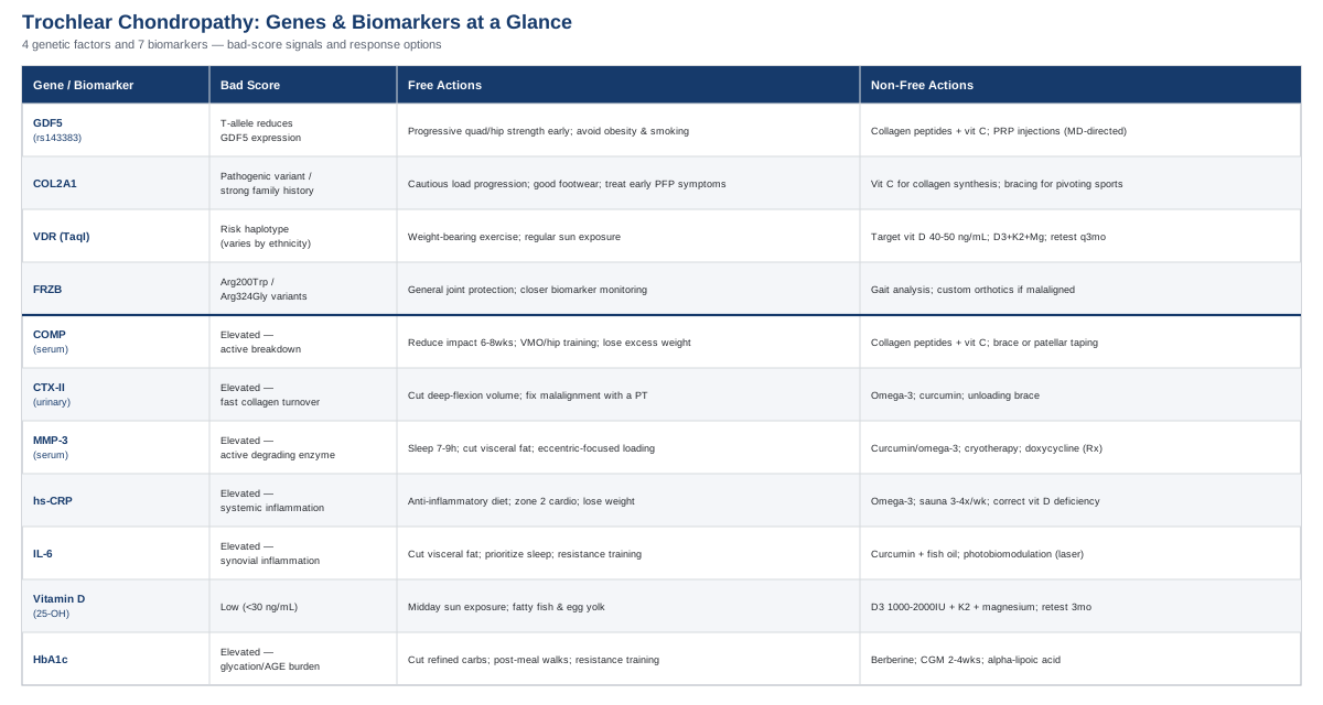
Biomarkers give the clearest, most actionable picture of what's happening in your cartilage right now, so that's where this article starts before moving into genetics, mobility research, and complementary care.
The 7 Biomarkers Worth Tracking For Trochlear Groove Chondropathy
Cartilage doesn't have nerve endings or blood vessels, so it can't tell you directly that it's breaking down — by the time pain shows up, the process is often well underway. Biomarkers are the closest thing to an early warning system: they're the breakdown products, enzymes, and inflammatory signals that leak into blood and urine as cartilage turns over. Tracking a handful of them, alongside imaging and symptoms, gives a much sharper picture than "does it hurt today."
The seven below cover the three main pathways that damage trochlear cartilage: direct structural breakdown, inflammatory acceleration, and metabolic stiffening. Peter Attia and Thomas Dayspring popularized this kind of layered biomarker thinking for cardiometabolic health; the same logic — measure the mechanism, not just the symptom — applies well to joint cartilage.
Serum COMP (Cartilage Oligomeric Matrix Protein)
COMP is a structural glycoprotein that helps hold the cartilage matrix together. When cartilage is actively degrading, fragments of COMP spill into the bloodstream, and serum levels track fairly closely with radiographic severity, pain, and disability in knee osteoarthritis, including patellofemoral involvement. Some studies have found it distinguishes OA knees from healthy ones with very high sensitivity, which is why it's one of the most studied cartilage-breakdown markers in the research literature (serum COMP as a potential biomarker for knee osteoarthritis).
How to measure it
A simple blood draw sent to a lab running an ELISA-based COMP assay. This isn't part of standard bloodwork — you'll need a sports medicine physician, rheumatologist, or a direct-to-consumer specialty lab willing to order it. Expect to pay roughly $70 to $200 out of pocket, since it's rarely covered by insurance for this indication.If the score is bad, the plan without supplements
Reduce mechanical overload while your knee is in an active breakdown phase: cut high-impact and deep-flexion loading for 6 to 8 weeks, and replace it with closed-chain quad and hip work in the 0-to-45-degree range, which loads the trochlear cartilage far less. Add structured VMO and hip-abductor strengthening 3 times a week — this specific combination has been shown to reduce patellar lateralization and instability risk compared with generic quad training (VMO-selective training versus general quadriceps strengthening). If you carry excess body weight, even modest loss meaningfully lowers patellofemoral joint force, since that joint sees several times body weight during stair descent and squatting. Retest COMP in 3 to 6 months.If the score is bad, the plan with supplements or equipment
Collagen peptides (10-15g daily, taken with 50mg vitamin C about 30-60 minutes before training to support collagen synthesis timing) are low-risk and have modest supporting evidence for cartilage-adjacent tissues; take them daily, no cycling needed, side effects are limited to mild GI upset in some people. A hinged knee brace or patellar taping during high-load activity can reduce shear on the groove while other changes take effect — use during aggravating activities only, not as a permanent crutch. Glucosamine/chondroitin has mixed trial evidence for structural change but may help symptomatically for some; if trying it, use 1500mg/1200mg daily for at least 8-12 weeks before judging effect, and avoid it if you have a shellfish allergy.Urinary CTX-II
CTX-II is a fragment released when type II collagen — the main structural collagen in cartilage — is broken down. Because it's collagen-specific, it's considered one of the better markers of the rate of cartilage degradation, and elevated levels have predicted both radiographic progression and future total joint replacement in longer-term cohort studies (CTX-II as a biomarker for radiological knee osteoarthritis: a meta-analysis, increased urinary CTX-II predicts cartilage loss on MRI).
How to measure it
A second-void morning urine sample, tested via ELISA (often marketed as "Urinary CTX-II" or "Cartilaps"). It's a research and specialty-lab test, not something your primary care doctor will order by default. Cost runs $100-250 depending on the lab.If the score is bad, the plan without supplements
Because CTX-II reflects collagen turnover rate rather than a single event, the fix is about reducing chronic overload, not chasing a single workout. Audit repetitive deep-flexion activities (kneeling, deep squatting, stair-heavy jobs) and reduce volume where possible. Address any lower-limb malalignment or patellar maltracking with a physical therapist, since uneven loading concentrates collagen breakdown in one part of the groove. Retest at 6 months minimum, since this marker moves slowly.If the score is bad, the plan with supplements or equipment
Omega-3s (2-3g combined EPA/DHA daily, taken continuously with food to reduce GI upset, caution if on blood thinners) have anti-catabolic, anti-inflammatory effects on cartilage in trial settings. Curcumin with piperine (500-1000mg daily, cycled 8-12 weeks on and 4 weeks off to reduce theoretical gallbladder strain with long-term high-dose use) is a reasonable adjunct. An unloading or offloading brace during unavoidable high-load activity (long hikes, physical jobs) reduces the shear component that specifically damages the groove.Serum MMP-3
Matrix metalloproteinase-3 is one of the enzymes chondrocytes and synovial cells produce that actively degrades cartilage matrix — collagen and proteoglycans both. Unlike COMP or CTX-II, which are downstream breakdown products, MMP-3 is closer to the actual degrading machinery, which is part of why it has prognostic value for how fast cartilage damage will progress (serum MMP-3 as a prognostic marker for progression of cartilage injury).
How to measure it
Blood draw, ELISA-based, typically through a rheumatology-oriented or research lab, $100-150.If the score is bad, the plan without supplements
MMP-3 tends to track with systemic inflammatory load, so reducing the upstream drivers matters more than any single knee exercise. Prioritize sleep (7-9 hours; poor sleep independently raises inflammatory enzyme activity), reduce visceral fat if present, and build in planned deload weeks after any flare rather than pushing through swelling. Eccentric-focused quad loading, progressed slowly under a physical therapist's guidance, tends to be better tolerated than concentric-heavy or plyometric work when MMP activity is elevated.If the score is bad, the plan with supplements or equipment
Curcumin and omega-3 (as dosed above) both have some evidence of dampening MMP activity indirectly through reduced inflammatory signaling. Cryotherapy (ice, 10-15 minutes after aggravating activity) is a low-cost, low-risk adjunct. Doxycycline has been studied as a prescription MMP-inhibitor in OA trials — this is a physician-directed, prescription-only option, not a self-directed supplement, and worth raising with your doctor only if MMP-3 stays elevated despite lifestyle changes.High-Sensitivity CRP (hs-CRP)
hs-CRP is a general marker of low-grade systemic inflammation, and in knee osteoarthritis specifically, elevated levels have been associated with faster joint space narrowing and greater osteophyte growth — even in early-stage disease, before major structural change is visible (hs-CRP and progression of knee osteoarthritis: the Yakumo study).
How to measure it
A standard, cheap blood test ($10-30), often already included in routine metabolic or cardiac panels — one of the easiest of these seven to get.If the score is bad, the plan without supplements
Shift toward an anti-inflammatory eating pattern: cut ultra-processed foods and added sugar, increase fiber and vegetables. Add 150 minutes a week of zone 2 cardio, which lowers systemic CRP over 8-12 weeks in most people. If you carry excess weight, this is one of the highest-leverage levers, since adipose tissue itself secretes inflammatory cytokines.If the score is bad, the plan with supplements or equipment
Omega-3s again show the most consistent CRP-lowering data among supplements. Regular sauna use (3-4x/week, 15-20 minutes) has been associated with modestly lower CRP in observational data — reasonable as an adjunct if accessible, avoid if you have uncontrolled cardiovascular disease. Correcting vitamin D deficiency if present (see below) can also help. Don't reach for a statin purely to lower CRP; that decision belongs to your cardiovascular risk profile, not your knee.IL-6
Interleukin-6 is a pro-inflammatory cytokine found at elevated levels in the synovial fluid of joints with focal cartilage defects, and it directly drives cartilage matrix breakdown while also sensitizing pain pathways — part of why inflamed knees often hurt more than the structural damage alone would predict (IL-6 elevated in synovial fluid of patients with focal cartilage defects).
How to measure it
Specialty serum ELISA, $100-200, not part of routine panels — usually ordered through a functional medicine or research-oriented lab.If the score is bad, the plan without supplements
Same core lifestyle levers as hs-CRP, since fat tissue is a major IL-6 source: reduce visceral adiposity, prioritize sleep, and add regular resistance training, which lowers chronic IL-6 over months even independent of weight change.If the score is bad, the plan with supplements or equipment
Curcumin and fish oil (dosed as above, cycle curcumin 8-12 weeks on/4 off) are the best-supported options. Photobiomodulation (low-level laser or red light therapy applied locally to the knee) has trial evidence for reducing joint inflammation and pain in knee OA and is covered in more detail later in this article.Vitamin D (25-OH)
This one deserves an honest caveat: while vitamin D receptor signaling plays a real role in chondrocyte regulation, randomized trials correcting vitamin D levels in people who already have knee osteoarthritis have not shown a reduction in cartilage volume loss — only possibly modest symptom benefit (vitamin D supplementation and cartilage volume loss: a randomized controlled trial, vitamin D and knee osteoarthritis: a meta-analysis). It's still worth knowing and correcting for general bone and muscle function around the joint, just without expecting it to reverse cartilage loss on its own.
How to measure it
Standard blood test, $30-50, widely available and usually insurance-covered.If the score is bad, the plan without supplements
15-20 minutes of midday sun exposure several times a week (adjusted for skin tone and latitude), and more dietary fatty fish and egg yolk.If the score is bad, the plan with supplements or equipment
Vitamin D3, 1000-2000 IU daily if deficient, paired with vitamin K2 (about 100mcg) and magnesium for proper utilization, retesting after 3 months and adjusting toward a 30-50 ng/mL target. Avoid sustained doses above 4000 IU/day without monitoring, since hypercalcemia is a real (if uncommon) risk with megadosing.HbA1c (as a proxy for glycation and AGE burden)
Chronically elevated blood glucose accelerates the formation of advanced glycation end-products (AGEs) that accumulate in cartilage, stiffen the matrix, increase chondrocyte-driven proteoglycan breakdown, and reduce new matrix synthesis — a slow, cumulative process that's more relevant than most people realize for cartilage that isn't aging gracefully (hyperglycemia-induced AGE accumulation promotes knee osteoarthritis).
How to measure it
Standard blood test, $10-20, part of most routine panels.If the score is bad, the plan without supplements
Reduce refined carbohydrate and added sugar intake, add a 10-15 minute walk after meals (meaningfully blunts post-meal glucose spikes), and add 2-3 resistance training sessions weekly to improve insulin sensitivity.If the score is bad, the plan with supplements or equipment
Berberine (500mg, 2-3x daily with meals, cycled 3 months on and 1 month off; GI upset is the main side effect, and it interacts with several medications, so check with your physician first) has glucose-lowering evidence comparable to some pharmaceuticals in trials. A continuous glucose monitor for 2-4 weeks is a useful equipment-based option to see exactly which foods spike you personally. Alpha-lipoic acid (300-600mg daily) may reduce AGE formation directly, with mild GI upset or rare skin rash as side effects.Biomarkers tell you what's happening inside the joint right now; genetics helps explain why some people are more or less prone to these patterns in the first place, which is where a shorter, complementary look is worthwhile.
What Your Genes May Be Telling You About Cartilage Resilience
Genetic variants don't determine whether you'll develop trochlear groove chondropathy, but they do shift the odds and help explain why identical training loads produce different outcomes in different people. Researchers like Ali Torkamani, who has written extensively on interpreting consumer genetic data, and clinicians like Gary Brecka, who popularized actionable gene-based health coaching, both emphasize the same principle that applies here: a "bad" variant is a starting point for compensation, not a diagnosis. The four genes below have the strongest human evidence for cartilage and joint health specifically.
GDF5 (rs143383)
GDF5 codes for a growth factor that's essential for cartilage formation and joint development. The common rs143383 variant sits in the gene's regulatory region, and the risk (T) allele reduces GDF5 expression — meaning carriers may have a somewhat reduced capacity for cartilage matrix repair. This is one of the most consistently replicated osteoarthritis-risk variants across populations, with meta-analyses confirming the association across both Asian and Caucasian cohorts (GDF5 gene-environment interaction and knee osteoarthritis risk).
If the gene is bad, the plan without supplements
Since the constraint is repair capacity rather than damage rate, the priority is minimizing unnecessary damage in the first place: build quad and hip strength progressively from early adulthood rather than reactively after pain starts, and avoid sudden volume spikes in high-impact training. Gene-environment studies specifically found that the GDF5 risk genotype's effect was amplified by obesity and smoking — removing those two factors meaningfully lowers the practical impact of carrying this variant.If the gene is bad, the plan with supplements or equipment
Collagen peptides with vitamin C provide raw material for matrix synthesis even when the signaling for that synthesis is somewhat blunted; take 10-15g daily, no cycling required, mild GI upset is the only common side effect. Platelet-rich plasma (PRP) injections are a procedural, equipment-based option some sports medicine physicians use for early cartilage changes; a typical protocol is 1-3 injections spaced about 4 weeks apart, with transient soreness and swelling as the main side effects — this should be discussed with a physician rather than self-directed, and evidence is still evolving rather than definitive.COL2A1
COL2A1 encodes type II collagen, the dominant structural protein in cartilage. Rare pathogenic mutations in this gene cause chondrodysplasias with early, severe osteoarthritis — these are uncommon and usually accompanied by a strong family history and skeletal abnormalities beyond just knee pain (COL2A1 mutation associated with spondyloepiphyseal dysplasia and precocious osteoarthritis). Most people don't carry these rare variants, but if you have a strong family history of early-onset OA across multiple joints, this is worth a genetic counseling conversation rather than guesswork.
If the gene is bad, the plan without supplements
Extra caution with joint-loading progression: avoid high-torque pivoting sports without solid conditioning first, pay close attention to footwear and surface on impact activities, and treat any early patellofemoral symptoms as a signal to modify load rather than push through.If the gene is bad, the plan with supplements or equipment
Vitamin C is a required cofactor for collagen synthesis, so ensure adequate dietary intake (citrus, peppers, berries) rather than relying on supplements alone; if supplementing, 500-1000mg daily is a reasonable, low-risk range. Collagen peptides timed 30-60 minutes before loading exercise, paired with vitamin C, has some evidence of enhancing collagen synthesis specifically around the training window. Bracing during high-risk pivoting activities is a sensible, low-cost equipment option. If family history suggests a pathogenic variant rather than common polymorphism, genetic counseling and referral to a specialist is more appropriate than a supplement plan.VDR (Vitamin D Receptor, TaqI polymorphism)
Certain VDR haplotypes have been associated with increased knee osteoarthritis risk, with one study finding a more than two-fold increased relative risk for a specific haplotype — though this evidence is inconsistent across ethnic populations, and a Japanese cohort found no significant association at all (vitamin D receptor gene polymorphism as a predictor of arthritis development). The proposed mechanism is that certain VDR variants blunt the receptor's sensitivity to vitamin D, so even normal serum levels may not translate into normal signaling at the cartilage level.
If the gene is bad, the plan without supplements
Regular weight-bearing exercise and sun exposure, since mechanical loading itself upregulates local vitamin D receptor activity independent of serum levels.If the gene is bad, the plan with supplements or equipment
Given reduced receptor sensitivity, some practitioners target the higher end of normal serum vitamin D (40-50 ng/mL) rather than the lower end, using the same D3 plus K2 and magnesium approach described in the biomarker section, with retesting every 3 months rather than annually. This is a reasonable adjustment, not a guarantee — the underlying trial evidence for vitamin D correcting cartilage outcomes remains mixed regardless of genotype.FRZB
FRZB encodes a Wnt-signaling antagonist, and specific variants (Arg200Trp, Arg324Gly) that impair this antagonism have been linked to hip osteoarthritis risk in women, with one study reporting a substantial odds ratio (FRZB variants associated with hip osteoarthritis in females). It's worth being direct here: replication across studies has been inconsistent, and the evidence is considerably stronger for hip than knee OA. This one belongs in the "early and uncertain" category rather than the well-established category, and it's included mainly because the underlying Wnt-pathway mechanism is biologically plausible for cartilage turnover generally.
If the gene is bad, the plan without supplements
There's no variant-specific protocol to point to here given the inconsistent evidence — the most useful response is general joint protection (progressive loading, addressing malalignment early) and closer biomarker monitoring rather than a distinct exercise plan.If the gene is bad, the plan with supplements or equipment
No supplement currently has meaningful human evidence for modulating Wnt-pathway activity in joints, so the honest recommendation is to lean on the biomarker tracking described earlier in this article rather than speculative supplementation. If gait or alignment issues are present, a gait analysis and custom orthotics are a reasonable, low-risk equipment investment.Genes and biomarkers explain the biology, but a growing body of mobility-focused research argues that how you actually move your joints day to day may matter just as much — which is the subject of a widely discussed podcast conversation worth summarizing here.
What A Major Mobility-Focused Podcast Gets Right About Knee Cartilage
Dr. Kelly Starrett's appearance on the Huberman Lab podcast on mobility and joint health has reshaped how a lot of people think about cartilage — not as a fixed structure that simply wears down with age, but as living tissue that actively responds to how, and how often, you move it. It challenges the passive "protect your joints by resting them" instinct that a lot of standard medical advice still leans on. Below are ten of the most useful, practically applicable ideas from that conversation and the mobility research behind it, each with direct relevance to a joint like the trochlear groove that depends on consistent, well-distributed tracking to stay healthy.
Cartilage has no blood supply, so movement is its nutrition delivery system
Because articular cartilage is avascular, chondrocytes get oxygen and nutrients almost entirely through diffusion from synovial fluid, and loading the joint helps drive that exchange. Research on loading-induced changes in synovial fluid found that post-exercise fluid actually enhanced cartilage matrix synthesis compared to pre-exercise fluid, with more growth factor activity present (loading-induced changes in synovial fluid affect cartilage metabolism). Sitting still doesn't protect cartilage — it starves it.Range of motion you stop using, you eventually lose
Joint capsules and surrounding tissue tighten to match the range you actually use regularly. For a trochlear groove that needs the patella to track smoothly through a full arc, losing terminal knee flexion or extension range shifts contact pressure to smaller, more concentrated areas of cartilage.Ten minutes daily beats one long session weekly
Frequency of joint loading matters more than total weekly volume for tissue adaptation. Short, consistent daily mobility work keeps synovial fluid turnover active far more reliably than a single long workout followed by days of inactivity.Controlled Articular Rotations are a useful joint self-check
Slow, deliberate rotations through a joint's available range — done attentively rather than fast — help identify exactly where range is limited or where discomfort shows up first, which is useful information before it becomes symptomatic damage.Strength at end-range prevents instability-driven wear
It's not enough to be strong in the middle of a movement; a joint that's weak at its outer range is more prone to uncontrolled, high-shear movements that concentrate load unevenly — exactly the kind of loading pattern linked to faster patellofemoral cartilage breakdown.Positional variety throughout the day matters more than people assume
Long stretches in one position (prolonged sitting with knees bent, for instance) followed by a single workout don't offset each other. Cartilage responds better to varied loading spread across the day than to a single bout after hours of stillness.Pain is protective information, not just an obstacle to push through
Reframing pain as a data signal rather than something to override changes how people modify training — which directly reduces the kind of repeated overload that damages a compromised trochlear groove.VMO-targeted strength work has a real mechanical basis, not just tradition
This dovetails with the biomarker section above: structured VMO and hip-abductor training measurably reduces patellar lateralization and instability risk compared to generic quad strengthening (VMO-selective training versus general quadriceps strengthening trial), giving a mechanistic reason why this specific exercise selection outperforms generic leg day.Test your baseline before you need to
Establishing a mobility and strength baseline while asymptomatic makes it much easier to notice small, early changes — the same logic behind tracking biomarkers before symptoms appear rather than waiting for pain to prompt testing.Small, consistent inputs compound over years
The cumulative argument across the conversation is that joint longevity isn't won by occasional intense effort but by daily, unglamorous consistency — a few minutes of targeted mobility and strength work, repeated for years, outperforms sporadic intense intervention.Mobility and mechanics research explains a lot about how cartilage responds to movement; the next section looks at complementary therapies with actual clinical trial data specifically in knee cartilage conditions.
Complementary Approaches With Real Evidence Behind Them
These aren't substitutes for addressing the biomechanics, biomarkers, or genetics discussed above — but each of the five below has genuine human clinical trial support specifically in knee osteoarthritis or patellofemoral pain, which is a meaningfully higher bar than most complementary options meet.
Tai Chi
Tai chi combines slow, controlled weight-shifting movements with balance and proprioceptive training — qualities that map directly onto the kind of controlled, evenly distributed loading that a compromised trochlear groove benefits from, since it avoids the ballistic, high-shear movements that concentrate cartilage stress.
A well-designed comparative effectiveness trial randomized 204 people with symptomatic knee osteoarthritis to either tai chi (twice weekly for 12 weeks) or standard physical therapy, and found tai chi was non-inferior to physical therapy for pain and functional outcomes at 52 weeks (comparative effectiveness of tai chi versus physical therapy for knee osteoarthritis).
Realistically, this works best as a twice-weekly practice sustained for at least 12 weeks before judging effect, ideally under instruction rather than self-taught from video, since form quality matters for the balance and loading benefits. It's very low risk, though people with significant balance impairment should start with a chair-assisted or slower variation.
Yoga
Yoga's combination of static holds through varied joint angles and controlled breathing addresses both the mobility and the stress/inflammation side of cartilage health, and unlike higher-impact exercise, most poses can be modified to avoid aggravating positions for a sensitive trochlear groove.
A 2024 systematic review and meta-analysis of randomized controlled trials found yoga meaningfully reduced pain and stiffness and improved physical function in knee osteoarthritis patients, though evidence for broader quality-of-life measures was weaker (impact of yoga on patients with knee osteoarthritis: a meta-analysis).
A reasonable, cautious approach is 40-60 minutes, 2-3 times weekly, avoiding deep unsupported knee flexion poses (deep lunges, full kneeling) until symptoms are well controlled, and working with an instructor who can modify poses around a patellofemoral-specific limitation rather than a generic class.
Massage Therapy
Massage around the knee and surrounding musculature can reduce the guarding and compensatory tension that often develops around a painful trochlear groove, and may modestly improve local circulation and range of motion.
The landmark randomized controlled trial by Perlman and colleagues assigned adults with radiographically confirmed knee OA to eight weeks of Swedish massage or delayed-intervention control, finding significant improvements in pain, stiffness, and function in the massage group (massage therapy for osteoarthritis of the knee: a randomized controlled trial).
A practical protocol mirrors that trial: twice weekly for the first month, then weekly for a second month, focusing on the quadriceps, IT band, and hip musculature rather than the joint line directly. It's low risk, though it should be avoided during an acute flare with significant swelling.
Biofeedback (EMG)
Electromyographic biofeedback gives real-time visual or audio feedback on muscle activation, which is particularly useful for retraining the VMO — a muscle notoriously hard to isolate voluntarily — into firing in better coordination with the vastus lateralis to keep the patella tracking centrally in the groove.
A 2022 randomized controlled trial in young adult athletes with patellofemoral pain syndrome combined EMG biofeedback-guided isometric quadriceps training at varied knee angles with patellar taping, and found significant improvements in quadriceps strength and functional performance compared to sham treatment (EMG biofeedback and patellar taping for patellofemoral pain syndrome: a randomized controlled trial) — this is one of the more directly relevant trials to trochlear groove issues specifically, since it targets the exact muscle imbalance implicated in patellar maltracking.
This requires access to an EMG biofeedback unit, typically through a physical therapy clinic rather than at home, for 4-6 weeks of guided sessions 2-3 times weekly. It's low risk and one of the more targeted options on this list for anyone whose chondropathy is driven by tracking issues rather than pure structural wear.
Low-Level Laser Therapy (Photobiomodulation)
Low-level laser therapy applies specific light wavelengths to reduce local inflammation and may support tissue repair processes, without the heat or tissue damage of higher-powered lasers — a plausible adjunct given how much of trochlear chondropathy is driven by local inflammatory activity.
A 2019 systematic review and meta-analysis of randomized, placebo-controlled trials found low-level laser therapy produced meaningful reductions in pain and disability in knee osteoarthritis patients meeting standard diagnostic criteria (efficacy of low-level laser therapy on pain and disability in knee osteoarthritis: a systematic review and meta-analysis).
In practice, this means a series of sessions (often 2-3 times weekly for 4-6 weeks) delivered by a clinic with a proper therapeutic-grade device — home units exist but vary widely in power output and evidence quality, so it's worth confirming device specifications match what the trials used. Side effects are minimal; the main caution is around directly irradiating over any suspected malignancy or during pregnancy over the abdomen.
Conclusion
Trochlear groove chondropathy responds better to specific information than to generic advice. The seven biomarkers here — COMP, CTX-II, MMP-3, hs-CRP, IL-6, vitamin D, and HbA1c — let you see whether your cartilage stress is mainly structural, inflammatory, or metabolic, which changes what's actually worth doing about it. The genetic layer (GDF5, COL2A1, VDR, FRZB) helps explain why the same load affects different knees differently, without turning any of it into a fixed sentence. And mobility research alongside targeted complementary therapies rounds out a picture where movement, not rest, is usually the more useful default.
None of this replaces a proper orthopedic evaluation, and no single number or gene should be acted on in isolation. The realistic next step is a modest one: get a baseline on two or three of the more accessible biomarkers (hs-CRP and HbA1c are cheap and routine; COMP or CTX-II if you want a more direct cartilage-specific signal), track your symptoms alongside them for a few months, and bring the results to a sports medicine physician or physical therapist who can connect them to what they see on your imaging and exam. That combination — your numbers, your imaging, and a qualified clinician's judgment — is a far better foundation for a knee-specific plan than any generic checklist.