This article contains AI generated content.
Tumor-Induced Osteomalacia: 5 Genes and 7 Biomarkers to Track
If you or someone you love has been dealing with unexplained bone pain, recurring stress fractures, and a slow loss of the ability to simply stand up from a chair without wincing, you already know how isolating this can feel. Many people with tumor-induced osteomalacia spend years being told it's fibromyalgia, chronic fatigue, depression, or "just getting older," while the real driver, a small and usually benign tumor quietly secreting a hormone called FGF23, stays hidden. The average path to diagnosis is measured in years, not weeks, and that delay is not a personal failing. It is a structural problem in how this disease is looked for.
Generic advice does not help much here. "Take more vitamin D," "do weight-bearing exercise," or "get more calcium" are reasonable things to say to almost anyone with bone concerns, but they miss the actual mechanism at play in this condition, a tumor driving phosphate out of the body faster than any diet or supplement can replace it. Treating the symptom without identifying the source rarely moves the needle, and in some cases it can even mask what is happening in the labs.
This article takes a more specific approach. Instead of general bone-health tips, it walks through the exact biomarkers that reveal what is happening physiologically, the genes and gene-related pathways that explain why this happens and how doctors tell it apart from look-alike inherited conditions, and the complementary approaches that have actual human evidence behind them for bone and pain-related conditions.
None of this replaces a physician, and nothing here is a cure claim. But accurate information changes decisions. Knowing which labs to ask for, what a Ga-68 DOTATATE scan actually does, and which gene changes explain a lifelong versus an adult-onset case can shorten a diagnostic odyssey that too often drags on far longer than it should.
Summary
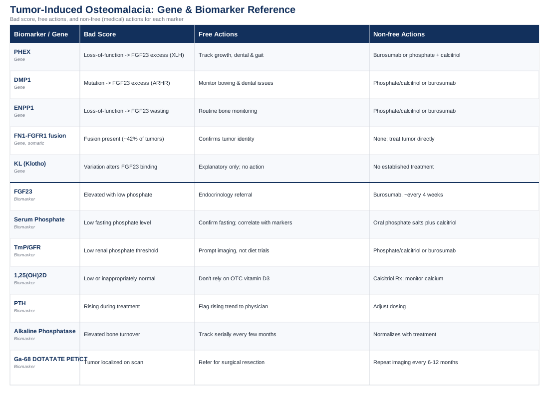
Tumor-induced osteomalacia is driven by one hormone, FGF23, but understanding it well enough to act on it takes more than a single lab value. Below, you'll find the seven biomarkers that, read together, tell the real story: which one flags the tumor's activity, which one distinguishes this from a dozen other causes of bone pain, and which one is actually a scan rather than a blood draw. You'll also see the five genes that matter here, some belonging to the tumor itself, some belonging to inherited look-alike conditions that get confused with this one, and why that distinction changes the entire treatment conversation. A bonus section pulls out ten practical lessons from a widely read longevity book that reframes how proactive bone and muscle tracking should be, and a final section looks at which complementary approaches actually have human evidence behind them for bone pain and fracture-related recovery. The diagram below maps how the tumor's signal moves through the body and where each biomarker sits along that path.
7 Biomarkers That Explain Tumor-Induced Osteomalacia
Tumor-induced osteomalacia, sometimes called oncogenic osteomalacia, is caused almost always by a small, slow-growing tumor called a phosphaturic mesenchymal tumor that secretes large amounts of fibroblast growth factor 23, or FGF23. This single hormone forces the kidneys to dump phosphate into the urine and blocks the kidneys from making active vitamin D, and the combination starves bone of the minerals it needs to harden properly. The result is osteomalacia: soft, painful, fracture-prone bone in adults, and rickets in children. The condition is described in detail in a recent systematic review of the disease mechanism and clinical picture (phosphaturic mesenchymal tumors and FGF23 excess).
The seven biomarkers below are the ones that, taken together, confirm the diagnosis, rule out mimicking conditions, and later track whether treatment (usually tumor removal) is working. None of them should be interpreted alone. A single low phosphate reading can happen for many benign reasons; the pattern across all seven is what tells the real story.
Intact FGF23
This is the central biomarker in the entire condition. FGF23 is a hormone made by bone cells that normally fine-tunes phosphate balance and calcitriol production, described in this review of its physiology (FGF23 and renal phosphate handling). In tumor-induced osteomalacia, the tumor produces far more FGF23 than the body needs, which is what drives everything else on this list.
How to measure it: a blood draw sent to a specialty reference lab (most routine hospital labs do not run this in-house). Expect a cost in the rough range of 200 to 400 US dollars out of pocket, or a standard lab fee if covered by insurance, with results typically available within one to two weeks.
If the score is bad, the plan without supplements or equipment: an elevated FGF23 in the context of low phosphate is the single strongest signal to pursue whole-body functional imaging and a referral to an endocrinologist experienced with FGF23-related disease, because the actual fix for an FGF23-driven tumor is locating and removing it, not a supplement regimen. In the meantime, pacing activity to avoid falls, keeping a simple pain and mobility diary, and avoiding high-impact exercise on painful bones are reasonable, cost-free steps.
If the score is bad, the plan with supplements or equipment: when surgery is not immediately possible, or the tumor cannot be found despite imaging, physicians may prescribe burosumab, a monoclonal antibody that blocks FGF23 directly. In a phase 2 trial, it raised serum phosphorus meaningfully within weeks of starting treatment (burosumab in tumor-induced osteomalacia). It's given as a subcutaneous injection roughly every four weeks, cycled based on phosphate response, and requires regular blood monitoring since hyperphosphatemia occurred in about 14 percent of trial participants. This is a prescription therapy requiring specialist supervision, not a self-directed supplement.
Fasting Serum Phosphate
Low blood phosphate is the hallmark finding that usually triggers the whole workup. Muscles, including the heart, and bone mineralization both depend on adequate phosphate, so chronically low levels explain much of the fatigue, weakness, and bone pain patients describe.
How to measure it: a simple fasting morning blood draw, typically 15 to 40 dollars as part of a metabolic panel, often already included in routine bloodwork.
If the score is bad, the plan without supplements or equipment: confirm the reading is a fasting, non-hemolyzed sample (falsely low results happen with improper handling), then correlate it with FGF23 and TmP/GFR rather than acting on phosphate alone. Reducing high-intensity impact activity temporarily while bones are soft, and prioritizing calcium-adequate meals (dairy, leafy greens, fish with bones), supports the skeleton without overcorrecting a hormonal problem with diet.
If the score is bad, the plan with supplements or equipment: physician-directed oral phosphate salts, typically split into three to six doses through the day because phosphate is cleared quickly by the kidneys, often paired with calcitriol. A large multicenter review found this combination used in roughly 40 percent and 26 percent of patients respectively, with complications including secondary or tertiary hyperparathyroidism in about 3 percent of long-term users and nephrocalcinosis in a smaller fraction (oral phosphate and calcitriol therapy outcomes). This regimen needs periodic kidney ultrasound and PTH checks and is never appropriate to self-dose.
TmP/GFR (Renal Phosphate Threshold)
This calculated value, derived from paired blood and urine phosphate and creatinine, tells you whether low blood phosphate is due to the kidneys inappropriately dumping it, which is exactly what happens under FGF23 excess, as opposed to poor dietary intake or a shift of phosphate into cells. It is one of the most useful values for separating renal phosphate wasting from other causes of low phosphate (TmP/GFR in the workup of hypophosphatemia).
How to measure it: requires a fasting blood sample and a second-void morning urine sample, both sent for phosphate and creatinine, then calculated with a standard nomogram. Cost is generally 50 to 100 dollars combined, since it uses the same assays as basic metabolic testing.
If the score is bad, the plan without supplements or equipment: a low TmP/GFR confirms renal phosphate wasting and should prompt imaging to find the source rather than repeat dietary phosphate trials, which will not correct a renal leak. Documenting symptom timing alongside this result helps a specialist judge disease activity over time.
If the score is bad, the plan with supplements or equipment: management mirrors the phosphate biomarker above, oral phosphate and calcitriol, or burosumab, decided jointly with an endocrinologist based on how low the value is and how quickly a source tumor can realistically be found.
1,25-Dihydroxyvitamin D (Calcitriol)
This is the paradox biomarker. When phosphate is low for ordinary reasons, the body normally responds by raising active vitamin D to pull more phosphate from the gut and bone. In tumor-induced osteomalacia, FGF23 blocks the kidney enzyme that makes this hormone, so calcitriol stays low or inappropriately "normal" despite low phosphate, which is itself a red flag (inappropriately low calcitriol in FGF23-mediated hypophosphatemia).
How to measure it: a dedicated blood test for 1,25-dihydroxyvitamin D, distinct from the common 25-hydroxyvitamin D test used for general vitamin D status. Expect roughly 150 to 220 dollars, since it requires a more specialized assay.
If the score is bad, the plan without supplements or equipment: recognize that ordinary vitamin D3 supplementation will not fix this, because the problem is the kidney's ability to activate it, not a dietary shortage. Safe, moderate sun exposure and dietary vitamin D intake are still reasonable general health habits but should not be relied on to correct this specific pattern.
If the score is bad, the plan with supplements or equipment: physicians prescribe active vitamin D directly, as calcitriol, bypassing the blocked kidney step, generally once or twice daily alongside phosphate. Monitoring blood calcium periodically is essential, since active vitamin D can push calcium up and increase kidney stone risk if overdosed, which is why this is dose-titrated by a specialist rather than taken as an over-the-counter supplement.
Parathyroid Hormone (PTH)
PTH is usually normal in tumor-induced osteomalacia, which is actually a useful diagnostic clue that helps rule out primary hyperparathyroidism or plain vitamin D deficiency as the cause of bone pain. Over the course of treatment, though, PTH can climb as a secondary response to long-term phosphate and calcitriol therapy, so it becomes a monitoring biomarker rather than just a diagnostic one.
How to measure it: a standard blood draw, typically 60 to 120 dollars, widely available at any lab.
If the score is bad, the plan without supplements or equipment: a rising PTH during treatment often reflects the therapy itself rather than a new problem, so the practical step is simply flagging the trend to the treating physician rather than adjusting anything independently.
If the score is bad, the plan with supplements or equipment: if PTH becomes persistently elevated (secondary, or eventually tertiary, hyperparathyroidism), physicians may space out phosphate dosing, adjust calcitriol, or in rare persistent cases consider parathyroid-directed treatment; this occurred in roughly 3 percent of patients on long-term therapy in the multicenter review cited above, underscoring why periodic monitoring matters during medical management.
Alkaline Phosphatase (Bone-Specific)
Alkaline phosphatase reflects how active the bone-remodeling process is, and it tends to run high in osteomalacia because the skeleton is working overtime trying, and failing, to mineralize properly. It is also one of the most convenient values for tracking whether treatment, especially after tumor removal, is working, since it should trend down toward normal as bone healing catches up.
How to measure it: total alkaline phosphatase is a routine, inexpensive test, roughly 20 to 30 dollars, included in most liver or bone panels; the more specific bone-specific isoenzyme test runs closer to 90 to 150 dollars and is useful when liver-related causes need to be excluded.
If the score is bad, the plan without supplements or equipment: an elevated ALP alongside low phosphate and high FGF23 supports the osteomalacia diagnosis and is best tracked serially, ideally every few months, to gauge whether the underlying tumor activity is stable, worsening, or responding to treatment.
If the score is bad, the plan with supplements or equipment: ALP normalizes as the underlying phosphate and calcitriol deficits are corrected, whether through medical therapy or surgery, so there is no separate treatment aimed at ALP itself; it is a trend to watch rather than a target to medicate directly.
Ga-68 DOTATATE PET/CT (Tumor Localization)
Because most phosphaturic mesenchymal tumors carry somatostatin receptors on their surface, this specialized nuclear medicine scan lights up the tumor far more reliably than conventional imaging. In one study, it successfully localized the tumor in just over half of patients, outperforming older scanning techniques (Ga-68 DOTATATE PET/CT for phosphaturic mesenchymal tumor localization). Given how often these tumors hide in unexpected places, sometimes a small bone in a finger or foot, this scan is often the turning point in a diagnostic journey.
How to measure it: performed at specialized nuclear medicine centers, generally costing 3,000 to 6,000 dollars or more depending on region and insurance coverage, and typically requires a formal referral plus prior imaging (MRI or CT) to justify it.
If the score is bad (tumor found), the plan without supplements or equipment: surgical referral for tumor resection is the definitive step, since complete removal is curative in most cases and biomarkers such as phosphate and FGF23 typically normalize within days of successful surgery.
If the score is bad (tumor not found or not resectable), the plan with supplements or equipment: ongoing medical management with phosphate, calcitriol, or burosumab as above, combined with periodic repeat imaging every six to twelve months, since these tumors can be very small and sometimes only become detectable as they grow.
Taken together, these seven values form a coherent picture: FGF23 explains the "why," phosphate and TmP/GFR confirm the renal wasting pattern, calcitriol reveals the paradox that separates this from ordinary vitamin D deficiency, PTH and ALP track disease activity and treatment side effects, and the DOTATATE scan turns a lab pattern into an actual treatment plan by finding the tumor itself.
What the Genetics of FGF23-Driven Bone Disease Reveal
Tumor-induced osteomalacia itself is not usually an inherited condition passed down through families; it is caused by a tumor that develops in adulthood. But the genetics around it matter for two distinct reasons: understanding the somatic mutation inside the tumor, and correctly telling this condition apart from inherited look-alikes that also raise FGF23 and cause similar bone disease from childhood. Getting this distinction right changes the entire treatment conversation, since a lifelong genetic condition is managed very differently than an adult-onset tumor.
Researchers and communicators such as Ali Torkamani, whose work on genomic interpretation emphasizes that a gene variant is a probability, not a verdict, and Gary Brecka, who has popularized the idea that actionable genetic information should change what you actually do day to day, both make a similar point that applies here: knowing a gene's status only matters if it changes your plan. For the genes below, that's exactly the test to apply.
PHEX
PHEX is the gene most people have actually heard of in this space, because loss-of-function mutations in it cause X-linked hypophosphatemia (XLH), the most common inherited cause of FGF23 excess and a frequent look-alike for tumor-induced osteomalacia. PHEX normally helps suppress FGF23 production in bone; when it is broken, FGF23 rises chronically from birth rather than from a tumor (PHEX and FGF23 regulation in XLH). The evidence here is strong and well established in humans, not preliminary.
If the gene is confirmed abnormal, the plan without supplements or equipment: since this is lifelong rather than tumor-driven, the practical value of genetic confirmation is avoiding unnecessary tumor-hunting imaging in a patient whose hypophosphatemia started in early childhood, and instead focusing on growth monitoring, dental health (XLH carries dental abscess risk), and gait/alignment tracking in a pediatric or adult metabolic bone specialist's care.
If the gene is confirmed abnormal, the plan with supplements or equipment: burosumab is now specifically approved for XLH as well as for inoperable tumor-induced osteomalacia, dosed by weight and given roughly every two to four weeks, with the same hyperphosphatemia monitoring described earlier. Conventional oral phosphate and calcitriol therapy remains an option, with the same nephrocalcinosis and hyperparathyroidism monitoring needs.
DMP1
DMP1 mutations cause autosomal recessive hypophosphatemic rickets (ARHR), another inherited FGF23-excess condition that mimics the acquired tumor form. DMP1 normally helps regulate bone mineralization and keeps FGF23 production in check within osteocytes; without it, FGF23 rises independent of any tumor (DMP1 mutations and FGF23 elevation in ARHR). Human evidence for this pathway is solid, based on multiple confirmed family case series, though it is a much rarer condition than XLH.
If the gene is confirmed abnormal, the plan without supplements or equipment: genetic confirmation again shifts the workup away from repeated tumor imaging and toward long-term orthopedic monitoring for bowing deformities and dental complications, typically starting in childhood.
If the gene is confirmed abnormal, the plan with supplements or equipment: management follows the same phosphate/calcitriol or burosumab framework described above, chosen and monitored by a metabolic bone specialist.
ENPP1
ENPP1 loss-of-function mutations cause a related hypophosphatemic rickets subtype by enhancing FGF23-mediated renal phosphate loss (ENPP1 mutations and FGF23-mediated phosphate wasting). This is a newer and less common finding than PHEX or DMP1, and the human evidence base, while real, is built on a smaller number of case reports and case series rather than large cohorts, so it should be treated as reasonably strong but still developing.
If the gene is confirmed abnormal, the plan without supplements or equipment: as with the other inherited forms, the value is diagnostic clarity, confirming this is a lifelong condition rather than a tumor, which changes follow-up from repeat scans to routine metabolic bone monitoring, and in ENPP1 specifically, awareness of an associated risk of soft-tissue and vascular calcification that some patients also carry.
If the gene is confirmed abnormal, the plan with supplements or equipment: the same phosphate, calcitriol, or burosumab framework applies, again managed by a specialist given the added vascular monitoring considerations specific to ENPP1-related disease.
FN1-FGFR1 Fusion (Somatic, Tumor Tissue Only)
This one is different from the three above because it is not inherited at all. It is a somatic mutation found only inside the phosphaturic mesenchymal tumor cells themselves, not in the rest of the body's DNA. Tumor sequencing studies have found this fusion, which drives abnormal growth-factor signaling inside the tumor, in a substantial share of cases, roughly 42 percent of tumors in one series (FN1-FGFR1 fusion in phosphaturic mesenchymal tumors).
If the gene is confirmed abnormal, the plan without supplements or equipment: this finding does not change day-to-day self-care at all; its main value is pathological confirmation of the tumor's identity when a biopsy is ambiguous, and it is an active area of research into whether targeted therapies against this fusion could eventually help in cases where surgery fails.
If the gene is confirmed abnormal, the plan with supplements or equipment: there is no supplement or device relevant to this finding specifically; management remains focused on locating and removing the tumor that carries it.
KL (Klotho)
Klotho is the co-receptor that FGF23 needs in order to bind its receptor effectively, increasing binding affinity more than tenfold (Klotho as an obligate FGF23 co-receptor). Its role here is mostly explanatory rather than currently actionable in clinical practice; there is early, mechanistic-stage human and animal evidence about Klotho variation influencing FGF23 sensitivity, but no established clinical test or treatment built around an individual's Klotho status in tumor-induced osteomalacia today. It is worth knowing about mainly because it explains why FGF23 levels alone don't always perfectly predict disease severity.
The Book That Reframes How You Think About Catching Disease Early: Lessons from Outlive
Peter Attia's book Outlive is not written about tumor-induced osteomalacia specifically, and it would be misleading to suggest otherwise. But its central argument, that most serious health problems should be caught and addressed years before they become a crisis, and that bone and muscle health deserve the same proactive tracking usually reserved for cholesterol or blood pressure, maps directly onto why a seven-biomarker panel like the one above matters more than waiting for a fracture to happen. Below are ten of the book's most relevant ideas for anyone dealing with, or recovering from, a bone-wasting condition.
Medicine 3.0 Means Acting Before the Crisis
Attia's core distinction is between reactive medicine, which waits for a diagnosis to appear on a scan, and proactive medicine, which tracks trends and intervenes early. For a slow-growing tumor that can take years to surface on imaging, this philosophy argues directly for repeat, structured biomarker tracking rather than a single test and a shrug.
The "Four Horsemen" Framework Applies Beyond Its Original Four
The book organizes late-life disability around four major disease categories, but its underlying lesson, that the years-long lead time before a diagnosable event is where the real leverage lives, applies just as well to a bone condition that spends years disguised as vague fatigue and pain.
Train for the "Marginal Decade"
Attia frames fitness around function in the last decade of life, not appearance or performance today. For someone whose bones have been weakened by years of phosphate wasting, this reframes rehabilitation goals around concrete physical tasks, carrying groceries, getting off the floor, climbing stairs, rather than abstract strength numbers.
Muscle Loads Bone, and Loaded Bone Remodels
A recurring theme is that resistance training is not just about muscle, it mechanically stimulates bone to remodel and strengthen. This is especially relevant during recovery after tumor removal, when bone mineralization is actively normalizing and gentle, progressive loading (once cleared by a physician) supports that process.
Zone 2 Training Builds the Engine Underneath Everything Else
Attia emphasizes low-intensity aerobic training as a foundation for metabolic health and recovery capacity. For patients who have been deconditioned by years of pain and limited mobility, this offers a low-impact, joint-friendly starting point before higher-intensity training is appropriate.
Stability Training Is a Fracture-Prevention Tool, Not an Afterthought
The book treats balance and stability work as seriously as strength work, specifically because falls, not just weak bones, are what cause fractures. For anyone whose bones have been softened by osteomalacia, this is arguably the single most protective and lowest-risk category of exercise available.
Nutrition Should Be Personalized, Not Generic
Attia pushes back against one-size-fits-all diet advice, arguing protein needs and nutrient priorities vary by individual context. For bone-disease recovery, this supports working with a dietitian familiar with phosphate and calcium needs rather than following generic "bone health diet" lists.
Sleep Is Part of the Repair Process, Not Separate From It
The book treats sleep as foundational infrastructure for recovery and metabolic regulation, not a lifestyle nicety. For a body actively remineralizing bone, adequate sleep is a low-cost, side-effect-free lever worth prioritizing during recovery.
Emotional Health Belongs in the Same Framework as Physical Health
Attia is candid that proactive medicine includes mental health, not just lab values. This matters directly for a condition known for years of dismissed symptoms and diagnostic frustration; acknowledging that toll, and addressing it, is part of a complete recovery plan, not a distraction from it.
Track Trends, Not Single Data Points
Perhaps the most directly applicable idea: Attia repeatedly stresses that one lab value means little without a trend line. This is precisely why the biomarker panel above is described as a pattern to track over time, phosphate, FGF23, ALP, calcitriol, together and repeatedly, rather than a single result to react to once.
Complementary Approaches Worth Considering
No complementary therapy treats the tumor or corrects FGF23 levels, and none of the following should be mistaken for a substitute for locating and removing the underlying tumor or for prescribed phosphate, calcitriol, or burosumab therapy. What they do have is real human evidence for bone density support, pain management, and recovery-related outcomes in related musculoskeletal populations, which makes them reasonable, low-risk additions once a physician has confirmed they're safe given someone's current fracture risk.
Yoga
Yoga combines gentle weight-bearing positions with controlled breathing and balance work, which makes it relevant for people managing fragile, undermineralized bone who still need some form of safe loading to support skeletal health. Because many yoga poses load the spine and hips through body weight alone rather than external resistance, it can be a gentler entry point than traditional strength training for someone recovering from osteomalacia-related fractures or pain.
A widely cited pilot study by Loren Fishman followed over 700 participants doing a 12-minute daily yoga routine and found meaningful bone mineral density gains in the spine, hip, and femur among those who stayed compliant, with no serious yoga-related injuries reported (12-minute yoga regimen and bone mineral density). This evidence comes from people with osteopenia and osteoporosis generally, not tumor-induced osteomalacia specifically, so it should be read as supportive rather than condition-specific.
In practice, this means starting only once a physician confirms current fracture risk is low enough for weight-bearing poses, choosing a slow, alignment-focused style (restorative or gentle hatha rather than power yoga), avoiding deep spinal flexion poses if vertebral fragility is a concern, and working with an instructor experienced with osteoporosis-safe modifications, ideally two to three short sessions a week rather than daily intensity from the start.
Tai Chi
Tai chi is a slow, low-impact practice combining weight shifting, balance, and controlled movement, which makes it particularly relevant for fall and fracture prevention in people with compromised bone strength, since falls, not just weak bone, are what usually cause the fractures that matter most.
A meta-analysis of tai chi in populations with low bone density found a statistically significant improvement in vertebral bone mineral density, though it did not find a significant fall-prevention benefit in that specific analysis (tai chi and bone mineral density in osteoporosis). As with yoga, this evidence comes from general low-bone-density populations rather than tumor-induced osteomalacia specifically, so the honest takeaway is "likely helpful for bone and balance broadly" rather than "proven for this exact condition."
Realistically, this looks like two to three 20 to 30 minute sessions per week, ideally through a class rather than solo video-following at first so form and balance cues are corrected in real time, and it's a good option specifically for patients whose pain limits more vigorous exercise, since the movement intensity is easy to scale down.
Mindfulness-Based Stress Reduction (MBSR)
Chronic bone pain and years of unresolved diagnostic uncertainty both take a measurable toll on stress and pain perception, and MBSR is one of the better-studied non-drug approaches for chronic pain management generally, making it relevant as a coping and pain-modulation tool alongside, not instead of, medical treatment.
A network meta-analysis covering 21 studies and nearly 2,000 participants found MBSR produced meaningful improvement in chronic pain outcomes, comparable in magnitude to cognitive behavioral therapy (MBSR for chronic pain: a network meta-analysis). This evidence spans chronic pain conditions broadly rather than tumor-induced osteomalacia specifically.
A standard approach is an 8-week structured MBSR course, roughly 30 to 45 minutes of practice most days, either in person or through a validated app-based program; there are no known physical side effects, though some people initially find sitting practice uncomfortable if pain is severe, in which case guided body-scan or lying-down variations are reasonable substitutes.
Massage Therapy
Musculoskeletal pain and muscle guarding are common in osteomalacia, both from the bone disease itself and from compensatory movement patterns people develop to avoid painful motions, and massage therapy has reasonably solid evidence for reducing musculoskeletal pain generally.
A meta-analysis of randomized controlled trials found massage therapy produced a statistically significant reduction in pain compared to sham treatment or no treatment (massage therapy for pain: a systematic review and meta-analysis). Again, this evidence is general to musculoskeletal pain populations, not specific to tumor-induced osteomalacia.
Given fragile bone is a concern in this condition, the practical guidance is to use gentle, low-pressure techniques rather than deep tissue work, disclose the osteomalacia diagnosis to the therapist beforehand so pressure and positioning are adjusted accordingly, and avoid direct heavy pressure over any bone with a known or suspected stress fracture, typically once every one to two weeks as a supportive addition to medical care.
Conclusion
Tumor-induced osteomalacia is unusual among chronic conditions in that it has a single, identifiable driver, an FGF23-secreting tumor, and a genuinely curative fix when that tumor is found and removed. The seven biomarkers above (FGF23, phosphate, TmP/GFR, calcitriol, PTH, alkaline phosphatase, and Ga-68 DOTATATE PET/CT) exist to shorten the path between years of unexplained symptoms and that specific answer, while the genetic picture helps rule out lifelong look-alike conditions that need a different long-term plan entirely. Complementary approaches like yoga, tai chi, mindfulness training, and massage have real evidence behind them for bone and pain-related outcomes, but they support recovery around the edges; they do not replace finding and treating the source.
If any of this resonates, the most useful next step is a concrete one: bring this list of seven biomarkers to your physician or endocrinologist, ask specifically about FGF23 and TmP/GFR testing if they haven't already been done, and if a pattern consistent with FGF23 excess turns up, ask directly about Ga-68 DOTATATE PET/CT referral. Accurate information doesn't replace medical care, but it can make the conversation with your care team sharper, faster, and considerably less frustrating than the years many patients spend before getting there.
Musculoskeletal Endocrine & Metabolic
Musculoskeletal: Bone Conditions