This article contains AI generated content.
· UpdatedTumoral Calcinosis: 4 Genes and 7 Biomarkers to Track
Introduction
Tumoral calcinosis occupies a strange corner of medicine: rare enough that most people wait years for an accurate diagnosis, yet specific enough that once you understand the underlying biology, the clinical picture becomes surprisingly clear. The calcium deposits forming around joints and in soft tissue are not random. They are the predictable end result of a disrupted phosphate regulation system, and that system has a genetic blueprint that can now be read directly.
Most generic advice about calcium disorders misses the mark entirely for this condition. Reducing dairy, taking standard calcium supplements, or following broad bone-health protocols often make no meaningful difference — and in some cases make things worse — because the core problem is not calcium intake but phosphate retention. The kidneys are failing to excrete enough phosphate, usually because a specific hormone called FGF23 is not functioning correctly. That is where the investigation needs to start.
Two layers of analysis make this condition genuinely manageable with precision. The first is genetic: knowing which mutation is present changes the interpretation of every lab result and shapes every management decision. The second is biomarker-based: a small panel of blood and urine tests can reveal where the metabolic disruption is most severe, how well it is being controlled, and whether the intervention approach is working.
This article covers the four genes most directly responsible for heritable tumoral calcinosis and translates each mutation into a practical plan. It then covers seven biomarkers that track the condition's activity most reliably, with guidance on measurement, interpretation, and response. The goal is not to replace a specialist — this condition requires one — but to give you the background to make those conversations more productive and the monitoring more targeted.
Summary
This article maps tumoral calcinosis from its genetic roots to its most trackable lab markers. The four genes — GALNT3, FGF23, KLOTHO, and SAMD9 — each disrupt phosphate metabolism in a distinct way, and each calls for a different management approach. Knowing whether a mutation sits in GALNT3 versus SAMD9 changes not just treatment but the expected lab signature entirely: one form drives rampant hyperphosphatemia; the other produces normal phosphate with calcinosis that looks identical clinically.
On the biomarker side, seven measurements stand out as the most clinically useful: serum phosphate, intact FGF23, calcitriol (1,25-dihydroxyvitamin D), PTH, the calcium-phosphate product, serum Klotho, and alkaline phosphatase. Together these markers tell a story that individual readings cannot — and each has actionable thresholds worth knowing. The article also covers what cutting-edge research on the phosphate-aging axis reveals about why this condition matters beyond the deposits themselves, and which complementary approaches have genuine human evidence for the specific burdens this disease creates.
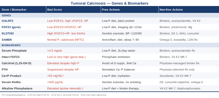
The Genetic Root of Tumoral Calcinosis: 4 Genes That Drive the Disease
Understanding which gene is disrupted is not just academic. In tumoral calcinosis, the mutation determines the entire metabolic signature — which biomarkers will be elevated, which interventions are likely to help, and which are likely to be irrelevant or harmful. Treating all forms of the condition identically is one of the most consistent clinical errors in managing this disease.
The FGF23-Phosphate Axis: Why Everything Connects Here
Three of the four genes discussed below disrupt a single physiological axis. FGF23 (Fibroblast Growth Factor 23) is a hormone produced primarily by bone cells (osteocytes) whose central job is to instruct the kidneys to excrete phosphate in urine rather than reabsorb it. When FGF23 works correctly, blood phosphate stays within a tight normal range — roughly 2.5 to 4.5 mg/dL in adults. When it fails, phosphate accumulates. At high enough concentrations, phosphate combines with calcium to form hydroxyapatite crystals that deposit preferentially in periarticular soft tissue, bursae, and skin, producing the characteristic masses of hyperphosphatemic familial tumoral calcinosis (HFTC).
Three elements are required for FGF23 signaling to work: the FGF23 protein must be produced intact (GALNT3 preserves this), the FGF23 protein must be structurally functional (FGF23 gene encodes it), and the kidney receptor complex must be able to receive the signal (Klotho enables this binding). A mutation in any one of these three components disrupts the same downstream effect — excess phosphate accumulation — but leaves a different laboratory signature that distinguishes one from another. That distinction is the key to targeted management.
SAMD9 operates through a completely different mechanism, producing calcinosis in the complete absence of hyperphosphatemia.
Gene 1: GALNT3 — The Most Common Genetic Cause
GALNT3 encodes an enzyme called polypeptide N-acetylgalactosaminyltransferase 3. This enzyme attaches a specific sugar group — a process called O-glycosylation — to the FGF23 protein, protecting it from proteolytic cleavage before it can reach the kidney. Without this protective glycosylation, FGF23 is broken down into inactive C-terminal fragments almost immediately after production. The kidney never receives a meaningful phosphate-excretion signal, phosphate retention begins, and calcinosis follows in susceptible periarticular tissue.
This is the most common genetic cause of HFTC. Inheritance is autosomal recessive — both alleles must carry a pathogenic variant for the full phenotype to manifest. Carriers with a single mutation are typically asymptomatic, though subtle phosphate-handling differences have been observed. The laboratory signature is distinctive: elevated C-terminal FGF23 with low or undetectable intact FGF23, accompanied by hyperphosphatemia and elevated calcitriol (1,25-dihydroxyvitamin D). This pattern helps confirm the diagnosis and distinguishes it from other genetic forms before sequencing results are available.
Topaz et al. first identified GALNT3 mutations as the cause of familial tumoral calcinosis in 2004, a landmark finding that shifted the understanding of the condition from idiopathic to mechanistic and opened the door to rational treatment.
If the gene is mutated: the plan without supplements
The most impactful first step is strict dietary phosphate restriction, targeting below 800 mg of dietary phosphate per day. This requires more than avoiding dairy. Phosphate additives — polyphosphates, disodium phosphate, phosphoric acid — are present in most processed foods, colas, packaged meats, and fast food, and they are absorbed at nearly 90-100% efficiency compared to 40-60% for organic phosphate in whole foods. Reading ingredient labels for any phosphate-containing additive is essential and makes a meaningful difference in absorbed phosphate load.
Protein sources matter more than most people realize. Plant proteins in legumes, tofu, and whole grains contain phosphate bound to phytate, which humans cannot fully hydrolyze. Animal proteins and commercial protein supplements deliver phosphate in a highly bioavailable inorganic form. Shifting protein intake toward plant-based whole food sources can reduce absorbed phosphate by 300-400 mg/day without reducing total protein intake.
Hydration supports urinary phosphate clearance: 2.5-3 liters of fluids daily, primarily water, maintains urinary flow and reduces the concentration gradient that favors crystal precipitation. It does not normalize phosphate in GALNT3-related HFTC — the tubular defect is too severe for hydration alone to correct — but it reduces the periarticular concentration driving deposit formation.
Physical activity should preserve joint mobility without loading inflamed or calcified areas. Targeted physical therapy focused on muscle groups adjacent to calcified joints prevents secondary contractures. Range-of-motion exercises in water (aquatherapy) reduce joint stress while maintaining meaningful function.
If the gene is mutated: the plan with supplements or medication
Phosphate binders are the pharmacological foundation and must be prescribed and monitored by a physician. Sevelamer carbonate binds dietary phosphate in the gut before absorption; standard dosing ranges from 800 to 2400 mg taken with each meal. Calcium carbonate can serve as a phosphate binder at doses taken with food, but requires caution to avoid compounding the calcium-phosphate burden. Aluminum hydroxide is effective but should be limited to short courses due to aluminum accumulation toxicity with prolonged use.
Acetazolamide (a carbonic anhydrase inhibitor) increases urinary phosphate excretion and has been used in documented HFTC cases at 250-500 mg twice daily. It requires regular monitoring of serum bicarbonate and potassium, as it promotes bicarbonate loss and can cause metabolic acidosis with prolonged use. It should never be initiated without physician oversight.
Vitamin K2 (MK-7 form) at 180-360 mcg daily activates matrix Gla protein (MGP), the most potent endogenous inhibitor of soft-tissue calcification. The evidence specific to HFTC is limited to case observations, but the mechanism is well-established in both animal and human vascular calcification studies. It can be taken continuously without cycling; no serious side effects have been documented at these doses. This is not the same as vitamin K1 (phylloquinone) — MK-7 specifically is required for MGP carboxylation.
An important caution: supplemental vitamin D in any form requires careful physician guidance in HFTC. Calcitriol (the active form) is typically already elevated due to failed FGF23 suppression of its synthesis. Adding cholecalciferol without monitoring 25(OH)D and serum calcium risks significantly worsening the disease state — this is one of the most common errors seen when generic supplementation advice is applied to this condition.
Gene 2: FGF23 — When the Signal Itself Is Broken
Mutations in the FGF23 gene itself cause a second, less common form of HFTC. Here, the FGF23 protein is either not secreted properly from osteocytes or is rapidly degraded regardless of GALNT3 protection. The downstream result — inadequate phosphaturic signaling, phosphate retention, and calcinosis — is identical to GALNT3-related disease, but the laboratory signature is different in a clinically important way.
In FGF23-mutation HFTC, both intact FGF23 and C-terminal FGF23 fragments are typically low or undetectable. This contrasts with GALNT3 mutation, where C-terminal fragments are elevated because only the intact form is degraded. The fragment pattern from the FGF23 assay is the fastest way to distinguish these two forms before genetic sequencing results return, and it should be ordered specifically with this distinction in mind.
Severity varies even within families carrying the same FGF23 mutation, suggesting that modifier genes — likely GALNT3, KLOTHO, or other phosphate regulators — influence the phenotype. This variability makes genetic testing more informative than clinical presentation alone for prognosis and family counseling.
If the gene is mutated: the plan without supplements
Dietary phosphate restriction remains the first-line lifestyle intervention with the same targets and food avoidance strategies described for GALNT3 mutation. An additional consideration here is the value of regular imaging surveillance every 6-12 months — CT or X-ray of affected regions — to track whether existing deposits are stable, regressing (which does happen with effective treatment), or actively enlarging. This information directly guides when to escalate pharmacological management and when to involve a surgeon.
Sleep quality is worth emphasizing here. Poor sleep increases pro-inflammatory cytokines (particularly IL-6 and TNF-α), and inflammation accelerates periarticular deposit formation in susceptible tissue. Targeting 7-9 hours of consolidated sleep with consistent timing is a practical and cost-free intervention that most disease management protocols do not mention.
If the gene is mutated: the plan with supplements or medication
The phosphate binder and acetazolamide strategies from GALNT3 management apply equally. One additional agent used in isolated HFTC cases is probenecid (1-2 g/day), a uricosuric drug with secondary phosphaturic effects at the proximal tubule. Its practical limitation is a long list of drug interactions; a full medication review is required before initiating.
Magnesium glycinate or malate at 300-400 mg of elemental magnesium daily is a worthwhile supportive supplement. Magnesium competes with calcium at certain renal tubular transporters, inhibits hydroxyapatite crystal growth directly, and supports overall renal function. Deficiency is common in people following restricted diets and worsens the calcium-phosphate dysregulation. It can be taken continuously; the primary dose-limiting side effect is loose stools at higher doses, which can be managed by splitting the dose between morning and evening.
Gene 3: KLOTHO — When the Receptor Cannot Listen
Klotho is a transmembrane protein expressed predominantly in the kidney's distal tubule. FGF23 cannot exert its phosphaturic effect without binding to a receptor complex that requires Klotho as a co-receptor. Even if FGF23 is produced normally and is structurally intact, it cannot signal without a functional Klotho partner. Mutations in the KLOTHO gene produce a rare but mechanistically instructive form of HFTC with a distinctive lab signature: markedly elevated intact FGF23 alongside hyperphosphatemia. The body senses the high phosphate and produces more FGF23 in response — the signal is being sent, but the receiver is broken. No other genetic form of HFTC produces this combination, making it a reliable pointer toward KLOTHO testing.
A pivotal case confirmed this mechanism: a 13-year-old with severe tumoral calcinosis, undetectable serum Klotho, and elevated intact FGF23, caused by a homozygous translocation disrupting the KLOTHO gene (Ichikawa et al., New England Journal of Medicine, 2007). This case established that Klotho deficiency is a cause of HFTC, not just a consequence of kidney disease.
What makes Klotho clinically important beyond its FGF23 co-receptor function is its role as an anti-aging, anti-fibrotic, and anti-inflammatory protein. Low Klotho is independently associated with accelerated vascular smooth muscle calcification, kidney disease progression, left ventricular hypertrophy, and increased all-cause mortality. In Klotho-mutation HFTC, the disease burden extends beyond periarticular deposits into a broader cardiovascular and renal phenotype that requires monitoring from diagnosis onward.
If the gene is mutated: the plan without supplements
Dietary phosphate restriction applies with the same principles and food avoidance strategies as the other HFTC forms. The critical added dimension is aggressive kidney function preservation. Klotho-deficient states accelerate renal tubular damage, and any nephrotoxic exposure compounds this risk. Avoiding long-term NSAID use for pain management, maintaining blood pressure below 120/80 mmHg, and staying well-hydrated are more urgent in Klotho-deficiency HFTC than in the other forms. Regular renal function monitoring — eGFR, creatinine, cystatin C — every 6 months is warranted from diagnosis.
Aerobic exercise at 150 minutes per week of moderate intensity has evidence in both animal models and small human studies for supporting endogenous Klotho expression in functional renal tubular tissue. A single functional Klotho allele, or remaining expression from affected tissue, may still respond to exercise-induced upregulation. Swimming and cycling are preferable to high-impact activities when periarticular deposits limit joint range of motion.
Cardiovascular monitoring — blood pressure, lipid panel, echocardiogram — should be integrated into annual follow-up given Klotho's role in vascular biology. This is not typically included in standard tumoral calcinosis protocols, but it is warranted in Klotho-mutation cases and changes the risk picture substantially.
If the gene is mutated: the plan with supplements or medication
Phosphate binders apply as in other HFTC forms. For Klotho-specific support, cholecalciferol (vitamin D3) at modest doses — 1000-2000 IU/day — has mechanistic support for upregulating Klotho expression in renal tissue at physiological vitamin D levels. This is the precursor form, not calcitriol. Unlike direct calcitriol supplementation, cholecalciferol at these doses carries lower risk of hypercalcemia when monitored with 25(OH)D and serum calcium every 3-6 months. Do not use without monitoring.
Curcumin with piperine (500 mg curcumin standardized to 95% curcuminoids, with 5 mg piperine, once daily) has demonstrated increased renal Klotho expression and reduced calcification markers in multiple rodent models with phosphate dysregulation pathology. Human evidence remains limited but the safety profile at this dose is well established across multiple clinical trials for other indications. A reasonable approach is 3-month cycles with a 4-week break between cycles. Potential interactions include mild antiplatelet activity and modest reduction in iron absorption.
Omega-3 fatty acids (2-3 g/day combined EPA+DHA from high-quality triglyceride-form fish oil) support vascular and renal inflammatory management — both particularly relevant given Klotho's role in cardiovascular biology and the accelerated vascular calcification risk in this variant. Continuous use is appropriate; a 2-4 week pause before any planned surgery is advisable due to mild antiplatelet effects.
Gene 4: SAMD9 — The Normophosphatemic Exception
SAMD9 encodes a growth-regulatory protein involved in cellular stress responses, innate immunity signaling, and endosomal trafficking. Mutations in SAMD9 cause normophosphatemic familial tumoral calcinosis (NFTC) — a form of the condition in which serum phosphate is completely normal, FGF23 levels are unaffected, and the entire FGF23-phosphate axis described above is intact and functioning correctly.
This is arguably the most important distinction in the genetics of tumoral calcinosis. A normal serum phosphate in a patient with periarticular soft-tissue deposits does not rule out a heritable cause — it points toward SAMD9. The mechanism is less fully characterized than the HFTC forms, but it likely involves dysregulation of TGF-β signaling and mTOR activity in periarticular connective tissue, resulting in ectopic mineralization through a pathway that does not require systemic phosphate elevation to drive crystal formation.
SAMD9 mutations in different positions on the gene are also associated with MIRAGE syndrome and other complex conditions, indicating that this gene has broad biological importance beyond calcinosis. Genetic counseling is particularly important for SAMD9-positive families.
If the gene is mutated: the plan without supplements
Because phosphate dysregulation is not the driver in NFTC, dietary phosphate restriction offers minimal benefit and need not be the primary focus. The more relevant lifestyle interventions target inflammation and local tissue stress. An anti-inflammatory dietary pattern — Mediterranean-style, rich in omega-3 fats, polyphenol-dense vegetables, extra-virgin olive oil, and low in processed foods and refined carbohydrates — reduces systemic cytokine burden that may contribute to periarticular deposit formation.
Sleep quality is relevant in a specific way here: SAMD9 is upregulated in response to cellular stress signals, and chronic sleep deprivation elevates pro-inflammatory cytokines that may amplify pro-calcification signaling in susceptible tissue. Targeting 7-9 hours of consolidated sleep with consistent timing is a practical, zero-cost intervention.
For pain management, non-NSAID approaches are preferable to protect kidney function over the long term: ice and heat cycling, topical diclofenac gel (lower systemic absorption than oral NSAIDs), targeted physical therapy, and transcutaneous electrical nerve stimulation (TENS) all provide meaningful symptom management without systemic anti-inflammatory risk.
Surgical removal of deposits remains the primary intervention when calcinosis significantly impairs function in NFTC as in HFTC. Recurrence rates post-surgery may differ from HFTC forms and should be discussed specifically with the operating surgeon and a geneticist familiar with SAMD9 biology.
If the gene is mutated: the plan with supplements or medication
Omega-3 fatty acids (EPA + DHA combined, 2-4 g/day from a high-EPA triglyceride-form supplement) are the most mechanistically relevant supplement here, given the TGF-β and inflammatory components of NFTC. Continuous use is appropriate; a brief break of 4 weeks every 6 months is a reasonable precaution around antiplatelet effects, particularly if other anticoagulant or antiplatelet medications are used.
Boswellia serrata extract (standardized to 65% boswellic acids, 300-500 mg twice daily) inhibits 5-lipoxygenase and reduces leukotriene production — an anti-inflammatory mechanism relevant to TGF-β-driven periarticular pathology. It is a well-tolerated alternative to long-term NSAIDs for periarticular pain and inflammation management in NFTC. A reasonable cycling approach is 8-12 weeks of continuous use, followed by reassessment. Side effects are minimal; occasional GI discomfort resolves when taken with food.
Low-dose naltrexone (1.5-4.5 mg at bedtime) has emerging evidence in inflammatory and innate immunity-related conditions through its modulation of TLR4 signaling. The evidence is early-phase and does not include SAMD9-specific studies, but given SAMD9's role in innate immune regulation, the mechanistic logic is plausible. This requires a prescription and physician supervision, and baseline liver function tests are recommended before initiation.
7 Biomarkers That Tell the True Story of Tumoral Calcinosis
Genetic diagnosis tells you why tumoral calcinosis developed. Biomarkers tell you how it is behaving right now and whether treatment is working. In a condition where therapeutic response is slow and clinical progression can be subtle for months, lab monitoring is not optional — it is the primary feedback system available between imaging appointments. These seven measurements, interpreted together, give the most complete and actionable picture of disease activity.
Biomarker 1: Serum Phosphate (Inorganic Phosphate)
Serum phosphate is the central metabolic marker in hyperphosphatemic forms of tumoral calcinosis. It reflects the net result of dietary phosphate intake, intestinal absorption, renal excretion, and cellular uptake — making it the direct readout of how well the FGF23-phosphate axis is (or is not) functioning. A persistently elevated phosphate above 4.5-5.0 mg/dL in adults indicates that the driving force for calcium-phosphate crystallization is active and that the current management strategy is not sufficient.
The timing of the blood draw matters critically. Phosphate follows a diurnal rhythm and rises significantly after meals — postprandial phosphate can be 20-40% higher than fasting values. Fasting morning specimens, drawn before 10 am after an 8-12 hour fast, are the only clinically comparable values across appointments. Random draws introduce so much variability that tracking treatment response becomes unreliable.
In SAMD9-related NFTC, serum phosphate will be normal — its value here is diagnostic (ruling out other conditions) rather than for ongoing monitoring.
How to measure it
Standard comprehensive metabolic panel or a dedicated phosphorus test at any clinical laboratory. Cost: $10-30 as a standalone test, usually included in comprehensive panels. Fasting sample drawn in the morning is required for meaningful comparison across visits. Monitoring frequency: every 3-6 months during active treatment adjustment; every 6-12 months when values are stable.
If the level is elevated: the plan without supplements
The dietary approach is the most immediate lever: eliminate all phosphate-additive foods from the diet, shift toward whole food plant protein sources, and increase daily water intake to 2.5-3 liters. This alone can reduce fasting serum phosphate by 0.5-1.0 mg/dL in patients with good adherence. Phosphate-reduction apps and food databases that flag phosphate additives by ingredient name are practically useful for tracking this.
If the level is elevated: the plan with supplements or medication
Phosphate binders (sevelamer carbonate, lanthanum carbonate, calcium carbonate used as a binder with meals) are the primary pharmacological tool, prescribed and dosed by a physician. Magnesium supplementation (300-400 mg elemental) provides secondary support. For refractory hyperphosphatemia, acetazolamide under physician oversight addresses the tubular reabsorption component.
Biomarker 2: Intact FGF23
Intact FGF23 is the active, unsplit form of the phosphate-regulating hormone. Its blood level answers a critical question: is FGF23 being produced and signaling effectively? The answer pattern varies by genetic cause, making FGF23 measurement as much a diagnostic fingerprinting tool as a monitoring marker.
In GALNT3 mutation: intact FGF23 is low (being degraded before it can signal); C-terminal FGF23 fragments are high (they accumulate). In FGF23 mutation: both intact and C-terminal FGF23 are low. In KLOTHO mutation: intact FGF23 is markedly elevated (the body produces more in response to persistent hyperphosphatemia, but the receptor cannot respond). This three-way pattern is the fastest pre-sequencing approach to narrowing the genetic diagnosis.
How to measure it
Intact FGF23 requires a specialized assay not available at all laboratories; it may require referral to a reference laboratory. Samples require careful handling — serum must be separated and frozen quickly, as FGF23 degrades at room temperature within hours. Both intact and C-terminal assays should ideally be ordered simultaneously for the diagnostic pattern. Cost: $80-250 depending on assay type and laboratory. Frequency: at diagnosis and every 6-12 months, or sooner if clinical status changes significantly.
If the level is abnormal: the plan without supplements
The dietary phosphate restriction approach targets the downstream consequence (elevated phosphate) rather than FGF23 directly, since the protein's dysfunction is genetically determined. Reducing phosphate load does, however, reduce the stimulus that drives compensatory FGF23 production, which can shift the intact/fragment ratio meaningfully over months of strict adherence.
If the level is abnormal: the plan with supplements or medication
No supplement directly restores FGF23 protein function. The management approach targets the pathway consequences: phosphate binders reduce phosphate burden; vitamin K2 MK-7 activates MGP to reduce soft-tissue deposit formation; and in KLOTHO-mutation cases specifically, curcumin and omega-3s support receptor pathway biology at adjacent points in the signaling chain. These do not fix the underlying mutation but reduce the measurable harm it causes.
Biomarker 3: 1,25-Dihydroxyvitamin D (Calcitriol)
Calcitriol is the active, hormonal form of vitamin D produced in the kidney. Its synthesis is normally stimulated by PTH and suppressed by FGF23. In HFTC, the failure of FGF23 signaling removes the usual inhibition of calcitriol synthesis. The result is elevated calcitriol despite normal or elevated phosphate — a combination that does not occur in normal physiology and is strongly suggestive of an FGF23-pathway disorder when seen together.
Elevated calcitriol drives increased intestinal absorption of both calcium and phosphate, compounding the phosphate retention already driven by impaired renal excretion. Calcitriol is therefore both a diagnostic marker and an active amplifier of the disease process, making its measurement essential for understanding the full metabolic picture in any suspected HFTC.
How to measure it
The specific test to order is 1,25-dihydroxyvitamin D, not to be confused with 25-hydroxyvitamin D (which measures vitamin D stores and should also be tracked but tells a different story). Both should be ordered. Cost: $50-150 depending on the laboratory. Frequency: at diagnosis and every 6-12 months; more frequently if vitamin D supplementation is being adjusted or if calcitriol-related symptoms (hypercalcemia, kidney stones) arise.
If the level is elevated: the plan without supplements
Avoid all supplemental vitamin D without explicit physician guidance specific to HFTC. Reduce calcium-rich food intake modestly if the calcium-phosphate product is also elevated. Limiting sun exposure to skin is rarely necessary but may be discussed with a physician if calcitriol is severely elevated and serum calcium is rising.
If the level is elevated: the plan with supplements or medication
No safe supplement lowers calcitriol directly. If calcitriol is significantly elevated and driving hypercalcemia, the intervention is pharmacological and physician-managed — primarily through strict phosphate restriction and binder therapy, which reduces the phosphate burden that sustains high calcitriol. In severe cases, physician-supervised pharmacological suppression of calcitriol synthesis may be considered as part of a specialist management plan.
Biomarker 4: Parathyroid Hormone (PTH)
PTH normally rises when serum calcium falls or phosphate rises, instructing the kidneys to both activate calcitriol and excrete phosphate. In HFTC, the phosphate-PTH relationship is distorted: phosphate rises, but PTH response is often blunted or paradoxically suppressed — partly because calcitriol is already elevated and calcitriol strongly suppresses PTH secretion. The result is a relatively low or inappropriately normal PTH in the setting of elevated phosphate and calcitriol, which is diagnostically helpful.
Tracking PTH across visits reveals whether the parathyroid-kidney-bone axis is responding appropriately to treatment. A rising PTH during phosphate binder therapy may indicate that treatment has swung too far, causing secondary hypocalcemia. A persistently suppressed PTH despite ongoing hyperphosphatemia suggests that calcitriol elevation is dominating the signal, which requires a different management adjustment than phosphate binders alone.
How to measure it
Intact PTH (iPTH) measured on a standard laboratory panel. Cost: $30-80. Should be drawn fasting, alongside serum calcium and phosphate from the same blood draw, for meaningful correlation. Frequency: every 3-6 months during active treatment adjustment; every 6-12 months when stable.
If the level is abnormal: the plan without supplements and with supplements
PTH management in HFTC is pharmaceutical and requires physician direction. The core principle is to normalize the calcium-phosphate balance through phosphate reduction rather than attempting to directly manipulate PTH through calcium or calcitriol supplementation. Aggressive calcium supplementation to suppress PTH worsens the calcium-phosphate product and accelerates deposits — a common pitfall when calcium-PTH relationships are managed without accounting for the HFTC context.
Biomarker 5: Calcium-Phosphate Product (Ca × P)
The calcium-phosphate product — calculated by multiplying serum calcium (mg/dL) by serum phosphate (mg/dL) — predicts the thermodynamic tendency for calcium phosphate crystals to precipitate in soft tissue. A product above 55 mg²/dL² is the threshold above which spontaneous periarticular deposition risk rises substantially. In active HFTC, the Ca×P product may reach 70-90 or higher during poorly controlled periods.
This single calculated value may be the most clinically actionable number in HFTC monitoring, because it directly represents the chemical driving force behind calcinosis progression. Reducing the Ca×P product — through any combination of phosphate restriction, phosphate binder therapy, or judicious calcium management — directly reduces the precipitation rate regardless of which specific gene mutation is causing the underlying disorder.
How to measure it
No additional test is required. It is calculated from a standard comprehensive metabolic panel: Ca (mg/dL) × Phosphate (mg/dL). Both values should be from a fasting, morning, same-draw specimen for accurate comparison. Cost: included in any comprehensive metabolic panel ($20-60). Frequency: every 3-6 months when values are above target; every 6-12 months when consistently below 55.
If the product is elevated: the plan without supplements
Phosphate restriction is the primary dietary lever since calcium is usually normal or only mildly elevated in HFTC. Avoid calcium-containing antacids, calcium-fortified foods in excess, and any supplemental calcium during periods when the Ca×P product is above 55. Daily hydration to 2.5-3 liters maintains the dilution that slows crystal nucleation.
If the product is elevated: the plan with supplements or medication
Sevelamer is preferred over calcium-based phosphate binders when the Ca×P product exceeds 55, since calcium-based binders add to the calcium load on top of an already elevated product. Vitamin K2 MK-7 (180-360 mcg/day continuously) activates MGP, which reduces crystal deposition in tissue independent of the Ca×P product level — it works downstream of the precipitation event, at the point where crystals would otherwise embed in tissue. This makes it a complementary rather than alternative approach to binder therapy.
Biomarker 6: Serum Klotho
Serum soluble Klotho measures the circulating form of the Klotho protein shed from kidney tubular cells into the bloodstream. It is emerging as a meaningful longevity and disease-trajectory biomarker across several conditions including chronic kidney disease, cardiovascular disease, and phosphate disorders. In KLOTHO-mutation HFTC, measuring it directly confirms the deficiency that the genetic mutation predicts. In other HFTC forms, a low Klotho may indicate secondary Klotho suppression from chronic hyperphosphatemia itself, suggesting greater risk of vascular and renal complications than would be predicted from phosphate values alone.
The approximate reference range for serum Klotho in adults is 400-700 pg/mL, though values vary across assay platforms and should be confirmed against the specific laboratory's reference range. A Klotho below 400 pg/mL in any form of tumoral calcinosis warrants more aggressive cardiovascular monitoring and may shift the risk calculus toward more aggressive phosphate management.
How to measure it
Serum Klotho is available through specialty reference laboratories; the IBL-Japan immunoassay is the most clinically validated platform. It is not part of routine panels and requires a specific, often physician-initiated order. Sample handling is important — it should be processed and frozen promptly. Cost: $150-350. This is an advanced biomarker, not essential for all patients from the outset, but important in KLOTHO-mutation HFTC and for those with clinical signs suggesting vascular complications. Frequency: annually or when assessing disease trajectory and cardiovascular risk.
If the level is low: the plan without supplements
Aerobic exercise (150 minutes/week of moderate intensity) is the most evidence-supported lifestyle approach for supporting Klotho expression in functional renal tissue. Phosphate restriction itself supports Klotho, because chronic hyperphosphatemia is directly toxic to the tubular cells that produce it. Smoking cessation is relevant and urgent: smoking markedly suppresses Klotho expression and compounds the vascular risk in Klotho-low states.
If the level is low: the plan with supplements or medication
Cholecalciferol (vitamin D3) at 1000-2000 IU/day, monitored with 25(OH)D and calcium levels every 3 months, has mechanistic support for Klotho upregulation in renal tubular cells at physiological concentrations. Curcumin with piperine (500 mg/5 mg once daily) and omega-3 fatty acids (2-3 g EPA+DHA/day) can be combined with cholecalciferol; monitor for additive mild antiplatelet effects from the combined omega-3 and curcumin regimen if other anticoagulants are also in use.
Biomarker 7: Alkaline Phosphatase and Bone-Specific ALP
Alkaline phosphatase (ALP) is an enzyme produced by osteoblasts, liver, intestine, and other tissues. Bone-specific ALP isolates the bone fraction and reflects osteoblast activity — essentially how actively the body is mineralizing tissues. In tumoral calcinosis, an elevated bone-specific ALP signals heightened mineralization activity that is not limited to bone and can occur in periarticular soft tissue simultaneously.
Tracking ALP over time provides indirect evidence of whether active calcinosis is in a growth phase or a quiescent phase. A falling bone-specific ALP in response to phosphate restriction and binder therapy is a favorable prognostic sign. A persistently elevated or rising ALP despite treatment suggests ongoing active mineralization that warrants escalation of the management strategy.
How to measure it
Total ALP is included in most comprehensive metabolic panels. Bone-specific ALP requires a separate order (immunoassay fractionation). Cost: total ALP $10-30 as part of a panel; bone-specific ALP $50-120. When ALP is elevated, ordering GGT or a liver panel simultaneously helps differentiate bone-origin elevation from hepatic causes — an important distinction before attributing elevation to calcinosis activity. Monitoring frequency: every 6-12 months.
If the level is elevated: the plan without supplements
In HFTC, consistent phosphate restriction combined with binder therapy that successfully reduces the Ca×P product toward normal will typically reduce bone-specific ALP over 3-6 months as the mineralization stimulus decreases. If total ALP is elevated with a disproportionately high GGT, investigate hepatic causes before attributing the elevation to calcinosis.
If the level is elevated: the plan with supplements or medication
Vitamin K2 MK-7 (180-360 mcg/day continuously) activates both osteocalcin and MGP, which can redirect active mineralization toward bone and away from soft tissue — directly relevant to the ALP elevation context. Bisphosphonates (prescribed and physician-monitored) reduce osteoclast activity and total ALP and have been used in selected HFTC cases; their role in reducing soft-tissue deposits specifically is less established than their bone effects.
What Cutting-Edge Research on Phosphate and Aging Reveals: 10 Things Worth Knowing
The connection between phosphate dysregulation and biological aging has emerged as one of the more surprising threads in longevity research over the past decade. Peter Attia's book Outlive: The Science and Art of Longevity covers the FGF23-Klotho axis in the context of kidney function and cardiovascular aging. His podcast, The Drive, has dedicated extended episodes to phosphate metabolism, FGF23, and Klotho as longevity markers — interviews that challenge the conventional view that phosphate is only a nutrition concern for people with advanced kidney disease. For someone managing tumoral calcinosis, this broader research provides a frame for understanding why certain interventions matter well beyond their effect on visible deposits.
1. Phosphate Is a Longevity Toxin at High Concentrations
Chronic elevation of serum phosphate — even within the upper range of conventional "normal" — accelerates vascular smooth muscle calcification and increases cardiovascular mortality in a dose-dependent way, as established in large CKD cohort studies. For tumoral calcinosis, this means the treatment goal should be the lower half of the normal range (2.5-3.5 mg/dL), not merely "within normal limits." Settling for a phosphate of 4.4 is not equivalent to 3.0.
2. FGF23 Is a Direct Cardiac Toxin in Excess
Elevated intact FGF23 — which occurs chronically in KLOTHO-mutation HFTC — has direct myocardial effects independent of phosphate. FGF23 promotes left ventricular hypertrophy through an FGFR4-dependent pathway in cardiomyocytes that does not require Klotho. This is not a phosphate effect — it is FGF23 acting directly on the heart. Periodic echocardiography is a rational addition to follow-up in KLOTHO-mutation HFTC.
3. Klotho Declines With Age Regardless of Genetics
Serum Klotho falls progressively from early adulthood, with accelerating decline after age 50. In individuals with KLOTHO mutations, the baseline is already impaired; normal age-related decline compounds this further over time. Strategies that preserve functional Klotho expression — exercise, phosphate restriction, avoiding smoking — become more important with each passing decade, not less.
4. Dietary Phosphate Additives Are Far More Harmful Than Organic Phosphate
Inorganic phosphate additives in processed foods are absorbed at 90-100% efficiency. Organic phosphate in whole foods (phytate-bound in legumes and whole grains) is absorbed at 40-60%. Switching from processed to whole food protein sources can reduce daily absorbed phosphate by 300-500 mg without reducing total food volume — a larger intervention than any single dietary restriction except eliminating an entire food group. This insight challenges the traditional advice to simply "watch phosphorus intake" without distinguishing additive from natural sources.
5. Exercise Releases Klotho Acutely and Measurably
Acute aerobic exercise reliably increases circulating soluble Klotho in healthy adults, with effects detectable within hours and persisting for 2-4 hours post-exercise. Whether this translates to protective benefit in genetically Klotho-deficient states remains unproven, but the mechanism supports aerobic exercise as a Klotho-adjacent intervention with plausible benefit even in mutation carriers.
6. Standard Vitamin D Advice Is Dangerous in HFTC
The failure of FGF23 signaling in HFTC removes the normal suppression of calcitriol synthesis, causing it to accumulate. Standard integrative medicine advice — increase vitamin D supplementation when levels look suboptimal — is directly contraindicated in active HFTC and can significantly accelerate disease. This is one of the most important and counterintuitive points in managing this condition and one that generic supplement guidance consistently gets wrong.
7. The Gut Microbiome Shapes Phosphate Bioavailability
Certain gut bacteria produce alkaline phosphatases that hydrolyze phytate and other organic phosphate esters, converting them to inorganic phosphate with dramatically higher bioavailability. A dysbiotic microbiome rich in these species can effectively increase absorbed dietary phosphate by hundreds of milligrams per day above what the food's label suggests — a hidden variable in phosphate management. Plant-forward, high-fiber diets support microbial compositions associated with lower phosphatase activity.
8. Matrix Gla Protein Is the Body's Main Guard Against Soft-Tissue Calcification
MGP is the most potent endogenous inhibitor of soft-tissue calcification currently known. It is activated through vitamin K2-dependent carboxylation. Low vitamin K2 intake is associated with higher arterial and periarticular calcification rates in general population studies; in conditions where calcification is metabolically driven, MGP activation through K2 supplementation represents a meaningful layer of protection that works independently of phosphate-reduction strategies.
9. Magnesium Is Consistently Undertreated in Calcification Disorders
Magnesium inhibits hydroxyapatite crystal nucleation and growth, competes with calcium at cellular entry points, and modulates PTH secretion. Deficiency is prevalent in people following restricted or whole-food diets and worsens calcium dysregulation. Standard serum magnesium testing is unreliable for detecting deficiency — it reflects a tiny fraction of total body magnesium. Red blood cell magnesium (RBC Mg) is a far more accurate indicator of tissue stores and is worth requesting as an adjunct test in anyone managing a calcification disorder long-term.
10. Late Diagnosis Is the Single Biggest Driver of Poor Outcomes
The median time from symptom onset to correct diagnosis in heritable tumoral calcinosis is measured in years. During this interval, deposits enlarge, surgical complexity increases, joint function deteriorates, and secondary complications develop. Genetic testing of first-degree relatives of any confirmed case — regardless of their current symptom status — is the highest-yield secondary prevention intervention available to affected families and should be actively offered at the time of the index patient's diagnosis.
Complementary Approaches With Meaningful Human Evidence
Tumoral calcinosis is primarily a metabolic-genetic disorder, and the core interventions are dietary and pharmacological. That said, the lived experience of the condition — persistent periarticular pain, progressive joint limitation, surgical recovery, and the psychological weight of managing a rare chronic disease — creates real openings for complementary modalities with documented human evidence. The three approaches below address specific dimensions of the condition rather than being generic wellness additions.
Mindfulness-Based Stress Reduction (MBSR)
Chronic periarticular pain in tumoral calcinosis often becomes centrally sensitized over time — the nervous system amplifies pain signals increasingly independent of the degree of active inflammation at the site. MBSR addresses this central sensitization dimension through structured attention training that changes how pain signals are processed, rather than simply suppressing them pharmacologically. For a condition managed over decades, MBSR also provides tools for navigating the psychological burden of an unpredictable rare disease that most clinicians have never encountered.
The evidence base for MBSR in chronic musculoskeletal pain is robust. A meta-analysis by Goyal et al. published in JAMA Internal Medicine (2014) found moderate-quality evidence for MBSR producing clinically meaningful reductions in chronic pain intensity and pain-related functional interference across heterogeneous pain conditions. The protocol involves 8 weeks of structured training, typically 2-2.5 hours weekly with guided home practice between sessions.
For practical application in tumoral calcinosis: the MBSR body scan practice is specifically useful for developing body awareness around calcified areas without the reactive bracing and guarding that worsens secondary muscle pain. The breathing components reduce the cortisol and sympathetic nervous system burden that contributes to systemic inflammatory tone. Many academic medical centers now offer 8-week MBSR programs; certified online programs through accredited MBSR instructors are a practical alternative for those with limited access. Evidence is strong for chronic pain broadly; it is extrapolated rather than condition-specific for tumoral calcinosis.
Low-Level Laser Therapy (Photobiomodulation)
Low-level laser therapy (LLLT) uses near-infrared or red-wavelength light at low intensities to stimulate mitochondrial energy metabolism (via cytochrome c oxidase), reduce local inflammation, and modulate fibroblast and phagocyte activity around affected tissue. For calcification-related conditions specifically, LLLT has documented clinical benefit: in calcific tendinitis — a condition involving periarticular hydroxyapatite deposits that is mechanistically similar to tumoral calcinosis at the tissue deposit level — multiple controlled trials have documented accelerated deposit resorption and pain reduction with LLLT.
A randomized controlled trial and subsequent meta-analyses on calcific shoulder tendinitis found statistically significant reductions in both deposit size on imaging and pain scores with LLLT applied at 810-980 nm wavelength, 4-8 J/cm² per session, 2-3 times per week for 6-8 weeks. The proposed mechanism for deposit resorption involves phagocyte activation in the perideposit tissue driven by mitochondrial ATP production enhancement. The evidence is strongest for calcific tendinitis; direct evidence in tumoral calcinosis is limited to case observations.
For practical application: LLLT should be delivered by a trained physiotherapist or rehabilitation physician using validated clinical parameters — 830 nm wavelength, 50-100 mW output, 4-8 J/cm² per point, 2-3 sessions per week for 8-12 weeks with direct application over or adjacent to calcified deposits. The safety profile at therapeutic doses is excellent. Cost per session ranges from $30-80 depending on provider. Given the extrapolated rather than condition-specific evidence base, it is best used as an adjunct to, not a replacement for, core medical management.
Microbiome-Directed Dietary Therapy
The gut microbiome influences phosphate metabolism in ways that only recently are being quantified in clinical research. Certain bacterial species express alkaline phosphatases capable of hydrolyzing dietary phytate and other organic phosphate compounds, converting them to inorganic phosphate with substantially higher intestinal bioavailability. A dysbiotic microbiome enriched with high-phosphatase species can effectively raise the absorbed fraction of dietary phosphate by several hundred milligrams per day above what the food's nutrient label would predict — a hidden variable that can undermine strict dietary phosphate restriction if not addressed.
Emerging research in gastroenterology and nephrology journals supports using dietary patterns that favor Lactobacillus and Bifidobacterium species (associated with lower phosphatase expression) over Gram-negative enteric bacteria (associated with higher phosphatase activity and greater phosphate liberation from organic substrates). High-fiber, plant-rich diets consistently favor the former microbial profile.
For practical application in tumoral calcinosis: a targeted approach combining high total fiber intake (at least 30 g/day from mixed soluble and insoluble sources), daily fermented foods (1-2 servings of kefir, plain yogurt with live cultures, kimchi, or sauerkraut), and an 8-week course of a multi-strain probiotic (Lactobacillus acidophilus plus Bifidobacterium longum, minimum 10 billion CFU/day) provides a well-tolerated and rationally motivated protocol. The evidence is indirect for tumoral calcinosis; the dietary approach is consistent with overall phosphate restriction goals and carries no meaningful risk. It is a sensible addition to the dietary framework rather than a standalone intervention.
Conclusion
Tumoral calcinosis is not a condition that yields to generic advice, but it is one where precision creates real and measurable benefit. Knowing which gene is disrupted — GALNT3, FGF23, KLOTHO, or SAMD9 — shapes every downstream decision, from which lab markers to monitor to which supplements help versus harm. The seven biomarkers described here provide a real-time feedback system for a slow-moving disease that rewards consistent, informed monitoring over reactive management of flares.
The most useful next step depends on where you currently stand. If genetic testing has not yet been done, requesting it through a metabolic bone specialist or clinical geneticist is the single highest-yield action available. If testing is complete and a mutation is identified, building the biomarker monitoring panel described here gives the data needed to track whether management is actually working. If both are in place, the complementary strategies — dietary microbiome optimization, photobiomodulation for periarticular deposits, and MBSR for central pain sensitization — add meaningful, low-risk dimensions to a comprehensive management approach.
Better information does not guarantee easier answers, but in a condition as mechanistically specific as tumoral calcinosis, it consistently leads to better decisions. The next productive conversation with your specialist starts with knowing exactly what you are tracking and why.