This article contains AI generated content.
· UpdatedUllrich Congenital Muscular Dystrophy Genes Biomarkers - 3 Genes And 6 Biomarkers To Track
Introduction
Living with Ullrich Congenital Muscular Dystrophy — or supporting someone who does — places you in a specific kind of solitude. UCMD affects fewer than 1 in 1,000,000 people, and the specialists you encounter have often seen only a handful of cases in their careers. The standard neuromuscular disease advice circulating online is built primarily around Duchenne muscular dystrophy, which has a very different mechanism, different progression, and different therapeutic logic from UCMD. Reading it with UCMD in mind often leads to more confusion than clarity.
The standard guidance — monitor breathing, do gentle exercise, see a specialist — is not wrong. It is just insufficiently precise. UCMD is driven by specific defects in collagen VI, a protein that anchors the structural scaffold around muscle cells. What most people do not know, because the research only became clear in the last decade, is that this is not just a structural problem. Collagen VI deficiency directly disrupts how mitochondria function inside muscle cells, triggers inappropriate cell death, and impairs the cellular recycling systems that keep muscle fibers alive. That mechanistic depth changes everything about what to track and what to consider doing about it.
This article is organized around two complementary approaches. The first focuses on six biomarkers worth measuring regularly in UCMD, with specific guidance on what each reveals, how to measure it affordably, and what to do when a value is off — with and without supplementation. The second examines the three collagen VI genes responsible for UCMD and what knowing your specific mutation means for which biological compensations are most relevant.
No claim of a cure appears anywhere in this article. What it does offer is more precision than most general resources provide. Better data leads to better decisions. For a disease this rare, that gap — between an informed management strategy and an uninformed one — is wider than almost anywhere else in medicine.
Summary
At the center of UCMD are three genes — COL6A1, COL6A2, and COL6A3 — whose defects do far more damage than a simple structural scaffold failure. Recent research has revealed a cascading biological story: absent collagen VI sensitizes a key mitochondrial channel, triggering inappropriate muscle cell death long before visible weakness appears. The 6 biomarkers covered here — serum creatine kinase, forced vital capacity, collagen VI ECM markers, inflammatory cytokines, LDH, and blood lactate/pyruvate ratio — each capture a distinct layer of this disease, from gross muscle damage to mitochondrial dysfunction, giving you a structured way to track what is actually happening and when to act.
Beyond the biomarker and genetics core, this article includes a synthesis of the mitochondrial paradigm shift that reshaped UCMD research, four evidence-supported complementary modalities with direct relevance to respiratory function and quality of life, and a practical conclusion with specific next steps. Whether you are newly navigating this diagnosis or years into it, the framework here is more mechanistically grounded than what most general resources offer — and that precision makes a real difference.
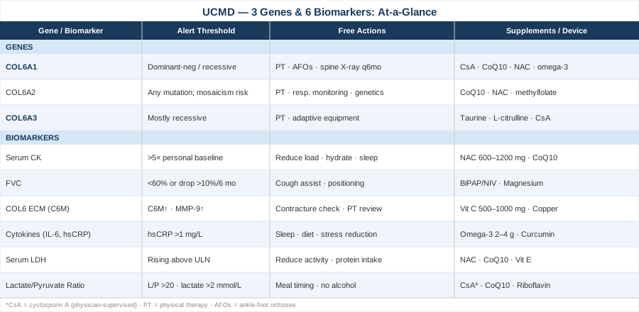
6 Biomarkers Worth Tracking in Ullrich Congenital Muscular Dystrophy
Biomarker monitoring in UCMD serves two distinct purposes: establishing a baseline to detect meaningful change over time, and providing objective evidence that a given intervention is — or is not — having an effect. The six biomarkers below are not equally accessible or affordable, but together they cover the most important biological dimensions of UCMD — muscle integrity, respiratory function, extracellular matrix health, inflammation, cellular damage, and mitochondrial efficiency. Beginning these measurements early, even before symptoms significantly progress, provides the reference points that make future data interpretable.
1. Serum Creatine Kinase (CK)
Why it matters
Creatine kinase is the first enzyme ordered in any suspected muscle disease. In Duchenne muscular dystrophy, CK climbs to 50–100 times the upper limit of normal. In UCMD, the picture is far subtler: most patients have CK values that are normal or mildly elevated, typically just 2 to 5 times normal. This does not reflect lower disease severity — it reflects the nature of collagen VI pathology, which disrupts the extracellular scaffold and mitochondrial function rather than causing the dramatic membrane rupture seen in dystrophin-deficient diseases.
This distinction matters enormously in practice. A "normal" CK has reassured both families and generalist clinicians into underestimating disease activity in UCMD, sometimes delaying respiratory monitoring, physical therapy referrals, and specialist consultations. Knowing what a normal CK actually means in this disease is the starting point.
How to measure it
CK is measured from a standard venous blood sample at any clinical lab. Cost: approximately $20–$60 in the United States, usually covered within a basic metabolic panel. Adult reference ranges: roughly 10–195 U/L in women and 10–250 U/L in men; pediatric ranges are age-specific. In UCMD monitoring, measure at diagnosis as a baseline, then every 3–6 months, and additionally whenever there is an acute illness, a change in activity level, or a perceived change in function.
If the score is elevated: the plan without supplements
A mild CK elevation in UCMD does not necessarily require pharmacological intervention. First rule out confounders: recent intense physical activity (CK peaks 24–72 hours after exertion), viral illness, statin medications, intramuscular injections, or the natural variability of serial measurements. If the elevation is genuinely unexplained and above 5x the individual's established baseline, reduce physical loading temporarily, ensure adequate hydration, prioritize 8–9 hours of sleep per night with respiratory support if FVC has declined, and recheck within 4–6 weeks. Avoid muscle-straining activities in the 48 hours before a planned measurement.
If the score is elevated: the plan with supplements or equipment
N-acetylcysteine (NAC) is one of the best-supported antioxidants for reducing oxidative stress in muscle tissue. It acts as a precursor to glutathione, the cell's primary endogenous antioxidant. Dose: 600–1,200 mg/day in two divided doses with meals. Generally very well tolerated; some patients report mild nausea at the higher dose range. No cycling required at standard therapeutic doses; reassess with a physician beyond 6 months of continuous use.
Coenzyme Q10 (ubiquinol form) supports mitochondrial electron transport and reduces free-radical leak from dysfunctional mitochondria. Dose: 200–400 mg/day with a fat-containing meal (it is fat-soluble; absorption drops dramatically without dietary fat). Strong safety record. Direct evidence in UCMD is limited, but the mechanistic rationale is sound given the established mPTP dysfunction.
2. Forced Vital Capacity (FVC)
Why it matters
Respiratory failure is the leading cause of premature death in UCMD. The respiratory muscles — primarily the diaphragm, intercostals, and accessory muscles — are affected by the same collagen VI deficiency that weakens limb muscles, and their progressive decline reduces the ability to take deep breaths, clear secretions, and maintain adequate overnight oxygenation. By the time daytime symptoms appear (breathlessness on mild exertion, morning headaches, fatigue), significant nocturnal hypoventilation has often already been occurring for months or years.
FVC, expressed as a percentage of the predicted value for age, sex, and height, is the primary functional parameter for tracking respiratory decline in UCMD. Catching a meaningful decline early — before the FVC falls below 50% predicted — is the most important action a clinical team can take to prevent respiratory crisis.
How to measure it
FVC is measured during a pulmonary function test (PFT) using a spirometer. The key addition in neuromuscular disease is measuring FVC in both the seated and supine (lying flat) positions. A drop of more than 10–15% between sitting and supine FVC is a reliable indicator of diaphragm weakness, which can be present even when seated FVC appears adequate. Cost: $100–$400 for a full PFT session depending on location and whether additional measurements (MIP, MEP, flow-volume loops) are included.
Monitoring frequency: every 3–6 months in pediatric patients and young adults, or at any symptom change. An FVC below 60% predicted warrants urgent pulmonologist consultation; rapid decline of more than 10% over six months requires immediate escalation regardless of absolute value.
If the score is declining: the plan without supplements
Initiate consultation with a pulmonologist or respiratory physiotherapist with experience in neuromuscular disease. Introduce an overnight pulse oximetry study or polysomnography to detect nocturnal desaturation before it becomes clinically severe. Learn and practice manually assisted cough technique (caregiver places hands over the lower chest and coordinated pressure is applied during forced expiration). Begin using a cough assist device (mechanical insufflation-exsufflation) if cough peak flow is below 270 L/min. Sleep positioning: elevate the head of bed 20–30 degrees to reduce the mechanical load on the diaphragm during sleep.
If the score is declining: the plan with supplements or equipment
Non-invasive positive pressure ventilation (NIV/BiPAP) is the single most evidence-based respiratory intervention available in UCMD. It is not a supplement — it is a piece of equipment that becomes part of daily management once FVC declines or nocturnal desaturations appear. Setting selection must be done by a pulmonologist. Starting NIV at the right time (not too early, not too late) requires regular monitoring.
Magnesium glycinate or magnesium malate supports respiratory muscle function and reduces neuromuscular excitability-related cramping. Dose: 300–400 mg elemental magnesium per day, taken in the evening. Very safe at these doses; excess intake (above 700 mg/day elemental) can cause loose stools. Deficiency is common and often undiagnosed in patients with limited dietary intake or gastrointestinal issues.
3. Collagen VI Levels and ECM Remodeling Markers
Why it matters
Collagen VI is not measured in routine clinical blood panels, but it is the central protein deficient in UCMD. Emerging assay technologies now enable detection of collagen VI degradation fragments in plasma — most notably the C6M assay, which measures a neoepitope released during MMP-mediated remodeling of collagen VI microfibrils. Elevated C6M reflects active matrix degradation. Paradoxically, even in patients who produce defective collagen VI, this assay can be informative about the rate of ECM turnover.
Surrogate markers of extracellular matrix dysregulation — including MMP-9 (matrix metalloproteinase-9), MMP-2, and TIMP-1 (tissue inhibitor of metalloproteinases-1) — reflect overall remodeling activity in muscle connective tissue and have been studied in multiple muscular dystrophy contexts. Their elevation indicates that the ECM is under active remodeling stress, which correlates with inflammation and fibrosis progression.
How to measure it
The C6M assay is available through Nordic Bioscience (primarily in Europe and via their research partnerships) at a cost of approximately $200–$500. It is not yet part of standard US clinical laboratory menus, but can be arranged as a specialty research order through academic medical centers. MMP-9 and TIMP-1 are available via ELISA through most major reference labs at $80–$200 each.
These markers are most useful as longitudinal tools: establish a baseline, then track at 6-month intervals. A single elevated value is difficult to interpret without context; a rising trend is the meaningful signal.
If markers are abnormal: the plan without supplements
Elevated ECM degradation markers suggest active matrix remodeling, which should prompt more frequent musculoskeletal review: assess for worsening contractures at elbows, wrists, hips, and knees; evaluate for scoliosis progression; review current physical therapy protocol for adequate joint range-of-motion maintenance. Serial photography of contractures (photographing joint position in standardized poses) provides low-cost documentation of change over time.
If markers are abnormal: the plan with supplements or equipment
Vitamin C (ascorbic acid): An essential cofactor for the hydroxylation of proline and lysine in the collagen triple helix. Without adequate vitamin C, newly synthesized collagen chains cannot properly fold into their helical structure. In collagen VI-deficient states where residual protein production is still occurring (especially in partial-loss or Bethlem-spectrum mutations), ensuring optimal vitamin C levels is mechanistically important. Dose: 500–1,000 mg/day. Very safe at these doses; above 2,000 mg/day may cause osmotic diarrhea.
Copper bisglycinate: Required for lysyl oxidase, the enzyme that cross-links mature collagen fibers for structural stability. Dose: 1–2 mg/day elemental copper. Take separately from zinc, as zinc and copper compete for intestinal absorption. Excess copper (above 10 mg/day) is hepatotoxic. At 1–2 mg/day, safety is well established.
4. Inflammatory Cytokines (IL-6, hsCRP, TNF-α)
Why it matters
Chronic low-grade inflammation is a feature of progressive muscular dystrophies that is both a consequence of ongoing muscle fiber damage and an independent driver of further damage. In UCMD, persistently elevated interleukin-6 (IL-6) and tumor necrosis factor-alpha (TNF-α) activate JAK-STAT and NF-κB signaling cascades that promote muscle protein breakdown, impair satellite cell regeneration, and accelerate replacement of functional muscle with fibro-fatty tissue. High-sensitivity C-reactive protein (hsCRP), a downstream marker of IL-6 activity, provides an accessible and reproducible proxy for chronic inflammatory state.
How to measure it
hsCRP is widely available and costs $20–$50. It requires two readings taken 4–8 weeks apart during a period of clinical stability (illness or injury will transiently spike CRP, making single readings unreliable). A hsCRP persistently above 1.0 mg/L in UCMD warrants attention; above 3.0 mg/L represents a genuinely elevated baseline that should be addressed. IL-6 and TNF-α can be measured through specialty cytokine ELISA panels at $100–$300 through most reference labs, and are more informative when run alongside hsCRP than as standalone tests.
If elevated: the plan without supplements
Address all modifiable inflammatory inputs systematically. Optimize sleep duration and quality (8–9 hours with respiratory support if indicated; fragmented sleep from nocturnal hypoventilation is itself a powerful driver of inflammatory cytokines). Reduce dietary ultra-processed food intake and refined sugars, which acutely elevate IL-6 through multiple mechanisms. Investigate and treat any chronic infections or dental disease. Manage psychological stress actively — cortisol chronically potentiates NF-κB activity and IL-6 production, which is why stress reduction is not peripheral but mechanistically relevant.
If elevated: the plan with supplements or equipment
Omega-3 fatty acids (EPA + DHA): Among the best-studied and most consistently effective anti-inflammatory supplements. EPA and DHA compete with arachidonic acid for cyclooxygenase and lipoxygenase pathways, reducing production of pro-inflammatory eicosanoids. Dose: 2–4 g/day of combined EPA and DHA from high-purity fish oil or algae-based sources. Monitor for blood-thinning effects above 3 g/day if the patient is on anticoagulants. No cycling required. Studies in inflammatory muscle disease have shown reductions in IL-6 and TNF-α with sustained supplementation.
Curcumin with piperine: Inhibits NF-κB transcription, reducing downstream production of IL-6, TNF-α, and IL-1β. Bioavailability of curcumin is poor without piperine (black pepper extract) or phospholipid formulations. Dose: 500–1,000 mg standardized curcumin with 5–10 mg piperine, twice daily with meals. Possible mild GI irritation. Long-term safety at these doses is well established.
5. Serum Lactate Dehydrogenase (LDH)
Why it matters
LDH is an intracellular enzyme released into circulation when cells are damaged or die. Multiple isoforms exist; the LDH-5 isoform is predominantly skeletal muscle in origin. In UCMD, where CK is often only mildly elevated, tracking LDH in parallel gives a complementary picture of cell death rate that can sometimes be more sensitive. LDH elevation can also reflect liver involvement, red blood cell hemolysis (from traumatic draws), or kidney disease, so it always requires clinical context before interpretation.
As a serial measurement in UCMD, a rising LDH trend — even within "normal" reference range — can signal increased muscle cell turnover before overt functional decline becomes apparent.
How to measure it
Serum LDH is included in many standard metabolic panels and costs $20–$40 as a standalone test. Normal range: approximately 105–333 U/L, varying by laboratory. If greater specificity is needed, LDH isoform fractionation (LD1–LD5 panel) can isolate the muscle-specific LD5 fraction at a cost of $80–$150. For UCMD monitoring, a standard LDH every 3–6 months as part of the same blood draw as CK provides a useful dual-marker picture of muscle integrity.
If elevated: the plan without supplements
Rule out confounders first: hemolytic sample, recent intense activity, concurrent liver or kidney disease. If none of these apply and LDH is trending upward, review the physical activity protocol (reduce intensity and duration), ensure adequate protein intake (1.5–2 g/kg body weight daily to support muscle protein synthesis), and review respiratory support adequacy (nocturnal hypoxia alone drives oxidative stress that elevates LDH). Recheck in 6–8 weeks under standardized conditions.
If elevated: the plan with supplements or equipment
The antioxidant approach serves double duty for both CK and LDH elevation: NAC (600–1,200 mg/day), CoQ10 (200–400 mg/day), and vitamin E as mixed tocopherols (200–400 IU/day). Mixed tocopherols are preferred over high-dose isolated alpha-tocopherol, which can paradoxically act as a pro-oxidant above 1,000 IU/day. Vitamin E's membrane-stabilizing properties have supporting evidence in muscular dystrophy models and are mechanistically relevant to reducing cell membrane fragility.
6. Blood Lactate / Pyruvate Ratio (Mitochondrial Function Proxy)
Why it matters
This is the most mechanistically central biomarker in UCMD and the one most frequently overlooked in clinical practice. Collagen VI deficiency does not just weaken the structural matrix outside muscle cells — it has been directly shown to dysregulate the mitochondrial permeability transition pore (mPTP). When collagen VI is absent, the mPTP — a critical regulator of mitochondrial integrity in the inner membrane — becomes pathologically sensitized. It opens too readily and stays open too long, triggering a cascade of mitochondrial swelling, cytochrome c release, and apoptosis in muscle fibers that would otherwise survive.
The blood lactate/pyruvate (L/P) ratio is a functional readout of mitochondrial oxidative phosphorylation efficiency. When mitochondria are dysfunctional, cells shift toward anaerobic metabolism, producing more lactate relative to pyruvate. A resting L/P ratio above 20–25 suggests clinically relevant mitochondrial dysfunction. It was precisely this biomarker pattern in UCMD patients, combined with the animal model data, that led Italian researchers to investigate cyclosporin A as a targeted intervention.
How to measure it
Blood lactate requires venous sampling at rest after at least 30 minutes of quiet inactivity (exercise will acutely elevate lactate and invalidate the measurement). Cost: $40–$80. Pyruvate measurement requires cold-transport sample handling (immediate acidification to stabilize the compound); most major reference labs can accommodate this with advance notice, at a cost of $80–$120. The L/P ratio is calculated from paired samples drawn simultaneously. A comprehensive mitochondrial assessment including respiratory chain enzyme activity in muscle biopsy tissue is available at specialized centers at $500–$1,500 and is appropriate if significant mitochondrial dysfunction is suspected.
For routine monitoring, resting blood lactate alone (target below 2.0 mmol/L at rest) combined with clinical context is a reasonable first-line approach.
If elevated: the plan without supplements
An elevated resting lactate in UCMD confirms that the mitochondrial dysfunction component is actively contributing to disease burden. Reduce all metabolic stressors: avoid prolonged fasting (maintain consistent meal timing every 4–6 hours, as fasting increases mitochondrial load); ensure adequate carbohydrate intake around any physical activity (pre-activity carbohydrate reduces the shift to anaerobic metabolism); completely eliminate alcohol (which directly inhibits complex I of the mitochondrial electron transport chain); and prioritize nighttime respiratory support, because sleep-related hypoxia independently worsens mitochondrial function.
If elevated: the plan with supplements or equipment
Cyclosporin A (physician-supervised only): The most direct pharmacological intervention targeting the mPTP dysfunction specific to UCMD. This is a prescription immunosuppressant, not a supplement. A clinical trial demonstrated improvements in mitochondrial function and motor outcomes with short courses at approximately 3.5 mg/kg/day over 30 days. Risks include nephrotoxicity, hypertension, gum overgrowth, and immunosuppression. It must be managed by a physician experienced in neuromuscular disease, with regular serum creatinine, blood pressure, and drug level monitoring. Cycling: typically 30 days on, followed by an off-period; long-term continuous dosing is not standard.
Coenzyme Q10 (200–600 mg/day) and riboflavin (vitamin B2) (100–200 mg/day) directly support electron transport chain function at complexes I and II respectively. Both have strong safety records and supportive evidence in mitochondrial dysfunction contexts. NAD+ precursors — NMN (500–1,000 mg/day) or NR (nicotinamide riboside, 300–1,000 mg/day) — support mitochondrial biogenesis through SIRT1/PGC-1α signaling and are being actively researched in muscle disease models. Well tolerated; evidence in UCMD is indirect but mechanistically grounded.
The Three Genes Behind UCMD: What Each Mutation Means for Your Strategy
Knowing that a patient has UCMD is necessary but not sufficient. Knowing which collagen VI gene is mutated, and whether the mutation is dominant-negative or recessive, changes both the prognostic picture and the logic behind specific therapeutic approaches. These three genes encode the three protein chains that must assemble into a functional heterotrimer before collagen VI can be secreted and integrated into the muscle basement membrane zone.
COL6A1 — The Alpha-1 Chain Gene
What the gene does
COL6A1, located on chromosome 21q22.3, encodes the alpha-1 chain of collagen VI. The protein contains a central triple-helical domain flanked by two von Willebrand factor A (VWA) domains at each end, which mediate both chain-chain assembly and interactions with fibronectin and heparan sulfate proteoglycans in the extracellular matrix.
Mutations in COL6A1 can be either dominant-negative or recessive. Dominant-negative mutations — most commonly glycine substitutions within the Gly-X-Y triple helix repeat — are particularly destructive because a single abnormal alpha-1 chain poisons the entire assembly process: the majority of collagen VI heterotrimers produced are defective and cannot integrate properly into the matrix. This means a heterozygous dominant glycine substitution in COL6A1 can produce severe UCMD despite only one mutated allele. Recessive COL6A1 mutations (frameshift, nonsense, large deletions) require both alleles to be affected to produce disease, but often allow some residual collagen VI production from structural differences in the mutation.
If the gene is bad: the plan without supplements
For dominant-negative COL6A1 mutations, the therapeutic priority is managing the downstream consequences of near-complete collagen VI absence. Structured physical therapy targeting joint range of motion is the backbone of non-pharmacological management — specifically, twice-daily passive and active-assisted stretching of at-risk joints (elbows, hips, knees, ankles). Night-time ankle-foot orthoses (AFOs) worn during sleep are strongly recommended to prevent equinus contracture progression. Serial casting is an option in younger children where contractures are actively worsening.
Scoliosis surveillance every 6 months with standing or sitting spine radiography is essential; early referral for spinal bracing when curves reach 20 degrees and surgical consultation at 40–50 degrees. Baseline echocardiography is recommended; although cardiac involvement is less prominent in UCMD than in dystrophinopathies, it should not be omitted. Respiratory monitoring should begin at diagnosis with the FVC/FVC-supine protocol described in the biomarker section.
If the gene is bad: the plan with supplements or equipment
For dominant-negative COL6A1, the mitochondrial mPTP dysfunction is typically severe, and this is the primary pharmacological target. Cyclosporin A at 3.5 mg/kg/day for 30-day cycles (physician-managed, with kidney function and blood pressure monitoring) addresses the core CypD-mPTP sensitization. Between cycles: CoQ10 (200–400 mg/day), NAC (600–1,200 mg/day), omega-3 fatty acids (2–4 g/day EPA+DHA) as a maintenance mitochondrial support stack.
Resveratrol (250–500 mg/day with a meal): Activates SIRT1 via allosteric binding and supports mitochondrial biogenesis through PGC-1α. Preclinical data in muscular dystrophy models are supportive. Generally well tolerated at standard doses; may interact with anticoagulants through CYP2C9 inhibition. No cycling required at standard doses.
Intermittent fasting (12–14 hour overnight fast, not extending into the day) activates autophagy and AMPK signaling. In UCMD models, autophagy reactivation has been shown to partially compensate for collagen VI loss by improving clearance of dysfunctional mitochondria. Adults with adequate nutritional status can try this approach; children should not fast without medical supervision and caloric needs must be maintained.
COL6A2 — The Alpha-2 Chain Gene
What the gene does
COL6A2 sits adjacent to COL6A1 on chromosome 21q22.3 in a head-to-head orientation, sharing regulatory elements. The alpha-2 chain has a similar domain architecture to alpha-1 but adds important molecular complexity through alternative splicing: two different C-terminal VWA domain isoforms (2a and 2b) are produced from COL6A2 via splice variation, and these isoforms have different matrix-binding properties that may partly explain phenotypic variability across the UCMD-Bethlem spectrum.
A clinically important feature of COL6A2 is its documented tendency toward somatic mosaicism. Apparently unaffected parents of children with UCMD have been found to carry COL6A2 mutations at low variant allele frequencies in blood, meaning the mutation arose in a subset of germline cells during parental development. This has direct implications for recurrence risk calculations — a parent who appears unaffected may have a meaningful recurrence risk for future pregnancies that standard testing might miss without deep-sequencing analysis.
If the gene is bad: the plan without supplements
The management framework for COL6A2 mutations mirrors that for COL6A1: structured physical therapy, respiratory monitoring with the sitting/supine FVC protocol, contracture prevention, and scoliosis surveillance. For cases with mosaic or milder phenotypes tending toward Bethlem myopathy, monitoring intervals may be extended in discussion with a specialist, but should not be abandoned. Genetic counseling is particularly important for COL6A2 — detailed discussion of mosaicism, recurrence risk, and prenatal testing options is warranted before any family planning decisions.
If the gene is bad: the plan with supplements or equipment
The core mitochondrial support stack (CoQ10, NAC, omega-3 fatty acids) applies here as with COL6A1. In addition, HDAC inhibition has been proposed as an epigenetic approach to upregulate residual COL6A2 expression: certain histone deacetylase inhibitors increase chromatin accessibility at the COL6A2 promoter region in cell culture models, theoretically increasing transcription from the functional allele in recessive or partial-loss cases. Valproic acid is one such agent occasionally used in other neuromuscular contexts, but its application in UCMD is not clinically established. This remains in early research.
Methylation support (folate 400–800 mcg/day as methylfolate, plus methylcobalamin B12 at 1,000 mcg/day) is inexpensive, broadly safe, and mechanistically relevant for maintaining appropriate epigenetic regulation of the COL6 loci. These are not UCMD-specific interventions, but represent a reasonable optimization for anyone with a gene-level defect where epigenetic factors may modulate residual expression.
COL6A3 — The Alpha-3 Chain Gene
What the gene does
COL6A3 is located on chromosome 2q37, a completely different chromosome from the COL6A1/A2 pair on chromosome 21. The alpha-3 chain it encodes is dramatically larger than the other two chains, containing approximately 10 N-terminal VWA domains and 2 C-terminal VWA domains, making it the primary "docking" chain responsible for integrating collagen VI microfibrils into the broader extracellular matrix network via interactions with fibronectin, perlecan, and decorin.
COL6A3 mutations are more commonly recessive in UCMD than those in COL6A1 and COL6A2. This is meaningful: recessive mutations require both alleles to be dysfunctional to produce disease, but they also potentially preserve some collagen VI production when the mutation allows even partial protein function. The large size of COL6A3 means it hosts a higher absolute number of mutations across its sequence, and mutations in different VWA domains can have meaningfully different functional consequences — some affecting matrix docking, others affecting helical assembly.
Whole-exome sequencing studies have increasingly identified COL6A3 variants in patients initially diagnosed with other connective tissue syndromes, confirming the importance of comprehensive collagen VI panel testing in diagnostically ambiguous presentations.
If the gene is bad: the plan without supplements
For recessive COL6A3 mutations where residual protein production is plausible, there may be a window for strategies aimed at upregulating remaining functional expression — a different therapeutic logic from dominant-negative COL6A1/A2 mutations where the defective chain must be suppressed. Physical management goals are identical across all three genes: joint mobility maintenance, respiratory monitoring, scoliosis surveillance. Adaptive equipment planning (power wheelchair, communication devices, seating support) should be introduced proactively when functional changes are detected, rather than reactively when a crisis occurs.
If the gene is bad: the plan with supplements or equipment
Taurine: An amino acid that stabilizes mitochondrial membranes, supports calcium handling, and reduces oxidative stress. Dose: 500–2,000 mg/day in divided doses. Extremely well tolerated, even at higher doses. Evidence in muscular dystrophy models is supportive; the mechanistic relevance to UCMD's mitochondrial pathology is clear.
L-citrulline malate (1,000–3,000 mg/day, taken before any physical activity): Supports nitric oxide synthesis and muscle blood flow, reducing metabolic stress during movement. Evidence is strongest in DMD; translational relevance to COL6 myopathies is reasonable but not established.
For COL6A3 recessive mutations, the cyclosporin A / CypD-mPTP strategy remains relevant; the mPTP dysfunction has been documented in both dominant and recessive collagen VI deficiency models. Physician supervision and kidney function monitoring remain mandatory if this approach is considered.
The Mitochondrial Paradigm Shift: 10 Insights That Reshaped How Researchers Think About UCMD
For most of its described clinical history, UCMD was understood as a disease of structural scaffolding — collagen VI was absent, the basement membrane zone around muscle fibers was compromised, and muscles gradually failed. The therapeutic logic was accordingly structural: replace the missing protein, reinforce the matrix. This view was not wrong, but it was incomplete. Beginning in the mid-2000s, a series of studies from the laboratories of Paolo Bernardi and Paolo Bonaldo at the University of Padova revealed that collagen VI deficiency sets in motion a cascading intracellular catastrophe that operates independently of — and in parallel to — the extracellular matrix failure. What follows are the ten most impactful insights from that body of work, each of which challenges and enriches the conventional picture.
1. Mitochondria Are Abnormal Before Muscle Fibers Die
The foundational observation was that muscle cells from UCMD patients and from Col6a1-null mice showed mitochondrial swelling, abnormal cristae morphology, and reduced membrane potential in fibers that were not yet visibly degenerating. The mitochondrial abnormality preceded cell death — it was not a downstream consequence of dying fibers releasing their contents. This repositioned UCMD from a passive structural failure to an active intracellular disease.
2. The mPTP Is the Central Mechanism
The mitochondrial permeability transition pore (mPTP) — a high-conductance channel that forms in the inner mitochondrial membrane under stress — was found to be abnormally sensitive in collagen VI-deficient muscle. Instead of transient, regulated opening, the mPTP in UCMD muscle opens too readily and stays open, dissipating the mitochondrial membrane potential and initiating the intrinsic apoptosis pathway. This explained why UCMD muscle cells die despite appearing structurally intact at the light microscopy level.
3. Cyclophilin D Is the Regulatory Target
Cyclophilin D (CypD), a peptidylprolyl isomerase in the mitochondrial matrix, directly regulates mPTP sensitivity. Genetic deletion of CypD in Col6a1-null mice normalized mitochondrial function and significantly reduced muscle pathology, providing causal proof — not just correlation — that CypD-mediated mPTP dysregulation is mechanistically central to UCMD. CypD became the first validated molecular target for drug repurposing in this disease.
4. Cyclosporin A Provides Proof of Concept in Humans
Cyclosporin A's known ability to bind and inhibit CypD made it an immediate candidate. Short courses in Col6a1-null mice produced striking improvements in mitochondrial morphology and grip strength. A clinical trial in UCMD patients subsequently demonstrated measurable normalization of mitochondrial function in treated patients, along with modest motor benefits — the first mechanistically guided clinical intervention in this disease. The risks of cyclosporin A (nephrotoxicity, immunosuppression) limit its long-term use, but the proof of concept opened the door to safer CypD-targeted therapies under development.
5. Autophagy Is Also Impaired, Independently of mPTP
A parallel discovery revealed that UCMD muscle accumulates damaged organelles and protein aggregates consistent with impaired autophagy — specifically, defective mitophagy (the clearance of dysfunctional mitochondria). Beclin-1 levels and LC3 lipidation patterns were abnormal in UCMD muscle, indicating that the cellular recycling system responsible for quality control was also compromised. Restoring autophagy — pharmacologically with rapamycin or through nutrient-sensing interventions like caloric restriction — partially rescued the muscle phenotype in animal models.
6. mTOR Suppression Activates Autophagy and Helps
Because mTOR (mechanistic target of rapamycin) is the primary suppressor of autophagy, its inhibition with rapamycin (sirolimus) was tested in UCMD models. Results showed improved autophagic flux, reduced accumulation of damaged mitochondria, and better preserved muscle architecture. This places UCMD in the growing category of diseases where mTOR dysregulation is pathological — alongside type 2 diabetes, some cancers, and age-related sarcopenia — and opens a pathway for repurposing mTOR inhibitors that are less immunosuppressive than cyclosporin A.
7. The Same Biology Applies Across the Full Collagen VI Spectrum
The mPTP/autophagy dysfunction has been documented not only in classic severe UCMD but also in Bethlem myopathy, the milder allelic disorder at the other end of the collagen VI spectrum. This tells researchers that the mechanism is not simply a consequence of the most severe protein absence — it operates even when some functional collagen VI is still present. Every patient on the collagen VI disease spectrum, regardless of severity, has this mitochondrial component as part of their biology.
8. Exercise Physiology Must Be Rethought
In healthy muscle, aerobic exercise is a potent stimulus for mitochondrial biogenesis — it induces controlled, transient mPTP opening that signals the cell to build more mitochondria. In collagen VI-deficient muscle with an already sensitized mPTP, uncontrolled aerobic exercise may tip the pore into prolonged opening, worsening apoptosis rather than inducing adaptation. Animal data suggest that the type and intensity of exercise in UCMD matter specifically because of this mPTP sensitivity: low-load, frequent activity is preferable to high-intensity effort.
9. There Is a Therapeutic Window Before Irreversible Loss
Because mitochondrial dysfunction is detectable before significant fiber loss occurs, there is a biologically meaningful window during which mPTP-targeted or autophagy-restoring interventions could protect fibers that would otherwise die. This is the strongest argument for early diagnosis, early biomarker monitoring (specifically the lactate/pyruvate ratio), and early consideration of targeted interventions before the disease is already well advanced at the functional level.
10. Gene Therapy Is Advancing Toward the Source
AAV-based delivery of functional COL6A1, COL6A2, or COL6A3 is in preclinical development at several major neuromuscular disease centers, including active programs at the NIH's Neuromuscular Diseases Group. Antisense oligonucleotide strategies targeting dominant-negative allele suppression — analogous to approaches used in DMD — are also in early exploration for specific COL6A1 and COL6A2 mutations. Staying connected to clinical trial registries (specifically clinicaltrials.gov with search terms "collagen VI" or "Ullrich muscular dystrophy") is the most practical way for families to track when these approaches enter human trials.
Complementary Approaches With Meaningful Evidence for UCMD
The following four modalities address real, measurable aspects of life with UCMD that standard pharmacological management cannot fully cover: respiratory muscle efficiency, joint tissue health, quality of life, and the psychological burden of a rare progressive disease. None of them replace medical care, and the evidence for UCMD specifically (rather than neuromuscular disease in general) is variable. Each is presented with its best available supporting evidence and a realistic protocol.
Breathing-Based Therapies
Breathing-based therapies encompass diaphragmatic breathing training, ventilation-assisted cough techniques, glossopharyngeal breathing ("frog breathing"), and structured respiratory physiotherapy. In UCMD, where progressive respiratory muscle weakness is the leading cause of life-limiting complications, these techniques are not complementary in the sense of being peripheral — they are directly disease-relevant. Regular diaphragmatic breathing practice maintains the muscular coordination and efficiency of remaining respiratory muscle function; glossopharyngeal breathing can augment tidal volume when diaphragm function is severely reduced; and practiced cough augmentation techniques (both manual and device-assisted) preserve the ability to clear secretions and prevent pneumonia.
A systematic review of respiratory management in progressive neuromuscular diseases published in Respiratory Care found that early introduction of structured airway clearance techniques, including manually assisted cough and mechanical insufflation-exsufflation, significantly reduced respiratory complications and hospitalizations in patients with progressive neuromuscular weakness. The physiological principles apply directly to UCMD's respiratory phenotype.
For practical implementation: work with a respiratory physiotherapist to design a daily 10–15 minute protocol including diaphragmatic breathing practice (5–10 slow, deep breaths focusing on belly expansion), manually assisted cough technique practice (3–5 repetitions with a trained caregiver), and if FVC is below 70% predicted, coordination of mechanical cough-assist device training. Reassess every 3–6 months and modify as respiratory function evolves. Sessions can be performed seated, semi-reclined, or in lateral positions — whatever maximizes comfort and full respiratory effort.
Massage Therapy
Therapeutic massage — specifically myofascial release, gentle passive mobilization, and connective tissue techniques — is directly relevant to UCMD's contracture biology. Progressive joint contractures at elbows, hips, knees, and ankles are among the most functionally limiting features of UCMD, and the underlying driver is stiffening of collagen-deficient periarticular connective tissue combined with reduced active movement. Regular gentle massage helps maintain tissue pliability, improve local circulation to chronically shortened tissues, and reduce musculoskeletal discomfort without imposing mechanical stress on fragile muscle.
Research on massage in neuromuscular conditions, including a controlled study published in the Journal of Pain and Symptom Management, has demonstrated significant reductions in pain, anxiety, and fatigue scores in patients with progressive muscle disease. While large-scale RCT data specific to UCMD do not exist, physiotherapy literature includes case series describing measurable improvements in joint range of motion following gentle passive mobilization protocols in children with collagen VI myopathies.
Practically: sessions of 30–45 minutes every 1–2 weeks with a massage therapist experienced in hypermobile and fragile connective tissue conditions (explicitly ask about neuromuscular disease experience before booking). Focus on lower extremity myofascial release, gentle elbow and shoulder mobilization, and lumbar support work. Avoid deep tissue pressure — the goal is circulation and pliability, not deep muscle manipulation. Parents of younger children can be trained by a physical or occupational therapist in simple home stretching and gentle massage routines that maintain these gains between clinical sessions.
Music Therapy
Music therapy delivered by a credentialed music therapist (MT-BC in the US) includes both receptive (listening, relaxation-oriented) and active (singing, playing instruments, rhythmic movement) components. For UCMD specifically, the most relevant application is the respiratory engagement involved in singing and wind instrument playing — both of which function as structured, enjoyable respiratory exercise, training expiratory muscle coordination in a way that does not feel like therapy. Secondary benefits include emotional expression, communication support, and quality of life maintenance during periods of increased medical burden.
A systematic review on music therapy in children and adults with chronic conditions found significant improvements in emotional wellbeing, social engagement, and — in studies including singing components — measurable improvements in respiratory parameters including expiratory muscle strength. For patients whose FVC is modestly reduced, even gentle, low-volume singing activates the full sequence of respiratory musculature in a coordinated pattern that other exercises do not easily replicate.
Practically: monthly or biweekly sessions with a certified music therapist who has experience in pediatric or chronic illness populations. For patients with limited breath support, harmonica or recorder at gentle volumes engages expiratory muscles with low exertion demand. For home practice: structured sessions of 20–30 minutes of singing or instrument playing, 3–4 times per week, adapted to the patient's current breath support. Avoid any breath-holding patterns or exertion that causes discomfort. Patients with FVC below 50% should use music engagement as gentle respiratory practice only, not as an exercise replacement.
Mindfulness Meditation and MBSR
Mindfulness-Based Stress Reduction (MBSR) is an 8-week structured program combining sitting meditation, body scan practices, and adapted gentle movement. For individuals living with UCMD — and for their caregivers — the psychological burden is substantial and frequently underaddressed. UCMD is rare, progressive, and poorly understood by most of the general medical community, creating chronic uncertainty and isolation. Unmanaged stress elevates cortisol, which directly potentiates NF-κB transcription and amplifies the inflammatory cytokine production (IL-6, TNF-α) that drives muscle inflammation and wasting. Stress reduction is not peripheral to UCMD biology — it is mechanistically connected to the inflammatory dimension of the disease.
A meta-analysis on MBSR in chronic illness found significant reductions in anxiety, depression, and pain with effect sizes comparable to pharmacological treatment for anxiety outcomes. For UCMD caregivers specifically, mindfulness-based programs have been shown to reduce burnout, improve emotional regulation, and reduce caregiver-reported stress markers in the chronic illness caregiving context.
In practice: the formal 8-week MBSR curriculum (developed by Jon Kabat-Zinn at the University of Massachusetts Medical Center) is available in-person in many cities and online through university hospital programs. The standard format requires approximately 2.5 hours per week of group practice plus 45 minutes of daily home practice. Shorter adapted programs (4-week formats, self-guided apps such as Insight Timer or Waking Up) provide effective entry points. For patients with significant physical limitations, all key MBSR practices can be done seated or lying down — the program was specifically designed with physically limited participants in mind. Begin with 10–15 minutes of daily practice and build from there.
Conclusion
UCMD is a rare disease, but it is not a disease without leverage points. The research of the past fifteen years has transformed the scientific understanding of what is actually happening inside muscle cells in collagen VI deficiency, and that transformation has direct practical implications for how the disease is monitored and managed. The six biomarkers in this article — CK, FVC, collagen VI ECM markers, inflammatory cytokines, LDH, and blood lactate/pyruvate ratio — give a structured, repeatable way to track the most important biological dimensions of this disease. The three gene profiles provide the mutation-level context that determines which compensating strategies are most relevant for a specific patient.
No single intervention will change the trajectory of UCMD dramatically. But the accumulation of informed decisions — the right biomarker tracked at the right interval, the right respiratory support introduced at the right time, the right mitochondrial support given the specific mutation pattern — compounds meaningfully over years. The difference between a management strategy that incorporates this precision and one that does not is real and measurable.
The next smart step is concrete: establish or update your biomarker baseline (starting with CK, FVC, and hsCRP as the most accessible three), confirm that your genetic result specifies the mutation at the protein level (not just "COL6A1 positive"), and bring this monitoring framework to your next specialist appointment. For families who have not yet connected with an expert neuromuscular disease center — including the programs at NIH, Newcastle upon Tyne, Padova, or similar — making that connection now puts you in the best position to participate in emerging clinical trials and access the most current management guidance.
Musculoskeletal: Muscle Conditions
Respiratory: Lung Conditions
Autoimmune: Inflammatory Conditions Connective Tissue Conditions