This article contains AI generated content.
· UpdatedUremic Arthropathy — 5 Genes And 7 Biomarkers To Track
Introduction
Living with uremic arthropathy means dealing with a particular kind of frustration: joint pain that arrives on top of an already demanding condition, treated by specialists who each address their corner of the problem without anyone quite connecting the dots. The nephrologist focuses on kidneys. The rheumatologist focuses on joints. And the patient sits in the middle, still hurting, still confused about what is actually driving the damage.
The difficulty is that uremic arthropathy is not one disease. It is a cluster of joint disorders driven by kidney failure — gout triggered by impaired urate clearance, beta-2 microglobulin amyloidosis depositing in joint spaces after years of dialysis, periarticular calcifications from mineral metabolism gone wrong, and hyperparathyroid-driven bone erosion near joint surfaces. Generic anti-inflammatory advice misses the point almost entirely. What helps one mechanism may do nothing for another.
What is shifting the picture is a more precise understanding of which biological signals actually govern these processes. Certain genes make some people far more likely to develop crystal arthropathy or respond poorly to standard treatments. Specific blood biomarkers reveal whether the underlying drivers are active, worsening, or in a range where intervention can still make a real difference. Neither genetic insight nor biomarker tracking replaces clinical care, but together they tell a story that generic protocols cannot.
This article is built around that precision. The core section covers the seven biomarkers most useful for monitoring uremic arthropathy — what each one reveals, what testing costs, and what can realistically be done when results are off. There is also a section on the five genes most relevant to this condition, a summary of the most actionable podcast content on uric acid and joint inflammation, and a review of complementary approaches with genuine clinical backing. Better information rarely cures anything by itself, but it consistently produces better decisions.
Summary
This article covers seven blood biomarkers — beta-2 microglobulin, serum uric acid, intact PTH, serum phosphate, hs-CRP/IL-6, cystatin C, and advanced glycation end products — with practical plans for each when results are unfavorable, both with and without supplements. The genetics section identifies five key genes (ABCG2, SLC22A12, HLA-B*5801, VDR, and RANKL-pathway variants) that shape individual risk and treatment response, with specific action plans. A Strategy 3 section summarizes the most impactful science on uric acid and joint-metabolic disease from Huberman Lab's episode with Dr. Rick Johnson. Four complementary modalities with real clinical evidence for this population round out the article. The goal is a concrete map of what to track, what the numbers mean, and which paths forward have the best evidence behind them.
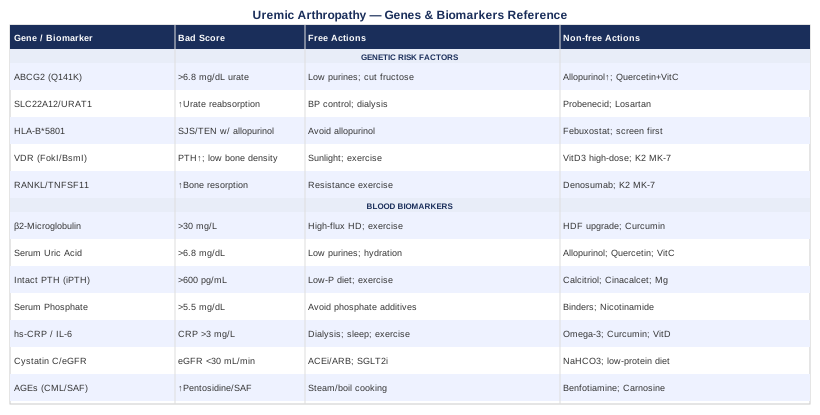
7 Biomarkers to Track in Uremic Arthropathy
Tracking biomarkers in uremic arthropathy is not about building a longer to-do list. It is about identifying which signals respond to which interventions, so that adjustments can be made and measured rather than guessed. The seven markers below were selected because each one reveals a distinct mechanism driving joint damage in kidney disease, and each one connects to specific, testable interventions.
1. Beta-2 Microglobulin (β2M)
Why it matters
Beta-2 microglobulin is a small protein shed continuously from the surface of nearly all nucleated cells. Healthy kidneys clear it efficiently. In chronic kidney disease — and especially in patients on long-term dialysis — β2M accumulates in the bloodstream and eventually deposits as amyloid fibrils in joint spaces, tendons, and the carpal tunnel. This is dialysis-related amyloidosis (DRA), one of the most debilitating and irreversible complications of long-term renal replacement therapy. Shoulders, wrists, and fingers are the most common targets, and once established, deposits do not resolve without a functioning transplant.
What it may reveal
Levels consistently above 30 mg/L indicate meaningful accumulation risk. Values above 50 mg/L have been associated with early amyloid deposition in joint tissue biopsies. The trend over six months is at least as important as the single reading — a steadily climbing β2M in the 25–35 mg/L range signals a trajectory worth addressing before symptoms solidify.
How to measure it
Serum beta-2 microglobulin is ordered by a nephrologist or rheumatologist. Cost: $30–$80 USD. It is part of routine dialysis follow-up in many centers but sometimes overlooked in pre-dialysis CKD. Frequency: every 3–6 months in dialysis patients; every 6–12 months in CKD stages 3–4.
If the score is bad, the plan without supplements
The most impactful intervention is dialysis modality optimization. High-flux hemodialysis membranes clear β2M measurably better than low-flux membranes. Hemodiafiltration (HDF), where available, achieves the highest β2M reduction per session. The ESHOL trial and similar European studies demonstrated that online HDF reduced β2M levels and was associated with lower all-cause mortality compared to standard high-flux hemodialysis. Increasing dialysis frequency (short daily or nocturnal sessions) further reduces accumulation between sessions. Exercise during dialysis sessions — even cycling on a bedside ergometer — has been shown to improve solute clearance including β2M. These are structural decisions that require nephrologist involvement.
If the score is bad, the plan with supplements or equipment
Curcumin (500–1000 mg/day with piperine for absorption) has anti-amyloid properties in cell models, though direct human evidence for DRA is still early-stage — it is a low-risk adjunct, not a primary strategy. Low-level laser therapy (LLLT) applied to affected joints has emerging evidence for reducing amyloid-related joint inflammation; this is covered in more detail in the complementary approaches section. No oral supplement reverses established β2M deposits; the priority is slowing accumulation through improved dialysis clearance.
2. Serum Uric Acid (Urate)
Why it matters
Impaired renal urate excretion is one of the earliest metabolic consequences of declining kidney function. As eGFR falls, uric acid accumulates in the blood. Above a certain concentration — approximately 6.8 mg/dL, the physiological solubility threshold — monosodium urate crystals can form and deposit in joints, producing acute and chronic gout. Gout in CKD is both more common and harder to treat than in the general population because first-line options like NSAIDs and full-dose colchicine carry real renal toxicity risks at lower eGFR. Beyond crystal-induced inflammation, uric acid itself has independent effects on endothelial function, tubulointerstitial inflammation, and CKD progression.
What it may reveal
Serum urate above 6.8 mg/dL represents supersaturation. Values above 9 mg/dL in CKD patients are associated with both faster kidney function decline and higher gout flare frequency. Asymptomatic hyperuricemia — elevated uric acid without obvious clinical gout — is increasingly recognized as a meaningful biological risk factor in this population, not merely a lab curiosity.
How to measure it
Standard serum uric acid, available at virtually any laboratory. Cost: $10–$30 USD. Measure fasting for most accurate results. Frequency: every 3–6 months in CKD patients with known hyperuricemia or gout history.
If the score is bad, the plan without supplements
Dietary purine restriction targets the highest-load sources: organ meats, red meat, shellfish, and especially high-fructose corn syrup in beverages. These changes can lower urate by 1–2 mg/dL reliably. Adequate hydration — targeting urine output above 2 L/day where renal capacity allows — supports urate excretion. Tart cherry consumption (240 mL/day of tart cherry juice or equivalent whole cherries) has modest but real evidence for reducing both serum urate and gout flare frequency, likely through combined urate-lowering and anti-inflammatory effects.
If the score is bad, the plan with supplements or equipment
Allopurinol, dose-adjusted for eGFR, is the standard urate-lowering therapy and is safe in most CKD patients when titrated carefully. Febuxostat is an alternative when allopurinol is contraindicated or not tolerated, though cardiovascular monitoring is recommended. Vitamin C (500–1000 mg/day) modestly lowers serum urate by increasing renal urate excretion — evidence from controlled trials shows reductions of approximately 0.3–0.5 mg/dL. Quercetin (500 mg/day) inhibits xanthine oxidase in preclinical models and has preliminary human data suggesting mild urate reduction; side-effect risk is low, but evidence remains limited. Cycle quercetin 8 weeks on, 4 weeks off to avoid tolerance.
3. Intact Parathyroid Hormone (iPTH)
Why it matters
Secondary hyperparathyroidism is nearly universal in advanced CKD. As kidney function declines, dysregulation of calcium, phosphate, and vitamin D drives the parathyroid glands into chronic overdrive, secreting excess PTH. Elevated PTH accelerates bone resorption, increases subchondral bone fragility, and promotes periarticular calcifications. This contributes directly to joint pain, pathological fractures near joint surfaces, and the calcific periarthritis frequently seen on imaging in dialysis patients. iPTH is the central marker for the mineral bone disorder (CKD-MBD) picture that underpins much of uremic arthropathy's skeletal component.
What it may reveal
KDIGO guidelines suggest iPTH targets for dialysis patients of roughly 2–9 times the upper limit of normal, typically 130–600 pg/mL depending on the assay used. Values persistently above this range indicate undertreated secondary hyperparathyroidism that is actively driving bone and joint damage. Values that are too low — suppressed by aggressive treatment — carry their own risk of adynamic bone disease, which also impairs joint-adjacent bone quality. Tracking the trend is essential.
How to measure it
Intact PTH blood test; requires an EDTA tube with rapid centrifugation. Cost: $40–$100 USD. Frequency: every 3 months in dialysis patients; every 6 months in CKD stages 3b–5 not yet on dialysis.
If the score is bad, the plan without supplements
Dietary phosphate restriction is the most actionable dietary lever — reducing processed foods, sodas, and food additives containing phosphate salts (inorganic phosphate, disodium phosphate) has a more meaningful impact on PTH than restricting naturally occurring phosphate in whole foods. Regular weight-bearing exercise, even adapted chair-based exercise in dialysis patients, has modest evidence for improving PTH regulation through mechanical loading signals on bone. Correcting metabolic acidosis (another common CKD complication) also reduces PTH-driven bone resorption.
If the score is bad, the plan with supplements or equipment
Activated vitamin D (calcitriol or analogues such as paricalcitol) directly suppresses PTH secretion and is standard nephrology practice. Calcimimetics like cinacalcet reduce PTH by sensitizing calcium-sensing receptors; they are typically reserved for patients who cannot achieve control through vitamin D analogues alone. Magnesium supplementation (glycinate or malate form, 200–400 mg/day) has evidence for modest PTH reduction, but must not be used in CKD without nephrology guidance given the risk of hypermagnesemia when renal excretion is impaired.
4. Serum Phosphate
Why it matters
Hyperphosphatemia is one of the most consistently damaging metabolic abnormalities in CKD. Elevated phosphate drives vascular calcification, worsens secondary hyperparathyroidism, and promotes calcium-phosphate crystal deposition in periarticular soft tissues — the mechanism behind the calcific periarthritis visible on imaging in long-term dialysis patients. Beyond joints, chronic hyperphosphatemia is independently associated with accelerated CKD progression and significantly elevated cardiovascular mortality. The calcium-phosphate product matters as much as phosphate alone.
What it may reveal
KDIGO guidelines target serum phosphate at 3.5–5.5 mg/dL in dialysis patients. Values consistently above 5.5 mg/dL in dialysis, or above 4.5 mg/dL in CKD stages 3–4, represent active risk for both articular and vascular calcification. A calcium-phosphate product (Ca × P) above 55 mg²/dL² is associated with ectopic calcification risk and should trigger immediate clinical review.
How to measure it
Standard serum phosphate (inorganic phosphorus) — must be measured fasting, as food intake causes transient elevation. Cost: $10–$30 USD; included in most basic metabolic panels. Frequency: monthly in dialysis patients; every 3–6 months in pre-dialysis CKD.
If the score is bad, the plan without supplements
The most impactful dietary change is targeting food additives containing inorganic phosphate — processed meats, fast food, flavored beverages, and packaged baked goods. Organic phosphate from whole foods (legumes, dairy, nuts) is absorbed at lower rates than inorganic additives. Reading ingredient labels for phosphate salts has more impact than blanket restriction of protein-rich whole foods. Adequate dialysis dose (Kt/V) also directly affects phosphate clearance between sessions.
If the score is bad, the plan with supplements or equipment
Phosphate binders taken with meals are standard: calcium carbonate, sevelamer (preferred in patients with vascular calcification), or lanthanum carbonate. The specific choice is a clinical decision based on the overall CKD-MBD picture. Nicotinamide (vitamin B3, 500–1000 mg/day) inhibits intestinal sodium-phosphate cotransporters and has evidence for modest phosphate reduction in dialysis patients; flushing and thrombocytopenia risk require monitoring during use. Iron-based binders (sucroferric oxyhydroxide) offer a lower pill burden as a newer option.
5. High-Sensitivity CRP and Interleukin-6
Why it matters
Chronic low-grade inflammation is both a driver and a consequence of uremic arthropathy. High-sensitivity C-reactive protein (hs-CRP) reflects the overall inflammatory burden generated by the liver in response to circulating cytokines — primarily IL-6. In CKD patients, persistently elevated hs-CRP predicts joint flare severity, accelerated bone erosion in inflammatory arthropathy, and cardiovascular mortality independent of traditional risk factors. IL-6 is the upstream cytokine driving CRP production and also directly mediates synovial inflammation and cartilage destruction through the JAK-STAT3 pathway. Peter Attia consistently emphasizes hs-CRP alongside metabolic markers as one of the most practical and underused inflammatory read-outs — a framing that applies with particular force in uremic arthropathy where uremic toxins themselves are constant inflammatory triggers.
What it may reveal
Optimal hs-CRP is below 1 mg/L. Values above 3 mg/L indicate elevated cardiovascular and inflammatory risk. In uremic arthropathy during non-infectious periods, values above 5–10 mg/L suggest active synovial inflammation contributing to joint damage. IL-6 above 3.1 pg/mL is considered elevated and, tracked alongside hs-CRP, gives a more mechanistic view of whether inflammation is cytokine-mediated or driven by other uremic processes.
How to measure it
hs-CRP: $10–$40 USD, widely available. IL-6: less routine, $50–$150 USD; useful when the picture is unclear or treatment response needs monitoring. Frequency: every 3–6 months, or with each nephrology review.
If the score is bad, the plan without supplements
The single most impactful non-supplement intervention for hs-CRP in this population is improving dialysis adequacy — uremic toxins themselves are the primary inflammatory trigger and their reduction directly lowers CRP. Sleep optimization (7–9 hours, consistent schedule) lowers CRP by 0.3–1.0 mg/L in chronic inflammatory conditions. Resistance exercise two to three times per week has well-documented CRP-lowering effects even in dialysis patients, with trials showing reductions of 0.5–2.0 mg/L over 12 weeks. A Mediterranean-pattern diet — rich in vegetables, olive oil, and oily fish, low in refined carbohydrates — lowers hs-CRP by 0.5–1.5 mg/L in controlled trials over three months.
If the score is bad, the plan with supplements or equipment
Omega-3 fatty acids (EPA+DHA, 2–4 g/day) have well-documented CRP and IL-6 reducing effects in dialysis patients specifically. Curcumin with piperine (500 mg curcuminoids, twice daily) reduces CRP by approximately 0.5–1.2 mg/L in multiple randomized controlled trials. Cycle it 8–12 weeks on, 4 weeks off. Vitamin D supplementation — if 25-OH-D is deficient, which is nearly universal in CKD — significantly reduces both CRP and IL-6 in deficient individuals. Correction of metabolic acidosis with sodium bicarbonate also lowers systemic inflammatory tone.
Omega-3 supplementation and inflammation markers in CKD and dialysis patients (PubMed 22854968)
6. Cystatin C and eGFR
Why it matters
Cystatin C is a protein produced at a constant rate by all nucleated cells and filtered by the kidneys, making it a more reliable marker of true filtration rate than creatinine-based eGFR — which is distorted by muscle mass, dietary protein intake, and the muscle wasting common in CKD patients. For uremic arthropathy, the trajectory of kidney function matters enormously: it determines how fast β2M, urate, and phosphate are accumulating, whether dialysis planning should be accelerated, and which medications are safe for joint management. A declining cystatin C-based eGFR is one of the earliest indicators that joint disease burden will increase in the near term.
What it may reveal
Cystatin C-based eGFR above 60 mL/min/1.73m² is relatively protective against uremic joint complications. Below 30 mL/min/1.73m² (CKD stage 4), most uremic arthropathy mechanisms accelerate simultaneously. The trajectory is as important as the absolute value — a consistent decline of more than 5 mL/min/year warrants proactive joint management planning rather than waiting for symptoms to escalate.
How to measure it
Serum cystatin C: $40–$80 USD; needs to be specifically requested as it is not always included in standard panels. CKD-EPI cystatin C equations provide a more accurate eGFR estimate than creatinine alone. Frequency: every 6 months in stable CKD stages 3–4; every 3 months in CKD stage 4–5 or rapidly progressing cases.
If the score is bad, the plan without supplements
ACE inhibitors or ARBs remain standard for slowing CKD progression through blood pressure and intraglomerular pressure reduction. SGLT2 inhibitors (empagliflozin, dapagliflozin) have transformed CKD management since the CREDENCE and DAPA-CKD trials demonstrated significant GFR protection even in non-diabetic CKD — worth raising specifically with a nephrologist. Blood pressure below 130/80 mmHg is a non-negotiable target. Supervised low-protein diet (0.6–0.8 g/kg/day) may slow GFR decline in pre-dialysis CKD under dietitian guidance.
If the score is bad, the plan with supplements or equipment
Sodium bicarbonate supplementation (500 mg–1 g three times daily) corrects the metabolic acidosis almost universal in CKD stages 4–5 and has evidence from recent trials for slowing GFR decline — it also directly reduces PTH-driven bone resorption at joints. Alpha-ketoacids combined with a very-low-protein diet reduce uremic toxin generation while maintaining nitrogen balance; they require careful clinical monitoring. These are medical-grade interventions requiring nephrology oversight, not purely self-directed strategies.
7. Advanced Glycation End Products (AGEs)
Why it matters
Advanced glycation end products are proteins and lipids modified by non-enzymatic sugar attachment through a process called glycation. In healthy individuals, the kidneys clear circulating AGEs efficiently. In CKD and ESRD, this clearance fails, and AGEs accumulate at concentrations three to five times higher than in age-matched controls. In joints, AGEs deposit in cartilage and collagen, stiffening the matrix, reducing its elasticity, and triggering inflammatory signaling through the RAGE receptor — receptor for advanced glycation end products. This creates a cycle of cartilage degradation and inflammation that is distinct from, and additive to, crystal-induced and amyloid-driven damage.
What it may reveal
Serum pentosidine and carboxymethyl-lysine (CML) are the most studied AGE markers. Elevated pentosidine correlates with joint stiffness severity, fracture risk, and overall uremic toxin burden. Skin autofluorescence (SAF), measured non-invasively with a dedicated device, provides a tissue-level AGE estimate that reflects long-term cumulative exposure rather than a single time-point snapshot — making it particularly useful for trend monitoring.
How to measure it
Serum CML or pentosidine: $80–$200 USD at specialized referral labs; not yet routine in most clinics. Skin autofluorescence via AGE Reader (DiagnOptics) or similar devices: available in some nephrology and diabetes clinics, non-invasive, takes under two minutes. Frequency: annually, or every 6 months during active monitoring.
If the score is bad, the plan without supplements
Cooking method modification is one of the most underrated interventions for AGE reduction. Boiling and steaming generate far fewer dietary AGEs than grilling, frying, or high-heat roasting. A study published in the American Journal of Clinical Nutrition found that reducing dietary AGE intake lowered serum CML and oxidative stress markers in CKD patients without changing caloric or protein intake. This is highly actionable and costs nothing.
If the score is bad, the plan with supplements or equipment
Benfotiamine (100–300 mg/day, a fat-soluble vitamin B1 precursor) reduces AGE formation by diverting glycolytic intermediates through the transketolase pathway — evidence is strong in diabetic neuropathy and extends plausibly to uremic AGE accumulation. Carnosine (500–1000 mg/day) acts as an AGE crosslink breaker in preclinical studies; human evidence remains preliminary but side-effect risk is low. Pyridoxamine (a specific form of vitamin B6, 250 mg twice daily) inhibits AGE crosslink formation and was investigated in diabetic nephropathy trials. Cycle AGE-targeted supplements 12 weeks on, 4 weeks off. Optimizing thiamine and riboflavin status supports the same protective pathways.
Genetics and Epigenetics: 5 Genes That Shape Your Risk
Individual responses to uremic arthropathy — who develops gout at low urate levels, who has a severe reaction to allopurinol, who loses bone density fastest — are partly determined at the genetic level. Understanding these variants does not change the diagnosis, but it significantly sharpens the intervention plan.
ABCG2 (rs2231142, Q141K Variant)
What it does: ABCG2 is a urate transporter expressed in the intestine and kidney tubules. The Q141K variant reduces transporter activity by roughly 50%, impairing urate secretion in both the gut and the kidney. Carriers have approximately a threefold higher gout risk compared to non-carriers. This is one of the best-established genetic risk factors for gout across multiple genome-wide association studies.
If the gene is bad, the plan without supplements: Aggressive dietary purine management matters more in Q141K carriers than in non-carriers because reduced transporter activity means the body cannot compensate for dietary purine load. Avoiding fructose-heavy foods is particularly important — fructose metabolism directly generates uric acid. Ensuring adequate hydration and limiting alcohol (which competes with urate excretion) have higher leverage in carriers.
If the gene is bad, the plan with supplements or equipment: Carriers often require lower urate targets (<5 mg/dL rather than the standard <6 mg/dL) and may need urate-lowering therapy at lower baseline serum urate levels. Quercetin and vitamin C as adjuncts can help bridge the gap. Allopurinol dose should be titrated with the genetic variant in mind — carriers may need higher doses to achieve target urate. Important: test for HLA-B*5801 before starting allopurinol (see below).
ABCG2 Q141K variant and reduced intestinal urate secretion in gout (PubMed 22592716)
SLC22A12 (URAT1, rs11602903)
What it does: SLC22A12 encodes URAT1, the primary urate reabsorption transporter in the renal proximal tubule. Loss-of-function variants dramatically lower serum urate (carriers are protected from gout); gain-of-function or high-expression variants increase urate reabsorption and raise risk. Most CKD-related hyperuricemia involves reduced URAT1-driven excretion as a consequence of the diseased tubular environment, amplified by any underlying genetic variant.
If the gene is bad, the plan without supplements: When tubular reabsorption is the dominant mechanism, improving residual kidney function matters more than dietary restriction alone. Maintaining blood pressure control, avoiding nephrotoxic medications, and optimizing dialysis adequacy all support whatever tubular function remains.
If the gene is bad, the plan with supplements or equipment: Probenecid (a uricosuric agent) increases tubular urate excretion and is occasionally used in CKD when GFR remains above 30 mL/min/1.73m². Losartan (an ARB) has a mild uricosuric side effect that can be clinically useful in hypertensive CKD patients with hyperuricemia — worth discussing with a nephrologist if an ARB is appropriate.
HLA-B*5801
What it does: This is not a gout-risk gene; it is a pharmacogenomic marker of severe hypersensitivity to allopurinol. Carriers of the HLA-B*5801 allele face a significantly elevated risk of Stevens-Johnson syndrome (SJS) and toxic epidermal necrolysis (TEN) when starting allopurinol — potentially fatal skin reactions. This allele is most prevalent in East Asian, Southeast Asian, and Korean populations (frequency up to 6–8%) but is found across all ethnic groups.
If the gene is bad, the plan without supplements: Allopurinol should be avoided entirely in confirmed carriers. Alternative urate-lowering therapy — febuxostat or pegloticase — should be used instead. Febuxostat does not share the SJS/TEN risk profile and is an effective alternative, though cardiovascular monitoring is warranted based on the CARES trial findings.
If the gene is bad, the plan with supplements or equipment: Given the safety implications, HLA-B*5801 testing ($50–$200 USD) before starting allopurinol is now recommended as standard practice in many nephrology and rheumatology guidelines, particularly in at-risk populations. This is a test worth requesting — the risk-benefit calculation strongly favors screening.
VDR (Vitamin D Receptor Variants: FokI, BsmI, TaqI)
What it does: The vitamin D receptor gene (VDR) has several common polymorphisms that affect transcriptional activity of VDR — the receptor through which activated vitamin D (calcitriol) exerts its effects on calcium absorption, immune modulation, and PTH suppression. In CKD, where vitamin D deficiency and PTH dysregulation are already nearly universal, VDR variants that reduce receptor sensitivity compound the problem. The FokI ff genotype and BsmI BB genotype have been associated with higher PTH levels, lower bone mineral density, and poorer responses to vitamin D supplementation.
If the gene is bad, the plan without supplements: Maximizing non-supplemental vitamin D exposure — appropriate sunlight where possible, and ensuring dietary sources of D3 (fatty fish, egg yolks, fortified foods) — supports baseline receptor signaling. Weight-bearing exercise upregulates VDR expression in osteoblasts even in carriers with reduced baseline receptor activity.
If the gene is bad, the plan with supplements or equipment: Carriers may require higher doses of vitamin D supplementation to achieve the same PTH-suppressing effect as non-carriers — this is a key reason why individual dose titration by 25-OH-D blood level matters more than following standard fixed-dose protocols. Vitamin K2 (MK-7 form, 100–200 mcg/day) works synergistically with vitamin D for bone matrix mineralization and is particularly relevant in patients with poor VDR response. Magnesium (required for vitamin D activation) should be co-supplemented when tolerated and monitored for safety in CKD.
TNFSF11 / RANKL Pathway Variants
What it does: TNFSF11 encodes RANKL (receptor activator of NF-κB ligand), the key cytokine that activates osteoclast-driven bone resorption. Its decoy receptor, osteoprotegerin (OPG, encoded by TNFRSF11B), counterbalances RANKL activity. In CKD-related bone disease, chronic uremia, metabolic acidosis, and hyperparathyroidism all shift the RANKL/OPG ratio toward increased bone resorption — and genetic variants that amplify RANKL signaling or reduce OPG expression accelerate both systemic bone loss and the subchondral bone erosion that destabilizes joints. This is one reason why joint damage in uremic arthropathy often has a structural erosive component beyond what crystal deposition alone would explain.
If the gene is bad, the plan without supplements: Resistance training — even low-intensity resistance exercise adapted for dialysis patients — is the most potent non-pharmacological OPG upregulator and RANKL suppressor. Trials in dialysis patients have shown that exercise programs increase OPG levels and reduce bone resorption markers. Correcting metabolic acidosis removes a direct RANKL-activating signal from the bone microenvironment.
If the gene is bad, the plan with supplements or equipment: Denosumab (a RANKL inhibitor antibody, brand name Prolia) is a clinical option for CKD-related osteoporosis with active resorption; it requires nephrology involvement due to hypocalcemia risk post-dose. Vitamin K2 (MK-7, 100–200 mcg/day) promotes OPG expression and inhibits osteocalcin-mediated calcification outside bone. Strontium ranelate was historically used but is now restricted due to cardiovascular concerns; it is not recommended as a general supplement. The exercise-first principle applies most strongly here.
What Rick Johnson's Work on Uric Acid Can Change About How You Think About This Condition
One of the most intellectually significant shifts in the understanding of uremic arthropathy's metabolic roots comes from Dr. Rick Johnson, a nephrologist and researcher whose work spans kidney disease, uric acid biology, and metabolic syndrome. His appearance on the Huberman Lab podcast — "Dr. Rick Johnson: How Fructose Causes Obesity & Accelerates Aging" — covers territory that most gout and kidney disease discussions entirely miss.
1. Uric Acid Is Not Just a Waste Product
Johnson's core reframing: uric acid is a biologically active signaling molecule, not merely an inert toxin to be cleared. Elevated uric acid activates a specific intracellular pathway (involving fructokinase and AMP deaminase) that shifts cellular metabolism toward fat storage and reduces ATP production — the same pathway activated by fructose metabolism.
2. Fructose Is the Upstream Driver Most Clinicians Ignore
Both fructose and its metabolite uric acid are generated simultaneously when fructose is metabolized. Unlike glucose, fructose bypasses the key regulatory step of phosphofructokinase and generates uric acid as a direct byproduct. CKD patients drinking even moderate amounts of fructose-sweetened beverages are feeding a metabolic loop that both raises uric acid and triggers inflammation independently of purine intake.
3. The Uric Acid Threshold Is Contextual, Not Fixed
The traditional threshold of 6.8 mg/dL treats all urate elevations the same. Johnson's research shows that even mildly elevated uric acid (6–7 mg/dL) causes meaningful metabolic and inflammatory effects in the presence of fructose metabolism — suggesting the threshold should be reconsidered for patients with high fructose intake.
4. Intracellular Uric Acid Matters Independently of Serum Levels
Serum uric acid underestimates intracellular uric acid activity, which can drive mitochondrial dysfunction and inflammation even when blood levels appear borderline. This explains why some patients with "normal" uric acid still show crystal-related joint changes on imaging.
5. The Switch From Energy Burning to Energy Storing
One of Johnson's most striking findings is that uric acid activates a metabolic switch — originally an evolutionary survival mechanism — that shifts the body from burning calories to storing them. This worsens the inflammatory and metabolic environment in CKD patients who are already metabolically stressed.
6. Allopurinol Has Anti-Inflammatory Effects Beyond Urate Lowering
Clinical trials using allopurinol in CKD show benefits on blood pressure, kidney function preservation, and endothelial function that exceed what urate reduction alone would explain. Johnson's mechanistic work suggests these benefits operate through the same fructokinase pathway — urate lowering interrupts an inflammatory signal, not just a crystal-formation process.
7. Dehydration Amplifies All Uremic Arthropathy Mechanisms
Johnson's research on how mild dehydration activates fructose production internally — through the conversion of glucose to fructose via the polyol pathway — is directly relevant. CKD patients on fluid restrictions face a structural vulnerability here that is rarely addressed in joint disease management.
8. Quercetin and Vitamin C Work Through Related Mechanisms
Both inhibit uric acid production through xanthine oxidase inhibition (quercetin) or increase renal urate excretion (vitamin C), but more importantly they interrupt parts of the same fructokinase signaling cascade. This mechanistic alignment explains why the combination tends to outperform either agent alone in clinical observations.
9. Uric Acid Drives Mitochondrial Dysfunction in the Kidney Itself
Johnson's lab work shows that elevated uric acid reduces mitochondrial ATP production in renal tubular cells — contributing to a self-reinforcing loop where kidney damage from urate impairs the organ's ability to clear more urate. This progressive deterioration has direct implications for how aggressively urate-lowering should be pursued in CKD patients, even before clinical gout appears.
10. The Intervention Hierarchy: Fructose First, Then Purines
The most counterintuitive takeaway from Johnson's work is that reducing fructose intake has a larger impact on urate and metabolic inflammation than reducing purines — yet standard dietary guidance for gout patients still emphasizes purine restriction over fructose restriction. For CKD patients, removing fructose from beverages (sodas, juices, sweetened teas) is the single highest-leverage dietary intervention, ahead of cutting meat.
Complementary Approaches With Real Evidence
The approaches below were selected because they have meaningful clinical evidence specific to joint pain, inflammation, or uremic disease — not because they are generally popular. Each section is honest about the strength of the evidence.
Tai Chi
Tai chi is a slow, weight-shifting movement practice that improves joint range of motion, balance, and proprioception with minimal cardiovascular and musculoskeletal load — making it well-suited to CKD and dialysis populations who tolerate high-intensity exercise poorly. The joint-specific benefits come from gentle repeated loading that stimulates cartilage nutrition and synovial fluid circulation without compressive impact. For uremic arthropathy patients, the combination of anti-inflammatory effects from regular movement and improved balance — reducing the microtrauma of falls and gait instability — makes it particularly relevant.
A 2020 randomized controlled trial published in Clinical Rehabilitation found that a 12-week tai chi program significantly reduced joint pain scores, fatigue, and depression in hemodialysis patients compared to a control group receiving standard care. Participants performed sessions three times per week, 45 minutes each, adapted for seated or partially supported practice. Improvements in joint pain and quality of life were maintained at 6-month follow-up.
Practically, chair-assisted tai chi is the most accessible entry point for patients with balance limitations or fatigue. The Yang style short form (24 movements) is widely taught in community and hospital settings and requires no equipment. Two to three sessions per week of 30–45 minutes is a realistic starting target. Patients on dialysis can practice on non-dialysis days or in a simplified form during outpatient sessions. A qualified instructor who understands CKD limitations — avoiding excessive squat depth and coordinating with nephrology on fluid status — makes the difference between a safe program and one that creates more problems.
Low-Level Laser Therapy (Photobiomodulation)
Low-level laser therapy (LLLT), also called photobiomodulation, uses near-infrared or red light at specific wavelengths (typically 630–1000 nm) and non-thermal power densities to stimulate cellular energy production, reduce inflammation, and accelerate tissue repair. In joint disease, LLLT penetrates into periarticular soft tissue and joint capsules, where it appears to reduce inflammatory cytokine expression, decrease synovial edema, and reduce local pain signaling. Its relevance to uremic arthropathy includes both direct anti-inflammatory effects on inflamed joints and the potential to reduce amyloid-associated joint stiffness by improving local tissue metabolism.
A Cochrane systematic review (Brosseau et al., updated trials through 2016) found LLLT significantly reduced pain and morning stiffness in rheumatoid arthritis compared to sham treatment, with a favorable safety profile. For dialysis-related amyloidosis joint involvement, direct RCT data is limited, but case series and pilot studies on shoulder and carpal tunnel deposition have shown pain reduction with 4–8 week courses. Cochrane review on LLLT for rheumatoid arthritis (PubMed 16235295)
Application protocol: 810–904 nm wavelength, 4–10 J/cm² per point, applied to affected joints 3 times per week for 4–8 weeks. Consumer-grade near-infrared panels (e.g., 660+850 nm combination devices) can provide accessible home use; sessions of 10–20 minutes over the joint area are typical. The key caution for CKD patients is avoiding high-power devices over arteriovenous fistulas used for dialysis access. Photobiomodulation is additive to, not a replacement for, dialysis optimization and medical management.
Mindfulness-Based Stress Reduction (MBSR)
MBSR is a structured 8-week program combining body-scan meditation, sitting and walking meditation, and gentle yoga. It was originally developed by Jon Kabat-Zinn for chronic pain patients. Its relevance to uremic arthropathy is not primarily direct tissue-level — it works through pain neuroplasticity, reducing the amplification of pain signals in the central nervous system, and through measurable reductions in inflammatory cytokines associated with psychological stress. CKD patients carry a high burden of psychological stress and sleep disruption, both of which raise CRP and IL-6 independently of uremic toxin load.
A 2023 pilot randomized trial in BMC Nephrology assessed mindfulness-based interventions in hemodialysis patients and found significant improvements in pain catastrophizing, fatigue, and hs-CRP at 8 and 16 weeks compared to usual care. A larger meta-analysis of MBSR across chronic pain populations consistently shows 0.4–0.8 standard deviation reductions in pain interference and improvements in sleep quality that translate to measurable inflammatory marker changes.
Practically, the standard 8-week MBSR format involves a 2.5-hour weekly group session and a 1-day intensive retreat, with 30–45 minutes of daily home practice. For patients with dialysis schedules, app-based adaptations (Insight Timer, Waking Up) with 15–20 minute sessions on non-dialysis days are realistic. The most direct joint benefit comes from body-scan practices that reduce pain catastrophizing around flare episodes, preventing the spiral from acute pain to chronic pain amplification. Results are typically evident by week four of consistent practice.
Microbiome-Directed Therapies
The gut-kidney axis has emerged as one of the more biologically significant contributors to uremic arthropathy that mainstream nephrology has been slow to incorporate. In CKD, disruption of the intestinal barrier and dysbiosis of the gut microbiome leads to increased bacterial translocation and the generation of protein-bound uremic toxins — primarily indoxyl sulfate and p-cresyl sulfate — which are absorbed from the gut, accumulate in the blood due to impaired renal clearance, and contribute directly to oxidative stress, endothelial damage, and systemic inflammation. These toxins also suppress the immune environment in synovial tissues, contributing to the chronic low-grade joint inflammation characteristic of uremic arthropathy.
A 2020 clinical trial in Clinical Journal of the American Society of Nephrology found that synbiotic supplementation (combined probiotics + prebiotics) reduced serum indoxyl sulfate and p-cresyl sulfate levels in hemodialysis patients compared to placebo over 3 months, with accompanying reductions in inflammatory markers. Probiotic strains with evidence in CKD include Lactobacillus acidophilus, Bifidobacterium longum, and Streptococcus thermophilus. Synbiotic supplementation and uremic toxins in CKD (PubMed 29963581)
The practical protocol involves a combined synbiotic containing multi-strain probiotics (minimum 10 billion CFU/day) with fermentable fiber (inulin, FOS) taken daily for 8–12 weeks, then reassessed. Dietary fiber from vegetables, legumes, and resistant starch supports beneficial microbial species and reduces gut-derived uremic toxin generation — an extension of the same principle. Patients on dialysis should avoid excessive potassium-containing fermented foods without dietitian guidance. A stool microbiome test (e.g., Viome, Biomesight) can provide a baseline profile, though clinical interpretation in the CKD context requires specialist input. This is an area of active research with early but genuinely promising human evidence.
Conclusion
Uremic arthropathy is a condition where biological precision matters more than most. The same joint pain can come from gout, amyloid deposition, periarticular calcification, or erosive bone disease — and the right intervention for one is often neutral or even counterproductive for another. Seven biomarkers can meaningfully narrow that picture: beta-2 microglobulin tells you about amyloid risk, serum urate about crystal disease, iPTH and phosphate about mineral metabolism, hs-CRP about active inflammation, cystatin C about the trajectory of underlying kidney function, and AGEs about cumulative glycation damage to joint tissue. Five genes shape how severe those risks become and how individual bodies respond to standard treatments.
None of this replaces clinical care — but it makes clinical conversations more specific and more useful. The next smart step is to identify which of these biomarkers you have not yet tracked, request them at your next nephrology appointment, and use the results to anchor a more targeted conversation about what is actually driving your joint symptoms. If genetics are accessible, HLA-B*5801 testing before any allopurinol prescription is a straightforward safety step worth insisting on. Small, precise adjustments based on what the numbers actually show will consistently outperform broad interventions aimed at a condition that is less uniform than it appears.
Musculoskeletal: Bone Conditions Joint Conditions
Autoimmune: Inflammatory Conditions
Urological: Kidney Conditions