This article contains AI generated content.
· UpdatedVon Willebrand Disease Genes and Biomarkers — 6 Genes and 6 Biomarkers to Track
Introduction
Living with von Willebrand disease means navigating a condition that most clinical workflows still treat as a single diagnosis, even though it spans three distinct types, multiple subtypes, and over 400 identified gene variants. The standard guidance — avoid NSAIDs, use DDAVP before procedures, follow up with a hematologist — is clinically valid but often too broad to explain why two people with the same Type 1 diagnosis can have dramatically different bleeding patterns, different responses to treatment, and different day-to-day challenges.
The gap between a diagnosis and a useful understanding of your own condition is wider than it should be. One person with VWD Type 1 may go years without significant symptoms; another with nearly identical lab values bleeds heavily with minor trauma. That difference almost always comes down to factors that standard panels do not fully capture: the specific VWF gene variant, the efficiency of VWF clearance from circulation, blood type, and whether the VWF being produced actually functions correctly once it is in the bloodstream.
This article approaches von Willebrand disease from a more precise angle. The primary section focuses on six biomarkers that give a much more complete picture of VWF behavior — not just how much is circulating, but whether it works correctly, how it is structured, and how your platelets respond under shear stress. A second section covers six genes that are most relevant to understanding why your biomarkers look the way they do and what targeted steps may help given each genetic profile. A summary of Dr. Malcolm Kendrick's framework in The Clot Thickens places VWF within vascular biology in a way that changes how you think about lifestyle inputs. And three complementary approaches round out the picture for managing the chronic burden that hematology appointments alone rarely address.
The goal here is not to replace specialist care — it is to help you arrive at those conversations better informed. More precise information leads to more targeted decisions, and that is where meaningful improvement becomes possible.
Summary
This article covers 6 actionable biomarkers that go well beyond a simple VWF level check — including why the ratio between two of them is often more diagnostic than either number alone, and which single test can distinguish a functional VWF defect from a purely quantitative one without any genetic testing at all. It also profiles 6 genes that influence VWF levels, structure, clearance rate, and platelet receptor interaction — with practical plans for what to do when a gene or marker comes back unfavorable, including which supplements and medications have evidence, their dosing, cycling schedules, and real side effects. Beyond the lab work, you will find a condensed framework from The Clot Thickens by Dr. Malcolm Kendrick, whose analysis places VWF at the center of vascular injury response in ways that have direct implications for lifestyle decisions. Finally, three complementary approaches supported by human evidence — mindfulness-based stress reduction, restorative yoga, and breathing-based autonomic training — are reviewed with honest caveats about what the evidence actually supports for this condition.
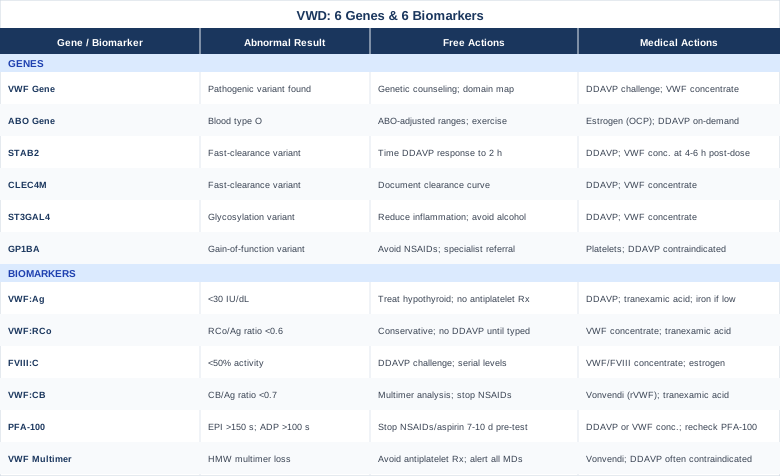
6 Biomarkers That Tell the Real Story of Von Willebrand Disease
Standard diagnostic workups for VWD often lead with a single VWF antigen level. That number matters, but it rarely tells the whole story on its own. The six biomarkers below represent a more complete panel — one that hematologists increasingly use to understand not just whether VWF is low, but whether it functions correctly, how your platelets respond to it under physiological shear conditions, and what its structural profile looks like. Together they can identify which VWD subtype you actually have, predict how well DDAVP is likely to work for you specifically, and reveal whether the underlying problem is primarily quantitative, functional, or structural.
Biomarker 1: VWF Antigen — How Much VWF Is Circulating
VWF antigen (VWF:Ag) measures the total amount of von Willebrand factor protein in plasma, regardless of whether it functions correctly. It is the starting point for every VWD workup and the number most people receive after a first hematology evaluation.
Normal range is typically 50 to 200 IU/dL, though this varies by laboratory. Values below 30 IU/dL strongly support a VWD diagnosis. Values between 30 and 50 IU/dL represent a gray zone — potentially significant in individuals with a clear personal or family bleeding history, but sometimes a normal low-end variant. Critically, blood type O individuals have physiologically lower VWF:Ag by 25–35% compared to non-O individuals, which means a value of 42 IU/dL in a type O person may carry less clinical weight than the same value in a type A or AB person. Many labs have not yet adopted ABO-adjusted reference ranges, making this an essential piece of contextual information to bring to your hematologist.
Testing is available through most hospital laboratories and reference labs. Out-of-pocket cost typically ranges from $50 to $150 depending on the provider and region.
If VWF:Ag is low — the plan without supplements
Before assuming a medication is needed, evaluate whether secondary factors are suppressing VWF. Hypothyroidism is a well-documented cause of low VWF:Ag and should be ruled out with a TSH measurement — this is inexpensive and frequently overlooked. Acute illness, recent estrogen withdrawal (postpartum period or stopping oral contraceptives), and chronic alcohol use all depress VWF. High-intensity endurance training can transiently reduce VWF:Ag as well. Addressing any of these upstream factors should precede any other intervention. Review your full medication and supplement list: high-dose fish oil (above 2 g EPA+DHA per day), ginkgo biloba, high-dose vitamin E, and garlic supplements will not change VWF:Ag but impair the broader hemostatic picture.
If VWF:Ag is low — the plan with supplements or medical options
DDAVP (desmopressin) remains the first-line medical intervention for low VWF:Ag in Type 1 VWD. It releases stored VWF from endothelial Weibel-Palade bodies, often doubling or tripling levels within 30–60 minutes. A formal DDAVP challenge with measurements at baseline, 1 hour, and 4 hours post-infusion is needed to establish your personal response profile. The nasal spray formulation (Stimate, 1.5 mg/mL) is self-administered for non-surgical bleeding events: one spray per nostril. Critical limitation: tachyphylaxis occurs — repeated doses over 24–48 hours progressively deplete endothelial stores and reduce the response. Limit use to three consecutive doses with at least a 24-hour rest period. Side effects include water retention and hyponatremia risk (most significant in young children; fluid restriction required), facial flushing, headache, and mild blood pressure elevation.
Tranexamic acid does not raise VWF:Ag but slows clot breakdown, making it a useful adjunct. Adult dosing is 1 gram three times daily (or 25 mg/kg) for acute mucosal bleeding, or starting 24 hours before and continuing 5 days after surgical procedures. Long-term daily use is generally avoided; cycling it for menstrual or procedural events is the standard pattern. GI side effects (nausea, cramping) are common; taking it with food helps. There is a theoretical thrombotic risk that warrants caution in patients with cardiovascular history.
Iron supplementation is relevant when repeated bleeding episodes have caused iron deficiency — very common in women with heavy menstrual bleeding. Ferrous bisglycinate 25–30 mg elemental iron on alternate days may improve absorption with fewer GI side effects than traditional ferrous sulfate. Recheck ferritin and hemoglobin at three months. Side effects include constipation and dark stools; staggered-day dosing reduces these considerably.
Biomarker 2: VWF Ristocetin Cofactor Activity — What VWF Actually Does
VWF ristocetin cofactor activity (VWF:RCo) — now more accurately called VWF glycoprotein Ib binding activity (VWF:GPIbR) in updated assays — tests the functional ability of VWF to bind to its platelet surface receptor, GPIbα. This interaction is what allows VWF to act as a molecular bridge between a damaged vessel wall and circulating platelets under high shear stress. A VWF molecule present in normal quantities but unable to bind GPIbα correctly will fail at precisely the moment it is most needed.
The VWF:RCo/VWF:Ag ratio is one of the most informative calculations in VWD assessment and one of the most underutilized. A ratio below 0.6 (with both values expressed in the same units) strongly suggests a qualitative defect — VWF is present but structurally or functionally compromised. This ratio is the primary way to distinguish Type 2 from Type 1 VWD at the functional level, and it directly changes treatment decisions because DDAVP can worsen certain Type 2B presentations by releasing dysfunctional VWF into circulation.
Testing is typically performed in the same panel as VWF:Ag. Combined cost at reference labs is usually $100–$250.
If the VWF:RCo/VWF:Ag ratio is low — the plan without supplements
A low ratio shifts management significantly and immediately. DDAVP may be contraindicated or of limited benefit depending on the specific subtype — VWF multimer analysis (Biomarker 6) is the next necessary step to characterize the defect precisely. Until subtype is confirmed, default to conservative management: strict avoidance of all antiplatelet substances, restriction from contact sports or high fall-risk activities, and direct communication with any dentist or surgeon that your VWD presentation requires pre-procedure specialist input. Do not self-administer DDAVP for a low VWF:RCo/VWF:Ag ratio without hematologist review.
If the VWF:RCo/VWF:Ag ratio is low — the plan with supplements or medical options
For confirmed Type 2 VWD, VWF concentrate therapy is the primary intervention. Products such as Humate-P and Wilate contain both VWF and FVIII in preserved multimer structure. Recombinant VWF (vonicog alfa, Vonvendi) contains no plasma-derived proteins and maintains the full high-molecular-weight multimer profile; it is increasingly preferred for younger patients. Tranexamic acid applies here as well for mucosal bleeding — same dosing as noted above. Estrogen-containing oral contraceptives may be considered in women with menorrhagia; while estrogen raises VWF:Ag, the VWF:RCo/VWF:Ag ratio itself typically does not normalize with estrogen alone, confirming that the qualitative defect is structural and not simply production-limited.
Biomarker 3: Factor VIII Coagulant Activity — The Hidden Passenger
Factor VIII coagulant activity (FVIII:C) measures the activity of coagulation factor VIII in the clotting cascade. The connection to VWD is mechanistically direct: VWF acts as a chaperone protein for FVIII in plasma, shielding it from premature proteolytic degradation. When VWF is low or absent, FVIII is cleared rapidly and its activity falls in proportion to the severity of VWF deficiency. Severe Type 3 VWD and a specific subtype — Type 2N (Normandy variant) — can present with FVIII:C levels in the mild hemophilia A range, low enough to cause deep tissue bleeding and hemarthroses in addition to the mucosal symptoms typically associated with VWD.
Normal FVIII:C range is 50–150%. Levels below 40% require careful perioperative planning. Levels below 10% are a medical emergency in the context of surgical procedures without prior prophylaxis planning.
If FVIII:C is low — the plan without supplements
When FVIII:C is low in the context of confirmed VWD, the immediate goal is to restore VWF since that is the vehicle protecting FVIII. Evaluate DDAVP responsiveness with serial measurements of both VWF:Ag and FVIII:C — a successful DDAVP challenge will raise both values together. Document the FVIII:C response profile: how high it rises and how quickly it returns to baseline. This profile is essential data for any surgeon or anesthesiologist managing a procedure. Type 2N should be distinguished from mild hemophilia A by genetic testing of both VWF and F8 genes, since the treatment approach differs.
If FVIII:C is low — the plan with supplements or medical options
VWF/FVIII concentrate (Humate-P, Alphanate) is strongly preferred over pure FVIII concentrate when VWD is the underlying diagnosis. Pure FVIII will be rapidly cleared in the absence of its VWF carrier, providing only transient benefit. For women with Type 2N who have heavy menstrual bleeding, estrogen-containing contraceptives raise VWF:Ag and secondarily stabilize FVIII:C — this is one of the clearest examples in VWD management where hormonal therapy produces a measurable downstream improvement in a second biomarker. DDAVP is appropriate when the underlying VWD is DDAVP-responsive, but confirm that FVIII:C rises adequately — in some Type 2N cases, the response is insufficient for surgical coverage.
Biomarker 4: VWF Collagen Binding Activity — How Well VWF Anchors to Damage
VWF collagen binding activity (VWF:CB) tests the ability of VWF to bind to collagen — the structural protein exposed at sites of vascular injury. This binding, mediated primarily by the A3 domain of VWF (and secondarily by the A1 domain for some collagen subtypes), depends almost entirely on the presence of high-molecular-weight (HMW) multimers. Smaller VWF multimers bind poorly to collagen and are far less effective at the critical first step of platelet adhesion at high shear. A low VWF:CB therefore reveals not just low VWF but structurally abnormal VWF that fails at the damage site.
The VWF:CB/VWF:Ag ratio — similar in concept to the VWF:RCo/VWF:Ag ratio — is the key calculation here. A ratio below 0.7 suggests selective loss of HMW multimers, the hallmark of VWD Type 2A. This single number predicts the treatment response to DDAVP: because DDAVP can release multimers that are then rapidly re-cleaved by ADAMTS13 before they can function, its benefit may be transient and insufficient for procedures in Type 2A patients.
Testing requires a laboratory running collagen binding assays alongside standard VWF panels. Some academic hemophilia treatment centers include this automatically; others require a specific order. Cost is typically $80–$180 as an add-on test.
If the VWF:CB/VWF:Ag ratio is low — the plan without supplements
A low VWF:CB/VWF:Ag ratio should directly trigger VWF multimer analysis to confirm the HMW multimer loss pattern. Do not proceed to any surgical or dental procedure without specialist consultation, as DDAVP response is likely to be limited and inadequate. In the interim, eliminate all substances that further impair platelet-collagen interaction: NSAIDs (stop 5–7 days before any procedure), aspirin (7–10 days), fish oil at anti-inflammatory doses (5–7 days). If iron deficiency is present from chronic blood loss, begin correction as described for Biomarker 1 — iron status independently affects platelet function.
If the VWF:CB/VWF:Ag ratio is low — the plan with supplements or medical options
VWF concentrate with intact HMW multimer profile is the standard of care for Type 2A presentations. Recombinant VWF (Vonvendi) is preferred when available because manufacturing preserves ultra-large multimers that are then physiologically processed to normal sizes after infusion. Tranexamic acid remains a useful adjunct for acute mucosal bleeding episodes between procedures. Platelet transfusion is reserved for major surgical procedures in severe cases where VWF concentrate coverage is deemed insufficient based on pre-operative PFA-100 measurements.
Biomarker 5: PFA-100 Closure Time — The Whole-System Test
The platelet function analyzer (PFA-100) — and its updated successor, the PFA-200 — is a point-of-care assay that measures how quickly platelets and VWF together close a collagen-coated aperture under conditions that simulate high arterial shear stress. It produces two closure time measurements: one using an epinephrine-coated membrane (EPI) and one using ADP-coated membrane. Normal EPI closure time is 62–150 seconds; normal ADP closure time is 62–100 seconds.
The PFA-100 EPI closure time is the most sensitive routine in-vitro screen available for von Willebrand disease. It is prolonged in most VWD types because effective platelet plug formation under shear requires both adequate VWF quantity and structural function. A normal PFA-100 in the context of a strong personal bleeding history does not rule out mild VWD — sensitivity is highest for more severe presentations — but it is a valuable initial screen and a practical monitoring tool for confirming treatment adequacy before elective procedures.
Cost is approximately $80–$150 at most hospital laboratories.
If PFA-100 closure time is prolonged — the plan without supplements
A prolonged PFA-100 in the absence of a known diagnosis should immediately prompt a full VWD panel (VWF:Ag, VWF:RCo, FVIII:C). If VWD is already confirmed, a persistently prolonged PFA before a planned procedure is actionable: it requires pre-treatment planning — either formal DDAVP challenge testing to confirm adequate response, or scheduling of VWF concentrate infusion with PFA recheck after dosing. Stop all substances that prolong platelet closure time before any testing or procedure: NSAIDs for at least 5–7 days, aspirin for 7–10 days, high-dose fish oil for 5–7 days. These windows are non-negotiable for accurate baseline testing.
If PFA-100 closure time is prolonged — the plan with supplements or medical options
If the prolonged PFA-100 reflects VWD and not isolated platelet dysfunction, DDAVP or VWF concentrate will typically normalize closure time in responsive patients — the post-treatment PFA-100 serves as confirmation of adequate hemostatic coverage before a procedure. This serial measurement approach (baseline, post-treatment) should be standard protocol. Tranexamic acid supports clot stability once platelet plug formation has been established by VWF replacement. For women, combined oral contraceptives raise baseline VWF:Ag over weeks, which can reduce background PFA-100 prolongation in Type 1 patients as a maintenance strategy separate from acute management.
Biomarker 6: VWF Multimer Analysis — The Structural Profile
VWF multimer analysis is the most technically demanding test in the VWD diagnostic panel and is typically performed at specialized hemophilia treatment centers or academic hemostasis laboratories. VWF circulates as a series of polymer chains of varying size — where the largest (high-molecular-weight) forms are by far the most hemostatically active because they have the greatest surface area for platelet and collagen binding. The analysis uses agarose gel electrophoresis to separate multimers by size and visualize their distribution.
The multimer pattern is diagnostic of VWD subtype with a precision that no single quantitative test can match: - Type 2A: Loss of both large and intermediate multimers, with only the smallest forms remaining - Type 2B: Selective loss of large multimers only, caused by a gain-of-function VWF variant that binds platelets spontaneously and is cleared faster as a result - Type 2M: Normal multimer distribution despite markedly impaired VWF:RCo activity — the multimers exist structurally but cannot interact with platelets correctly
This single test directly determines the treatment approach and contraindications. It is not a routine monitoring tool but a characterization test, ideally performed at diagnosis and repeated only if the clinical picture changes substantially. Cost ranges from $200–$400 at reference labs.
If the multimer pattern is abnormal — the plan without supplements
No lifestyle modification changes the underlying multimer pattern — this is a structural feature of the VWF being produced, determined by the VWF gene variant. However, managing bleeding triggers aggressively is critical: strict avoidance of aspirin, NSAIDs, and antiplatelet supplements; restriction of high-impact and contact activities; and proactive communication with all healthcare providers so that procedures are never scheduled without prior hemostasis planning. Ensure your hematology team documents the multimer pattern and VWD subtype explicitly in your medical record in a way that is accessible to emergency providers.
If the multimer pattern is abnormal — the plan with supplements or medical options
For Type 2A and confirmed Type 2B: DDAVP is relatively or absolutely contraindicated. In Type 2B specifically, DDAVP releases abnormal VWF that binds spontaneously to platelets, potentially worsening thrombocytopenia in addition to providing only transient multimer coverage. Recombinant VWF (Vonvendi) is the cleanest therapeutic option for HMW multimer replacement. For Type 2M, DDAVP may raise VWF antigen but the functional problem persists — concentrate is preferred for procedure coverage. Tranexamic acid remains a useful adjunct for mucosal bleeding across all subtypes. Platelet transfusion is used as salvage therapy in cases where VWF concentrate coverage is insufficient.
Having covered the biomarkers that characterize VWF behavior in the living body, it is worth stepping back to understand what shapes those numbers at the biological baseline — which is precisely where the genetic picture becomes essential.
The Genetic Landscape of Von Willebrand Disease — 6 Genes to Know
The biomarkers above describe what is happening in the bloodstream; the genes below explain much of why. Not all of these genes directly cause VWD — some are modifiers that shift the severity spectrum, treatment response, or clearance rate in ways that can make the same diagnosis look very different across individuals. Understanding which genetic context applies can reframe management decisions, make sense of apparently inconsistent lab values, and identify family members at risk before symptoms appear.
Gene 1: The VWF Gene — Where Most Answers Begin
The VWF gene on chromosome 12p13.3 encodes von Willebrand factor directly and is the primary genetic target in VWD (NCBI Gene: VWF). Over 400 pathogenic or likely pathogenic variants have been catalogued in international databases. Types 1 and 2 are caused by heterozygous variants, while Type 3 typically requires homozygous or compound heterozygous variants — meaning both copies of the gene are affected. A comprehensive clinical review by Leebeek and Eikenboom in the New England Journal of Medicine provides one of the most complete summaries available (Leebeek FW et al., NEJM 2016).
The functional domain affected by the variant predicts the clinical pattern with considerable precision. Variants in the D3 domain impair VWF multimerization, typically causing Type 2A. A1 domain variants can cause either Type 2M (lost GPIbα binding despite normal multimers) or Type 2B (gain-of-function binding that depletes platelets). D'/D3 domain variants cause Type 2N by disrupting the FVIII binding site on VWF, creating a hemophilia-A-like coagulation profile in a person with otherwise typical VWD symptoms.
If the VWF gene variant is pathogenic — the plan without supplements
Genetic counseling is the first clinical step. Understanding the inheritance pattern matters practically: autosomal dominant variants mean children have a 50% chance of inheriting the condition; recessive Type 3 variants mean both parents are carriers and siblings should be offered testing. Map the specific variant to its functional domain to predict treatment response before any procedure — an A1 domain variant indicates possible Type 2B and DDAVP caution, while a D3 variant points toward Type 2A and concentrate preference. Carry a patient-held medical summary card specifying the variant, subtype, DDAVP responsiveness status, and preferred treatment product — this information is critical in emergency settings where your regular hematologist is not available.
If the VWF gene variant is pathogenic — the plan with supplements or medical options
For Type 1 with a VWF gene variant: conduct a formal DDAVP challenge and establish your personal response parameters. Target: a 3-fold rise in VWF:Ag with normalization of PFA-100 EPI closure time. If DDAVP-responsive, nasal spray (Stimate 1.5 mg/mL, one spray per nostril) is effective for self-managed bleeding events. Cycling limit: no more than 3 doses over 48 hours due to tachyphylaxis; observe fluid restriction (1 liter per 24 hours) and avoid in children under 2 years due to hyponatremia risk. For DDAVP non-responsive variants or Types 2 and 3: intermediate-purity VWF/FVIII concentrates or recombinant VWF are the treatment backbone, dosed by your hemostasis team based on weight and severity of the event.
Gene 2: ABO Blood Group Genes — The Overlooked Modifier
ABO blood group is determined by the ABO gene on chromosome 9q34.2. Blood group O individuals have measurably lower VWF levels than A, B, or AB individuals — typically 25–35% lower — not because VWF production is reduced, but because VWF molecules lacking A and B oligosaccharide antigens are cleared from circulation faster by lectin-mediated hepatic receptors. This relationship is one of the most robustly documented non-VWF genetic influences on VWF levels. Jenkins PV and O'Donnell JS (Transfusion, 2006) demonstrated that ABO blood group is among the strongest single determinants of baseline plasma VWF levels across the general population.
The clinical implication is directly actionable: a type O person with VWF:Ag of 44 IU/dL may be phenotypically comparable to a type A person with VWF:Ag of 65 IU/dL. Many laboratories still apply a single reference range, potentially over-diagnosing VWD in type O individuals and under-appreciating the significance of borderline values in type A individuals.
If blood type O modifies your VWF — the plan without supplements
No intervention changes ABO blood type. The actionable response is interpretive and advocacy-based: request that your hematologist explicitly account for ABO blood type when interpreting your VWF:Ag values, and seek labs that provide ABO-stratified reference ranges. Beyond interpretation, regular moderate aerobic exercise maintains endothelial VWF stores through repeated low-level Weibel-Palade body stimulation, supporting a physiologically higher steady-state VWF availability over time. Stress management has direct hemostatic relevance: chronic sympathetic activation drives catecholamine-mediated VWF release that progressively depletes storage pools, leaving less reserve for genuine bleeding events.
If blood type O modifies your VWF — the plan with supplements or medical options
Estrogen is the most evidence-based pharmacological modifier of baseline VWF levels for type O individuals with borderline-low VWF:Ag. Combined oral contraceptive pills raise VWF:Ag by 20–50% depending on the estrogen component — for women with VWD-associated menorrhagia, this provides both hemostatic and gynecological benefit simultaneously. The tradeoff is an increased VTE risk, which must be assessed individually against the bleeding burden. DDAVP on-demand for procedures remains the most reliable acute intervention regardless of blood type, but the baseline from which DDAVP launches its response is lower in type O individuals — document this when establishing your challenge test parameters.
Gene 3: STAB2 — The Clearance Factor
STAB2 (Stabilin-2) encodes a scavenger receptor expressed on liver sinusoidal endothelial cells that mediates endocytosis of circulating VWF, effectively acting as a timer that determines how long VWF molecules remain in plasma before being captured and eliminated. Variants in STAB2 that increase receptor activity lead to faster VWF clearance and lower plasma VWF:Ag — without any structural defect in VWF itself and without any mutation in the VWF gene. The result is a person with low VWF:Ag, normal VWF:RCo/VWF:Ag ratio (because what VWF is present functions normally), and no identifiable VWF gene variant — a diagnostic picture that can be genuinely confusing without genetic context.
The CHARGE Consortium genome-wide association study identified STAB2 variants as among the most significant genetic determinants of plasma VWF levels in the general population (Smith NL et al., Circulation 2010).
If STAB2 variants accelerate VWF clearance — the plan without supplements
When VWF:Ag is persistently low with a normal VWF gene, STAB2 (and CLEC4M, below) are worth investigating through targeted genetic testing panels. The treatment implication is practical: because VWF production and storage mechanisms are intact in STAB2-driven low VWF, DDAVP typically elicits a brisk initial response — but the duration of effect may be shorter than in a VWF gene-driven case because the released VWF is cleared at the same accelerated rate. Document the response curve out to 2 hours post-infusion, not just the 1-hour peak, to characterize your clearance rate. Plan procedures earlier in the DDAVP response window.
If STAB2 variants accelerate VWF clearance — the plan with supplements or medical options
DDAVP is first-line and often highly effective in terms of peak response. For surgical procedures, transition to VWF concentrate at 4–6 hours post-DDAVP (before the DDAVP effect wanes below hemostatic threshold) is a common protocol if the procedure window is longer than 4 hours. No supplement-level intervention meaningfully slows STAB2-mediated VWF clearance. Estrogen raises VWF production continuously enough that it can partially offset the faster clearance — this is likely why women with STAB2-related low VWF often improve substantially with estrogen-containing contraceptives beyond what would be predicted from ABO type alone.
Gene 4: CLEC4M — A Second Clearance Pathway
CLEC4M (C-type lectin domain family 4, member M — also known as L-SIGN or DC-SIGNR) is expressed in liver sinusoidal cells and captures VWF through its lectin carbohydrate-recognition domain. Like STAB2, it acts as a VWF clearance receptor, but through a mechanistically distinct pathway. Common variants in CLEC4M were independently associated with lower plasma VWF and factor VIII levels in the CHARGE Consortium GWAS and have since been replicated in multiple European cohorts.
The practical significance of CLEC4M is that it represents a parallel clearance system operating alongside STAB2. A person carrying unfavorable variants in both genes can have cumulative clearance acceleration that substantially lowers baseline VWF:Ag even with a structurally normal VWF gene and a non-O blood type — creating a VWD-like phenotype entirely driven by clearance genetics.
If CLEC4M variants affect your VWF clearance — the plan without supplements
Clinical management mirrors the STAB2 approach: confirm DDAVP responsiveness with timed measurements to characterize your personal clearance curve. The presence of both CLEC4M and STAB2 unfavorable variants combined with blood type O creates a particularly challenging constellation where VWF:Ag may be 35–45% lower than population average with no VWF structural abnormality. This combination deserves explicit documentation in your hemostasis care plan, and any treating physician should understand that the lower baseline does not reflect a quantitatively equivalent deficit to VWF gene-driven Type 1 in terms of structural VWF quality.
If CLEC4M variants affect your VWF clearance — the plan with supplements or medical options
The treatment framework is the same as STAB2: DDAVP on-demand with careful attention to response duration, and transition to VWF concentrate for prolonged or surgical coverage needs. Estrogen-containing contraceptives help by increasing VWF production output sufficiently to offset clearance in many women. There is emerging preclinical interest in whether pharmacological inhibition of CLEC4M could raise VWF levels, but no clinical tools exist yet. Tranexamic acid remains useful as an adjunct for mucosal bleed management across all clearance-gene-driven presentations.
Gene 5: ST3GAL4 — Sugar Coating and VWF Survival
ST3GAL4 encodes a sialyltransferase enzyme responsible for adding sialic acid residues to glycan chains on the VWF protein surface. This "sugar coating" protects VWF from recognition by the asialoglycoprotein receptor (ASGPR) in the liver — a receptor that captures glycoproteins with exposed, desialylated sugars and routes them for elimination. When ST3GAL4 function is reduced, VWF molecules are undersialylated, ASGPR binds them more readily, and plasma VWF:Ag falls through accelerated hepatic clearance without any defect in synthesis or structural integrity.
Variants in ST3GAL4 have been identified in GWAS studies as contributors to VWF level variation in the population, representing a mechanistically distinct pathway from STAB2 and CLEC4M — the VWF itself is produced normally and the receptor system is intact; it is the molecular surface signal on the VWF protein that is suboptimal.
If ST3GAL4 variants affect VWF glycosylation — the plan without supplements
Lifestyle factors that broadly support glycan metabolism and endothelial function may have modest background-level relevance: reducing chronic systemic inflammation (which alters glycan transfer enzyme expression), adequate dietary intake of glycan precursors found in whole food sources (eggs, meat, dairy contain N-acetylneuraminic acid), and avoiding chronic alcohol (which disrupts glycosyltransferase activity). These are not evidence-based VWD interventions specifically, but they align with general endothelial health and avoid compounding the glycan deficit. Given the intact storage mechanism, document DDAVP responsiveness as a priority.
If ST3GAL4 variants affect VWF glycosylation — the plan with supplements or medical options
No approved supplement or medication specifically restores ST3GAL4-mediated sialylation of VWF in clinical practice. Research in this area remains preclinical. DDAVP and VWF concentrate are the clinical mainstays. Because the clearance mechanism affects VWF sialylation rather than production, the released VWF from DDAVP is equally undersialylated — the response curve should be characterized by serial measurements to identify the practical treatment window. Estrogen helps through production upregulation. For any planned procedure, the principle is the same as with other clearance-gene presentations: characterize your personal DDAVP response window, then work within it.
Gene 6: GP1BA — When the Receptor Is the Problem
GP1BA encodes glycoprotein Ibα, the platelet surface receptor that VWF binds to during the initiation of platelet adhesion at sites of vascular injury. In all other VWD types discussed, the problem lies with VWF itself. In GP1BA gain-of-function variants, the receptor is hyperactive — it binds VWF more readily than normal, causing spontaneous platelet-VWF aggregates that deplete both large VWF multimers and platelets from circulation. This condition, called platelet-type (pseudo) von Willebrand disease, mimics VWD Type 2B on virtually every standard diagnostic test, but it is caused by a platelet receptor defect, not a VWF defect.
The practical consequence is serious: DDAVP and VWF concentrate behave dangerously differently in pseudo-VWD compared to true VWD. Distinguishing the two requires mixing studies with normal platelet-rich plasma or cryoprecipitate, or direct sequencing of the GP1BA gene — neither of which is part of a standard VWD panel.
If GP1BA variant causes pseudo-VWD — the plan without supplements
Identifying pseudo-VWD immediately changes management at every level and makes referral to a specialized hemostasis center non-negotiable. DDAVP is generally contraindicated — releasing additional VWF into circulation worsens platelet binding, potentially causing acute thrombocytopenia. All platelet-depleting factors (NSAIDs, alcohol) must be scrupulously avoided. Carry a medical summary that specifically identifies this diagnosis, names it as pseudo-VWD distinct from true Type 2B, and notes that DDAVP is contraindicated — this information is critical in emergency care settings.
If GP1BA variant causes pseudo-VWD — the plan with supplements or medical options
Platelet transfusion is the cornerstone of treatment for significant bleeding in pseudo-VWD — supplying normal platelets with non-hyperactive GPIbα receptors restores the platelet component of hemostasis. VWF concentrate can be used cautiously in some clinical protocols but its effectiveness is variable because the problem is at the receptor, not the ligand. Tranexamic acid is a useful adjunct for mucosal bleeding and may reduce the frequency of platelet transfusion requirements for minor events. Genetic identification of the GP1BA variant allows cascade testing in first-degree relatives, which is particularly important given that pseudo-VWD is regularly misdiagnosed as true VWD Type 2B for years before genetic clarification.
Knowing the genetic context behind your biomarkers changes not just what the numbers mean, but which treatment paths are safe, effective, or potentially dangerous. The next section offers a different kind of framework — one that places VWF within the broader picture of vascular biology and lifestyle physiology.
What "The Clot Thickens" Gets Right About VWF
Dr. Malcolm Kendrick's 2021 book The Clot Thickens: The Enduring Mystery of Heart Disease is primarily about atherosclerosis and cardiovascular disease, but it contains one of the most accessible and biologically grounded discussions of von Willebrand factor available outside specialist hematology literature. Kendrick's central argument — that vascular disease is primarily a disease of endothelial injury and clotting response, not lipid accumulation — places VWF at the center of vascular biology in a way that has real practical implications for how someone with VWD manages their condition. The following ten points have direct relevance beyond bleeding management.
1. VWF Is the First Responder, Not Just a Clotting Factor
Kendrick reframes VWF as the body's primary vascular first-response protein — the first molecule released when an endothelial cell is injured. This framing situates VWD not just as a bleeding disorder but as a condition affecting the fundamental tissue repair mechanism of the circulatory system, which changes how you think about what endothelial health means for your condition.
2. Endothelial Health Determines Your VWF Reserve
The health of endothelial cells directly determines how much VWF is stored in Weibel-Palade bodies and therefore how much can be released when needed. Chronic endothelial injury — from smoking, hyperglycemia, oxidative stress, or repeated inflammatory insults — progressively depletes VWF storage capacity. For VWD patients, endothelial health maintenance is not abstract wellness advice.
3. Stress Raises VWF — Every Single Time, and the Mechanism Is Quantified
Epinephrine and cortisol both trigger VWF release from Weibel-Palade bodies within minutes through adenylate cyclase signaling. Kendrick emphasizes that this is not a subtle background effect — it is a major acute stimulus. Chronic psychological stress maintains a low-grade state of elevated VWF demand that progressively depletes storage pools and creates more variable lab values between measurements.
4. Estrogen Is a VWF Amplifier with a Documented Dose-Response
Estrogen upregulates VWF gene expression and raises plasma VWF:Ag substantially. This explains why VWD symptoms improve reliably during pregnancy and worsen sharply in the postpartum period when estrogen falls. Kendrick notes this as one of the clearest examples of an endogenous hormone acting as a direct VWF modulator.
5. Hypothyroidism Is a Silent and Reversible VWF Depressor
Thyroid hormone supports endothelial VWF synthesis at the transcriptional level. Subclinical hypothyroidism can depress VWF:Ag by 10–30% — a magnitude that can push a borderline-low VWD presentation into symptomatic territory. Kendrick treats this as an underappreciated example of a hormone system that operates upstream of VWF, with implications for any workup that finds persistently low VWF without a clear VWF gene explanation.
6. ADAMTS13 Is VWF's Natural Counterweight
The enzyme ADAMTS13 cleaves oversized VWF multimers, preventing them from triggering excessive platelet aggregation. Kendrick frames the VWF/ADAMTS13 balance as the fundamental dial controlling platelet plug formation intensity. In VWD, this balance is already shifted toward insufficient VWF; inflammatory states that simultaneously reduce ADAMTS13 activity can complicate the picture further.
7. Aerobic Exercise Releases VWF from Endothelial Stores Within Minutes
Moderate aerobic exercise triggers acute VWF release from Weibel-Palade bodies through shear-stress sensing by endothelial cells. This release is physiologically normal and beneficial. Kendrick's framing positions regular cardiovascular exercise as a VWF reserve management practice — repeatedly stimulating low-level endothelial VWF release without depleting it in the way that injury or stress does.
8. Inflammation Raises VWF:Ag — But Not Hemostatically Useful VWF
IL-6 and other inflammatory cytokines stimulate endothelial VWF production, which can temporarily elevate VWF:Ag into normal range during illness. This inflammation-driven increase is misleading in the context of VWD monitoring — it does not represent a genuine improvement in hemostatic capacity and will fall when the inflammatory episode resolves. Avoid testing VWF levels during acute illness or infection.
9. Blood Type O Has an Evolutionary Context That Changes How You Interpret Low VWF
Kendrick discusses how blood group O — the most ancestrally ancient ABO type — may have provided resistance advantages to certain infections while coincidentally producing faster VWF clearance. This positions blood type O with lower VWF not as inherent pathology but as evolved biology intersecting with modern diagnostic thresholds — a reframe that is clinically useful when interpreting borderline lab values.
10. VWF Is a Cardiovascular Risk Marker as Well as a Bleeding Marker
Elevated VWF is an independent marker of endothelial injury and is associated with increased cardiovascular event risk in population studies. Kendrick argues that VWF should be tracked broadly as an endothelial health indicator. For VWD patients on the lower end of the VWF spectrum, cardiovascular implications are less immediately pressing, but understanding VWF as an endothelial health proxy — not just a bleeding parameter — has real value for integrated management.
With a clearer picture of biomarkers, genetics, and biological context established, the following section addresses three evidence-informed approaches that can complement medical management for the quality-of-life dimensions that standard hematology care often does not adequately address.
Complementary Approaches Worth Considering
The evidence base for complementary therapies specifically in von Willebrand disease is limited — VWD is a relatively rare condition and most research infrastructure has gone toward pharmacological treatment. The approaches below have meaningful human evidence in closely related contexts: chronic bleeding disorders including hemophilia, chronic pain and inflammatory conditions, and autonomic regulation that is directly relevant to the VWF physiology described throughout this article. They are not alternatives to hematological care but adjuncts that address dimensions the medication cannot.
Mindfulness-Based Stress Reduction
Mindfulness-based stress reduction (MBSR) is an 8-week structured program that combines body scanning, sitting meditation, and gentle movement practices to cultivate present-moment awareness and reduce physiological and psychological stress reactivity. Its relevance to VWD is dual: chronic psychological stress drives catecholamine-mediated VWF release that progressively depletes endothelial storage reserves, as discussed above; and the anxiety and hypervigilance burden of managing a chronic bleeding disorder creates a sustained sympathetic state that MBSR specifically targets.
Research in the Haemophilia journal (Elander J et al., 2015) found that psychosocial interventions including mindfulness-based approaches significantly reduced anxiety and improved pain-related functioning in adults with hemophilia — the most closely studied population to VWD. The autonomic calming effect of MBSR reduces catecholamine-driven VWF baseline demand, which may contribute to more stable and reproducible VWF:Ag measurements between clinical visits.
Practical application: the standard 8-week MBSR program can be completed through hospital-affiliated programs or validated online formats with comparable outcomes. Daily formal practice of 20–45 minutes is the protocol; informal mindfulness during routine activities supplements this. There are no contraindications relevant to VWD. Treat MBSR as managing the chronic illness burden and stress physiology — not the VWF deficiency itself — and expectations stay calibrated appropriately.
Gentle and Restorative Yoga
Yoga as a broad category encompasses highly variable formats. High-impact, weight-bearing, or contact-risk practices are inappropriate for VWD and should not be attempted. Restorative and Yin yoga — formats that use props, hold passive poses for extended periods with minimal muscular effort, and emphasize parasympathetic activation — are a mechanistically distinct category and are relevant for VWD management through autonomic regulation, gentle joint mobility maintenance (particularly important for individuals with prior hemarthrosis from severe VWD), and long-term stress modulation.
A systematic review by Cramer H et al. examining yoga in chronic pain and inflammatory conditions broadly (published in Evidence-Based Complementary and Alternative Medicine, 2014) documented improved quality of life, reduced anxiety, and improved pain perception outcomes. VWD-specific trials do not exist; the evidence base for bleeding disorders comes primarily from hemophilic arthropathy research, which shares the joint burden but not all hemostatic features.
For practical application: 2–3 sessions per week of restorative or Yin yoga at 45–60 minutes each. Use an instructor familiar with joint protection protocols. Inform the instructor of your VWD diagnosis so they can modify any inversion or joint-pressure poses around areas with prior bleeding history. The goal is parasympathetic activation and tissue flexibility without any bleeding risk — not fitness or strength development.
Breathing-Based Therapies
Controlled, slow breathing techniques — including resonance frequency breathing, diaphragmatic pacing, and the physiological sigh — have demonstrated measurable autonomic regulatory effects across multiple clinical contexts. Their relevance to VWD lies in the same catecholamine pathway discussed throughout: psychological stress, pain anticipation, and anxiety during bleeding events all trigger epinephrine-mediated VWF release, and chronic autonomic dysregulation maintains a low-level hypervigilant state that adds physiological load to an already challenged hemostatic system. Breathing practices are among the most direct tools available for shifting this balance toward parasympathetic dominance.
A randomized controlled trial by Huang TS et al. (2018) in Applied Psychophysiology and Biofeedback demonstrated that resonance frequency breathing training at approximately 6 breaths per minute significantly increased heart rate variability and reduced resting sympathetic activity, with carryover effects on autonomic tone between sessions. This research was conducted in a general population cohort, not VWD patients, but the mechanism is directly applicable to the catecholamine-VWF connection.
Practical application: 10 minutes of controlled breathing daily at 5–6 breaths per minute (approximately 5 seconds of inhalation and 5 seconds of exhalation). No equipment is required; however, heart rate variability biofeedback devices paired with resonance frequency apps can sharpen training precision and provide measurable feedback on autonomic recovery over weeks. Begin with 2 weeks of daily sessions before reducing to a maintenance schedule of 3–4 sessions per week. There are no contraindications relevant to VWD, and the practice requires no physical exertion — making it one of the most accessible and mechanistically well-matched adjuncts available for this condition.
Conclusion
Von Willebrand disease is not a single condition with a single solution. It is a spectrum of molecular dysfunctions — quantitative, qualitative, structural, and clearance-driven — each producing a slightly different bleeding phenotype and requiring a meaningfully different clinical approach. The six biomarkers covered here — VWF:Ag, VWF:RCo, FVIII:C, VWF:CB, PFA-100 closure time, and multimer analysis — provide a layered picture of VWF behavior that no single number can capture. The six genetic variables — the VWF gene, ABO blood type, STAB2, CLEC4M, ST3GAL4, and GP1BA — explain much of why that picture looks the way it does for any given individual.
The most practical next step is to audit what has actually been measured in your workup. If you have only VWF:Ag on file, there is meaningful room to expand the panel. If you have never had a formal DDAVP challenge with timed serial measurements, that is a significant gap in your treatment planning data. If your genetic context includes blood type O, possible clearance-gene variants, or an uncharacterized low VWF with a normal VWF gene, that context deserves an explicit conversation with your hematologist — not as an academic exercise, but because it changes how your numbers should be interpreted and which interventions are most likely to work for you specifically.
Better information, pursued systematically and without urgency, leads to better decisions. That is the most grounded reason to go deeper than the standard panel.
Women's Health Cardiovascular Endocrine & Metabolic
Cardiovascular: Vascular Conditions
Autoimmune: Inflammatory Conditions