This article contains AI generated content.
· UpdatedWeill-Marchesani Syndrome Genes Biomarkers: 4 Genes And 6 Biomarkers To Track
Introduction
If you or someone you love has been diagnosed with Weill-Marchesani syndrome, you already know the particular fatigue that comes with a rare condition — the scarcity of specialists who truly understand it, the frustration of guidance that rarely goes beyond surveillance, and the sense that most medical frameworks were not built with you in mind. WMS is not simply a label. It reshapes daily life through stiff joints, limited hand function, challenging vision, and the quiet awareness that standard protocols are perpetually a step removed from your reality.
What makes WMS especially complex is that it sits at the intersection of genetics and multiple organ systems simultaneously. The same mutation that compresses your fingers also dictates whether your lens stays in place, whether your eye pressure climbs silently, and how your connective tissue holds up over decades. Telling someone with WMS to "eat well and stay active" is not wrong — it is just incomplete in a way that matters. A person whose WMS is driven by a variant in FBN1 faces different vulnerabilities than someone with ADAMTS10-related disease. That distinction changes what to monitor, what to support, and what risks to take seriously.
The science behind WMS has advanced meaningfully over the past two decades. Researchers have identified four key genes responsible for different inheritance patterns of this syndrome, mapped the biological pathways each one disrupts, and found that some of those pathways intersect with factors that are genuinely modifiable. Not curable — but adjustable in ways that can reduce the burden and slow progression in specific domains.
This article takes two complementary approaches to give you a more complete picture. The first goes deep into the four genes implicated in WMS — what each one does, what happens when it malfunctions, and what evidence exists for targeted support. The second turns to six measurable biomarkers that act as a live dashboard, showing what is actually happening in your body right now. Additional sections cover an eye-health framework from one of the most research-grounded science communicators working today, and four complementary modalities with real clinical backing. Better information does not guarantee better outcomes — but it makes better decisions possible.
Summary
Weill-Marchesani syndrome stems from mutations in four genes — ADAMTS10, FBN1, ADAMTS17, and LTBP2 — each disrupting the extracellular matrix through a slightly different mechanism. Which gene carries the mutation in your case changes what risks to prioritize and what targeted support makes biological sense. Beyond genetics, six measurable biomarkers provide a live picture of how the condition is evolving: intraocular pressure, IGF-1, TGF-beta1, high-sensitivity CRP, serum zinc, and ocular biometry each reveal a different dimension of WMS biology. The article then covers a Huberman Lab episode that reframes the visual system in ways directly relevant to WMS-related myopia and ocular stress, followed by four complementary approaches — including the De Angelis vision method, adapted yoga, photobiomodulation, and mindfulness — with real clinical evidence and condition-specific protocols. Each layer adds practical tools, not just information, and together they build a framework for smarter decisions between and during specialist appointments.
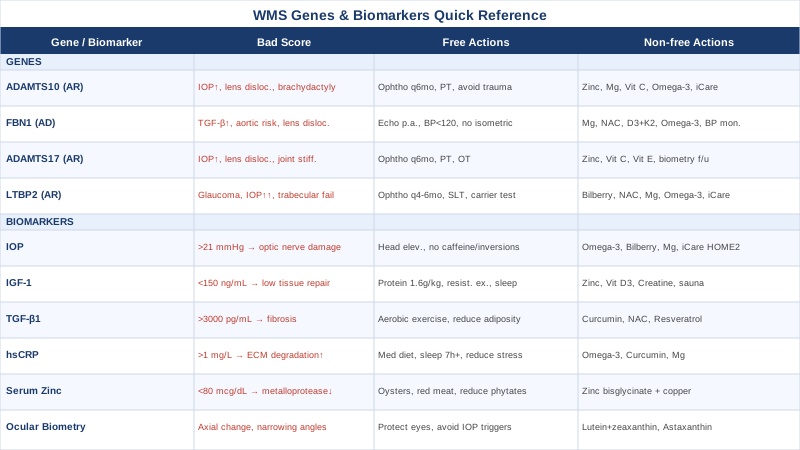
The 4 Genes Behind Weill-Marchesani Syndrome — And What You Can Do About Each
Weill-Marchesani syndrome is not caused by a single gene. At least four distinct genetic loci have been confirmed across both autosomal recessive and autosomal dominant inheritance patterns, each affecting a different component of the extracellular matrix assembly pathway. Knowing which gene carries the mutation is the starting point for any genuinely targeted management plan — because the downstream biology, and therefore the interventions that make scientific sense, differ meaningfully across the four.
Researchers like Ali Torkamani at the Scripps Research Translational Institute have emphasized that rare disease genetics is entering a new era. Variants once considered entirely unactionable are increasingly tied to biological pathways where modifiable factors — cofactor availability, inflammatory load, oxidative stress, and lifestyle — play measurable roles. That does not mean supplements replace surgery or specialist oversight. It means there is a growing scientific basis for building a supportive layer around the genetic reality that each patient carries.
Gene 1: ADAMTS10 — The Recessive Zinc-Dependent Metalloprotease
ADAMTS10 encodes a secreted zinc-dependent metalloprotease whose primary function is to assist in the assembly of fibrillin-1 microfibrils in the extracellular matrix. Fibrillin microfibrils are not simply structural scaffolding — they are the organizing framework of connective tissue, giving lens zonules, tendons, ligaments, and skin their ordered tensile properties. When biallelic mutations render ADAMTS10 non-functional, this assembly process fails from early development onward, and the characteristic WMS phenotype emerges: proportionate short stature, brachydactyly with stiff interphalangeal joints, microspherophakia, and elevated risk of secondary glaucoma through lens-induced angle changes.
This is the autosomal recessive form — both copies of the gene must carry a pathogenic variant for the condition to manifest. Carriers of a single mutation are typically clinically normal. The foundational study establishing ADAMTS10 as the causal gene was published in The American Journal of Human Genetics in 2004 (Dagoneau et al.), confirming mutations across five consanguineous families and establishing the molecular basis for recessive WMS. It remains the cornerstone reference for genetic diagnosis.
If ADAMTS10 is the causative gene: the plan without supplements
No lifestyle measure can restore a non-functional enzyme, so medical and therapeutic management carries the primary weight here. The priorities are:
- Ophthalmologic evaluation every six months, including intraocular pressure measurement and slit-lamp assessment of lens position — lens dislocation in ADAMTS10-WMS can shift from stable to surgically urgent without clear warning symptoms - Physical therapy targeting hand and finger mobility; joint stiffness in brachydactyly progresses if not actively countered - Occupational therapy to build functional adaptations for fine motor limitations - Avoidance of contact sports, martial arts, or any activity with meaningful risk of blunt ocular trauma — lens dislocation in microspherophakia can become an acute ophthalmologic emergency - Annual musculoskeletal review in children; in adults, periodic joint function assessment to track any deterioration
If ADAMTS10 is the causative gene: the plan with supplements and equipment
ADAMTS10 is structurally a zinc metalloprotease. Zinc is embedded in the catalytic domain of all ADAMTS-family enzymes — it is not optional cofactor chemistry, it is load-bearing architecture. While supplemental zinc cannot restore a non-functional enzyme, the broader ADAMTS-family enzymes that remain functional depend on adequate zinc status. There is a biologically sound argument that zinc optimization supports the surrounding metalloproteinase ecosystem even when one member of the family is lost.
- Zinc bisglycinate or zinc picolinate: 15–25 mg/day with food. Chelated forms have significantly superior absorption compared to zinc oxide or zinc sulfate. Cycle: continuous use for up to three months, then a four-week break; retest serum zinc before restarting. Always pair with 1–2 mg copper to prevent copper depletion from long-term zinc supplementation. Side effects below 40 mg/day are minimal; nausea occurs when taken on an empty stomach. - Magnesium glycinate: 300–400 mg/day, taken at night. Magnesium is a cofactor for hundreds of enzymatic pathways relevant to extracellular matrix homeostasis. The glycinate chelate minimizes the laxative effect common with magnesium oxide or citrate at similar doses. No cycling required; long-term use is safe in the absence of significant kidney disease. - Vitamin C (ascorbic acid): 500–1000 mg/day with food, split into two doses. Vitamin C is directly required for prolyl and lysyl hydroxylase activity — the enzymes that stabilize collagen triple helix structure. This supports the broader ECM environment where fibrillin microfibrils assemble. No cycling needed; long-term use at these doses is safe. - Omega-3 fatty acids (EPA+DHA): 2–3 g/day from triglyceride-form fish oil or algae-based sources. Reduces matrix metalloproteinase-driven ECM degradation and decreases the inflammatory milieu that accelerates connective tissue breakdown. Monitor for blood-thinning effects if combined with anticoagulants or antiplatelet medications. - iCare HOME2 tonometer: A rebound tonometer validated for patient self-measurement of intraocular pressure (approximately $500–800, sometimes loanable through ophthalmologists). For ADAMTS10-WMS patients with elevated glaucoma risk, trending IOP at multiple time points over weeks — including early morning, when IOP peaks — provides clinically far more useful data than a single midday clinic reading.
Gene 2: FBN1 — The Dominant Fibrillin-1 Variant
FBN1 encodes fibrillin-1, one of the most structurally critical glycoproteins in human biology. It forms 10–12 nanometer microfibrils that constitute the backbone of elastic fibers in the aorta, skin, lung, lens zonules, and virtually every connective tissue in the body. FBN1 is the same gene mutated in Marfan syndrome — but WMS-causing mutations are typically different in type and chromosomal location, producing what looks like the opposite of Marfan in several ways: short stature rather than tall, stiff joints rather than hypermobile, and a spherically compact lens rather than a stretched one.
The autosomal dominant form of WMS arises from heterozygous mutations that act through dominant-negative interference or haploinsufficiency. A critical downstream consequence is dysregulated TGF-beta signaling: fibrillin-1 normally sequesters latent TGF-beta in the extracellular matrix, holding it in reserve. When fibrillin-1 is structurally abnormal, TGF-beta is released in excess, driving tissue fibrosis, altered skeletal growth plate signaling, and pathological ocular changes. This TGF-beta excess is not just a molecular curiosity — it has been therapeutically targeted in Marfan syndrome with measurable success, and that knowledge is increasingly informing management discussions for FBN1-related WMS.
If FBN1 is the causative gene: the plan without supplements
- Annual or biannual echocardiography: Cardiac involvement in FBN1-WMS is generally less severe than in Marfan syndrome, but it cannot be assumed absent — aortic root dimensions should be established at diagnosis and trended over time - Ophthalmologic evaluation every six months with attention to lens position, IOP, and zonular integrity - Blood pressure optimization: Target below 120/80 mmHg; elevated blood pressure accelerates mechanical stress on the aortic root in any FBN1 condition - Avoid maximal isometric exercise and heavy weightlifting; prefer aerobic, moderate-intensity movement that does not produce large spikes in systolic blood pressure - Discuss losartan with your cardiologist or geneticist: in Marfan syndrome, losartan has been evaluated in multiple randomized trials for its ability to blunt TGF-beta signaling and slow aortic root dilation; its applicability in FBN1-WMS has not been established in dedicated trials but is mechanistically plausible and worth specialist discussion - Physiotherapy or physiatry for joint management, adapted for the specific stiffness pattern of FBN1-related WMS
If FBN1 is the causative gene: the plan with supplements and equipment
- Magnesium glycinate: 400 mg/day. Preclinical and observational evidence suggests magnesium has modest TGF-beta-modulatory properties; it also supports vascular smooth muscle function, which matters for aortic wall health in any FBN1 condition. Long-term use is safe; no cycling required. - NAC (N-acetylcysteine): 600 mg twice daily. A glutathione precursor with well-documented ability to reduce oxidative stress in connective tissue environments. Oxidative stress activates latent TGF-beta — reducing it is a reasonable adjunct strategy. Cycle: three months on, one month off. Rare GI discomfort; avoid in active asthma without physician guidance. - Vitamin D3 + K2: 3000–5000 IU D3 daily, paired with 100–200 mcg MK-7 K2. K2 directs calcium into bone matrix and away from vascular and soft-tissue calcification — directly relevant when ECM structure is already compromised. Monitor serum 25(OH)D; target 50–70 ng/mL. Long-term use is safe with periodic monitoring. - Omega-3 fatty acids: 3 g/day EPA+DHA. Anti-inflammatory support for ECM integrity, with mild additional cardioprotective properties relevant to the aortic considerations in FBN1-WMS. - Wrist or upper arm blood pressure monitor (validated home device, e.g., Omron series): home BP trending provides far more actionable data than isolated clinic readings, especially for a condition where aortic safety depends on consistent pressure control across the full day.
Gene 3: ADAMTS17 — The Related Metalloprotease With Its Own Profile
ADAMTS17 is a structural paralog of ADAMTS10 — closely related in sequence, functionally overlapping in its role in fibrillin microfibril assembly, and similarly classified as a zinc-dependent metalloprotease. Mutations in ADAMTS17 cause a WMS-like syndrome with features that overlap substantially with ADAMTS10-related disease, though sometimes with distinctive severity distributions across clinical features. The molecular distinction matters for recurrence risk counseling and will increasingly matter as gene-specific therapeutic approaches are explored.
Research on ADAMTS17 is less extensive than on ADAMTS10. Its role as a WMS-causing gene was established by Morales et al. in The American Journal of Human Genetics in 2009, confirmed in additional families since then. The shared enzymatic architecture with ADAMTS10 — including zinc-dependency and involvement in the same ECM assembly pathway — means that much of the biological rationale developed for ADAMTS10-WMS applies here as well, though with the caveat that ADAMTS17-specific human intervention studies do not yet exist.
If ADAMTS17 is the causative gene: the plan without supplements
The clinical management protocol largely mirrors the ADAMTS10 approach: - Ophthalmologic surveillance every six months, emphasizing IOP measurement and lens position assessment - Physical and occupational therapy for joint mobility and adaptive function - Avoidance of ocular trauma risks, particularly in work and sport contexts - Annual musculoskeletal review in growing children; periodic assessment in adults for any progression of joint stiffness
If ADAMTS17 is the causative gene: the plan with supplements and equipment
Given the shared zinc-dependent metalloprotease mechanism: - Zinc bisglycinate: 15–25 mg/day with food, cycling every three months. The cofactor rationale is identical to ADAMTS10 — zinc supports the activity of the remaining functional ADAMTS-family enzymes. Pair with 1–2 mg copper for long-term use. - Vitamin C: 500–1000 mg/day; supports the collagen hydroxylation and broader ECM environment in which both ADAMTS17 and its related proteins operate. - Vitamin E (mixed tocopherols): 100–200 mg/day. Mixed tocopherol formulations — rather than alpha-tocopherol alone, which can interfere with gamma-tocopherol at high doses — provide antioxidant protection relevant to ECM oxidative stress. Long-term use at these doses is safe. Avoid synthetic dl-alpha-tocopherol-only products. - Optical biometry follow-up: Coordinate with your ophthalmologist to obtain serial biometric measurements (axial length, anterior chamber depth, lens dimensions) at each visit and document them for trend comparison — the same monitoring rationale applies to ADAMTS17-WMS as to ADAMTS10.
Gene 4: LTBP2 — The TGF-beta Gatekeeper and Glaucoma Risk Gene
LTBP2 (Latent TGF-beta Binding Protein 2) occupies a functionally distinct position among the WMS genes. It is not a metalloprotease — it is an extracellular matrix scaffolding protein that simultaneously supports elastic fiber assembly and controls the spatial availability of TGF-beta in tissues. This dual function places LTBP2 at the center of two WMS-relevant biological processes: structural connective tissue integrity and ocular pressure regulation.
The clinical significance of LTBP2 mutations extends to primary congenital glaucoma — the same gene has been identified in families with isolated early-onset glaucoma, pointing to how critical LTBP2 is for the trabecular meshwork, the aqueous drainage structure whose dysfunction underlies elevated IOP. Mutations in LTBP2 were described in WMS-spectrum families by Désir et al. in Human Molecular Genetics (2010), establishing the gene's role in a phenotype that includes microsphaerophakia, WMS features, and glaucoma risk — often with significantly earlier and more severe IOP elevation than other WMS gene subtypes.
If LTBP2 is the causative gene: the plan without supplements
- Ophthalmologic evaluation every four to six months, with particular emphasis on IOP and trabecular meshwork health — the glaucoma risk in LTBP2-WMS is not a secondary possibility but a primary feature that warrants dedicated glaucoma-specialty consultation, not just general ophthalmology - Early discussion of laser trabeculoplasty (selective laser trabeculoplasty, SLT) if IOP trends upward, before pharmacological escalation - If glaucoma medications are initiated — prostaglandin analogs, topical beta-blockers, carbonic anhydrase inhibitors — strict adherence is essential; the structural TGF-beta dysregulation driving the pressure elevation does not resolve with intermittent treatment - Physical therapy for musculoskeletal stiffness - Carrier testing for siblings and parents; LTBP2-related WMS follows autosomal recessive inheritance, meaning siblings carry a one-in-four risk
If LTBP2 is the causative gene: the plan with supplements and equipment
- Bilberry extract (standardized to 36% anthocyanins): 160–240 mg/day in divided doses. Anthocyanins have demonstrated support for trabecular meshwork cell viability and anti-inflammatory effects in ocular tissue studies. No established cycling protocol; long-term use at these doses appears safe. Source from standardized pharmaceutical-grade extracts, not generic berry powders. - NAC: 600 mg twice daily. In addition to general antioxidant support, NAC has been studied in glaucoma models for neuroprotective and IOP-modulatory effects. Cycle: three months on, one month off. - Magnesium glycinate: 400 mg/day. Vasodilatory effects may support optic nerve perfusion in glaucomatous conditions; observational data link lower serum magnesium with higher IOP in population studies. - Omega-3 fatty acids: 3 g/day EPA+DHA — meta-analyses in healthy populations suggest modest IOP reduction of approximately 1–2 mmHg with sustained supplementation; a meaningful adjunct when every millimeter of mercury counts. - iCare HOME2 tonometer: The glaucoma risk in LTBP2-WMS makes home IOP monitoring especially important. Early-morning IOP — when pressure peaks due to the physiological circadian pattern — is routinely missed in standard clinic measurements. Daily or near-daily home monitoring with trend reporting at every ophthalmology visit gives your physician the data needed to make truly informed treatment decisions.
With the genetic picture established, the next step is connecting it to what is measurable in real time — the biomarkers that act as a live dashboard of WMS biology.
6 Biomarkers That Give Weill-Marchesani Syndrome a Dashboard
Genetics tells you the underlying risk architecture. Biomarkers tell you what is actually happening right now — and whether it is moving in the right or wrong direction. For a condition like WMS where complications accumulate insidiously across multiple organ systems, tracking the right numbers creates an early warning system that no amount of clinical intuition alone can replicate.
The six biomarkers below were selected because they are measurable in standard or near-standard clinical settings, directly tied to the biological mechanisms driving WMS complications, and actionable — meaning there are real things to do when a number is off. Several are standard clinical tests most physicians will recognize; others sit at the edge of what a general practitioner will order spontaneously, and knowing what to ask for matters.
Biomarker 1: Intraocular Pressure (IOP)
Why it matters: Elevated IOP is the leading modifiable risk factor for glaucomatous optic nerve damage — and in WMS, the risk arrives through multiple anatomical doors simultaneously. The small, spherical microspherophakic lens can mechanically block aqueous outflow, narrow angles predispose to closure glaucoma, trabecular meshwork dysfunction (particularly in LTBP2-WMS) reduces drainage capacity, and lens dislocation can produce acute pressure emergencies. Optic nerve damage from chronically elevated pressure is irreversible. This is the single most consequential number to track in WMS-related ocular disease.
How to measure it: The gold standard is Goldmann applanation tonometry at a slit-lamp examination, typically included within a comprehensive ophthalmologic evaluation ($50–150 depending on insurance and practice). Rebound tonometry (iCare principle) is available in many clinics and does not require eye drops. For home measurement, the iCare HOME2 has been clinically validated for patient self-use ($500–800 to purchase; sometimes available for loan through glaucoma practices). Normal IOP: below 21 mmHg. Values consistently above 22 mmHg warrant close monitoring; acute elevation with eye pain, rainbow halos around lights, or blurred vision requires emergency ophthalmologic assessment.
If IOP is elevated: the plan without supplements - Elevate the head of the bed 20–30 degrees during sleep: IOP follows a circadian pattern and peaks during the recumbent position — this simple change reduces overnight pressure buildup - Eliminate all sustained head-down positions: inversions in yoga, prolonged Valsalva maneuvers, tight collar clothing, and breath-holding during exercise all raise IOP meaningfully - Reduce or eliminate caffeine: each caffeinated beverage produces a transient IOP rise of 1–3 mmHg; in a pressure-sensitive eye this is not trivial - Discuss selective laser trabeculoplasty (SLT) with your glaucoma specialist as a first-line option before escalating to medications in appropriate cases
If IOP is elevated: the plan with supplements and equipment - Omega-3 fatty acids (EPA+DHA): 3 g/day — pooled data from supplementation studies suggest a modest but consistent IOP reduction of approximately 1–2 mmHg in supplemented individuals; long-term safe, check for anticoagulant interactions - Bilberry extract: 160 mg twice daily — anthocyanins support trabecular meshwork cell function and vascular health of the optic nerve; limited direct IOP trial data in WMS but strongly mechanistically supported given the trabecular pathology - Magnesium glycinate: 400 mg/day — observational studies link adequate magnesium intake with lower IOP, and magnesium's vasodilatory properties support optic nerve blood flow - iCare HOME2 for daily or twice-daily measurement; bring multi-week trend reports to every ophthalmology visit — spot readings at midday in the clinic systematically miss early-morning pressure peaks that may be driving silent damage
Biomarker 2: IGF-1 (Insulin-like Growth Factor 1)
Why it matters: IGF-1 is the primary downstream mediator of growth hormone's anabolic effects across tissues — bone, muscle, and connective tissue included. In WMS, short stature is a core feature, and monitoring IGF-1 provides an objective window into how the growth hormone axis is functioning. In children, suboptimal IGF-1 may point to a correctable axis insufficiency on top of the primary gene-driven growth limitation. In adults, IGF-1 remains a marker of tissue maintenance and anabolic signaling. Critically, IGF-1 interacts with TGF-beta pathways — the central mechanism in WMS — making it a systemically relevant indicator beyond growth alone.
How to measure it: Standard serum blood draw, ideally in the morning and without specific fasting requirements. Cost: $50–150 at commercial labs. Reference ranges are age- and sex-specific; a pediatric endocrinologist should interpret values in growing children. In adults, frameworks informed by Peter Attia's longitudinal work target IGF-1 in the 150–250 ng/mL range as consistent with tissue maintenance — not just absence of deficiency, but true functional optimization.
If IGF-1 is suboptimal: the plan without supplements - Adequate dietary protein: 1.6–2.2 g per kg of body weight daily is consistently associated with higher IGF-1 production; this is achievable through whole-food protein sources (meat, fish, eggs, legumes) without supplementation - Resistance exercise adapted for WMS: even mild progressive resistance training — using bands, water resistance, or machines rather than heavy free weights — acutely and chronically elevates IGF-1; adapt for hand and joint limitations - Sleep quality: the majority of growth hormone — and therefore IGF-1-stimulating pulses — occurs during deep sleep; 7–9 hours with consistent sleep timing and minimal light exposure creates the hormonal environment where IGF-1 is optimized without any supplementation - In children: endocrinology consultation if IGF-1 is significantly below the age-expected range, as growth hormone therapy may be appropriate in some cases
If IGF-1 is suboptimal: the plan with supplements and equipment - Zinc bisglycinate: 15–25 mg/day — zinc deficiency is a documented suppressor of the GH/IGF-1 axis; correcting suboptimal zinc status can normalize signaling. Cycle: three months on, one month off, with serum zinc testing. - Vitamin D3: 3000–5000 IU/day — low vitamin D is consistently associated with reduced IGF-1 bioavailability in epidemiological data; correcting deficiency restores IGF-1 toward normal. Monitor 25(OH)D to target 50–70 ng/mL. - Creatine monohydrate: 3–5 g/day — the most rigorously studied ergogenic compound for resistance training adaptation; supports IGF-1-dependent muscle protein synthesis particularly when combined with resistance exercise. No cycling needed; long-term safe. - Sauna (traditional or infrared): repeated sauna sessions (four sessions per week, approximately 20 minutes each at 75–85°C) have been documented to produce large acute elevations in growth hormone. Long-term IGF-1 effects are less rigorously characterized, but the protocol is low-cost and widely accessible. For patients with FBN1-related WMS and any cardiac consideration, confirm suitability with a cardiologist before use.
Biomarker 3: TGF-beta1 (Transforming Growth Factor Beta 1)
Why it matters: TGF-beta1 is not a peripheral player in WMS biology — it is mechanistically central to the pathology driven by FBN1 and LTBP2 mutations. Fibrillin-1 normally sequesters latent TGF-beta in the ECM; LTBP2 scaffolds its controlled release. When either protein is dysfunctional, TGF-beta is liberated in excess, driving tissue fibrosis, altered skeletal development, trabecular meshwork dysfunction, and potentially accelerated connective tissue remodeling. Serum TGF-beta1 is not yet part of standard clinical WMS monitoring, but it is measurable and provides a meaningful signal about how active this pathway is systemically.
How to measure it: ELISA-based serum assay available through specialized commercial labs (including LabCorp, Quest Diagnostics, and European equivalents). Cost: $100–250. Pre-analytical variables significantly affect results — sample handling, platelet activation during clotting, and lab-to-lab differences mean comparisons across different labs should be made cautiously; track trends with the same lab over time. General reference: serum TGF-beta1 in the range of 1000–2500 pg/mL is typical; values consistently above 3000 pg/mL in the right clinical context are worth specialist discussion.
If TGF-beta1 is elevated: the plan without supplements - Address the inflammatory load that activates latent TGF-beta: adipose tissue is a major TGF-beta1 source; poor sleep, ultra-processed food intake, and sedentary behavior all elevate it - Aerobic exercise: 4–5 sessions per week at moderate intensity — documented to reduce circulating TGF-beta1 over 3–6 months in multiple populations - Discuss losartan with your physician: the strongest human evidence for pharmacological TGF-beta pathway blunting in FBN1-related conditions comes from Marfan syndrome trials, where losartan showed benefit in slowing aortic dilation; applicability to FBN1-WMS is mechanistically plausible and merits expert conversation
If TGF-beta1 is elevated: the plan with supplements and equipment - Bioavailable curcumin (with piperine or liposomal formulation): 500–1000 mg/day. Multiple randomized controlled trials across fibrosis-driven conditions have demonstrated curcumin's ability to reduce TGF-beta1 at this dose range. Cycle: three months on, one month off; piperine-containing preparations interact with several medications including blood thinners and immunosuppressants — verify with a pharmacist. - NAC: 600 mg twice daily — reduces oxidative stress that activates latent TGF-beta1 in connective tissue. Cycle: three months on, one month off. - Resveratrol: 250–500 mg/day — shown to modulate TGF-beta signaling in fibrosis models including cardiac and renal; human evidence for WMS-specific applications is early-stage. Cycle: three months on, one month off. Side effects at doses below 1 g/day are rare; high doses may affect estrogen metabolism.
Biomarker 4: High-Sensitivity CRP (hsCRP)
Why it matters: Systemic inflammation accelerates extracellular matrix degradation through upregulation of matrix metalloproteinases — the same enzyme superfamily that includes ADAMTS10 and ADAMTS17. An inflammatory environment is hostile to already-compromised connective tissue, lowering the threshold for structural failure in tissues that are genetically predisposed to it. High-sensitivity CRP is inexpensive, standardized across labs, and reliable as a signal of background inflammatory load. Peter Attia and Thomas Dayspring consistently rank hsCRP among the highest-priority biomarkers for long-term tissue and cardiovascular health — and for WMS patients, the connective tissue dimension makes it even more relevant than it already is in the general population.
How to measure it: Standard blood test; cost: $10–40 depending on whether ordered alone or as part of a panel. Target for WMS patients: below 1 mg/L; ideally below 0.5 mg/L. Values above 3 mg/L represent elevated inflammatory burden actively worsening connective tissue homeostasis. Retest after ruling out acute illness — CRP is an acute-phase reactant that spikes dramatically even with minor infections.
If hsCRP is elevated: the plan without supplements - Identify and address root causes: excess adiposity, disrupted sleep (less than seven hours consistently raises CRP within days), ultra-processed food diet, and chronic psychological stress are the four largest modifiable drivers - Mediterranean-style dietary pattern: fatty fish three or more times per week, abundant vegetables, olive oil as the primary fat, minimal refined carbohydrates — replicated hsCRP reductions of 20–40% in controlled dietary trials - Sleep extension: correcting chronic short sleep to seven or more hours consistently lowers inflammatory markers, including CRP, within two to four weeks without any other intervention
If hsCRP is elevated: the plan with supplements and equipment - Omega-3 fatty acids (EPA+DHA): 3–4 g/day — multiple meta-analyses show 20–30% hsCRP reduction with sustained omega-3 supplementation over three to six months; this is among the most replicated supplement-biomarker effects available - Bioavailable curcumin: 500–1000 mg/day — well-replicated hsCRP reductions in RCTs across metabolic, inflammatory, and connective tissue conditions; cycle three months on, one month off - Magnesium glycinate: 400 mg/day — consistently lower CRP is associated with higher dietary magnesium intake across large population studies; the mechanistic link involves NF-kB pathway modulation - Retest hsCRP at eight to twelve weeks after initiating interventions to verify that the protocol is producing measurable benefit before committing to it long-term
Biomarker 5: Serum Zinc
Why it matters: ADAMTS10 and ADAMTS17 are both zinc-dependent metalloproteases — zinc is structurally embedded in their catalytic domains, not merely a peripheral cofactor. The activity of these enzymes, and of related zinc-metalloproteinases involved in matrix remodeling, is directly and proportionally tied to zinc availability. In the context of WMS, where one or two copies of these genes are already functionally compromised, ensuring that every remaining functional metalloproteinase in the system has full cofactor support is one of the most biologically coherent support strategies available. Even modest zinc insufficiency — not clinical deficiency, but suboptimal status — measurably reduces enzyme activity.
How to measure it: Serum zinc blood test; cost: $30–60 at commercial labs. Plasma zinc is marginally more reliable if both options are offered. Optimal target: 80–110 mcg/dL. Values below 70 mcg/dL indicate deficiency in most frameworks; values in the 70–80 mcg/dL range represent suboptimal status worth correcting. Critical caveat: zinc is an acute-phase reactant that drops significantly during infection or inflammatory stress — always test when clinically well to obtain a meaningful baseline.
If serum zinc is suboptimal: the plan without supplements - Increase dietary zinc through the highest-bioavailability sources: red meat, shellfish (oysters are by far the most concentrated food source), pumpkin seeds, and hemp seeds; animal-source zinc absorbs 2–3 times more efficiently than plant-source zinc - Reduce phytate competition: soaking, fermenting, or sprouting legumes and grains reduces phytate content and significantly improves zinc absorption from plant sources - Address gut inflammation if present: dysbiosis and intestinal permeability reduce zinc absorption broadly alongside other minerals
If serum zinc is suboptimal: the plan with supplements and equipment - Zinc bisglycinate or zinc picolinate: 15–25 mg/day with food. These chelated forms absorb significantly better than zinc sulfate or zinc oxide. Cycle: three months on, then a four-week break with retesting; restart if levels fall. Always co-supplement with 1–2 mg copper to prevent copper depletion — high-dose zinc depletes copper over time, and copper deficiency causes its own neurological and hematological problems. Side effects below 40 mg/day: minimal; above that threshold, nausea and metallic taste become common. - Retest serum zinc after eight to twelve weeks on supplementation to confirm target range is reached.
Biomarker 6: Ocular Biometry — Axial Length and Lens Dimensions
Why it matters: For WMS, the eye is often where the most clinically urgent — and most irreversible — complications occur. Microspherophakia creates a measurable biometric signature: an unusually spherical lens with abnormally high refractive power, narrow anterior chamber angles, and zonular fragility that predisposes to spontaneous or trauma-triggered dislocation. Serial ocular biometry is not just a baseline measurement — it is a monitoring tool that detects whether lens position is stable or drifting, whether myopia is progressing through axial elongation, and whether the angle anatomy is changing in ways that elevate glaucoma risk.
How to measure it: Optical biometry instruments — the Lenstar LS 900 or IOLMaster 700 — measure axial length, keratometry, anterior chamber depth, and lens thickness with high reproducibility. These are available in most academic ophthalmology departments and increasingly in private practice. Cost: typically included within a comprehensive evaluation or billed discretely at $100–300. Anterior segment OCT adds detailed angle anatomy and is particularly important for monitoring angle-closure risk. Ultrasound A-scan biometry provides a fallback when optical methods are obstructed by dense lens opacity.
Serial comparison is more important than any single measurement: request that all biometric values be recorded at each visit and explicitly ask for trend comparison at every appointment.
If biometry shows concerning trends: the plan without supplements - If axial length is changing rapidly in a child with WMS-related myopia: discuss urgency of optical management and, if indicated, surgical planning with your ophthalmologist — progressive axial change in the context of microspherophakia raises dislocation risk - If anterior chamber angles are narrowing: laser peripheral iridotomy (LPI) may be recommended to reduce acute closure risk before it becomes an emergency - Avoid all activities producing sustained IOP elevation: heavy lifting with breath-holding, inversions, and impact sports all increase mechanical stress on already-fragile zonular fibers - Protective eyewear in all moderate-to-high-risk environments — structurally compromised zonules cannot withstand the same impact forces as a normal eye
If biometry shows concerning trends: the plan with supplements and equipment - Lutein and zeaxanthin: 20 mg lutein plus 4 mg zeaxanthin daily — macular carotenoids with the strongest evidence base in ocular nutrition; relevant for long-term photoreceptor protection in eyes already under anatomical stress. Safe long-term; fat-soluble, take with a meal. - Astaxanthin: 6–12 mg/day — a carotenoid with potent antioxidant and anti-inflammatory properties in ocular tissues, with some evidence for retinal and optic nerve cytoprotection. Long-term use appears safe; no established cycling protocol. - Blue-light-filtering spectacle lenses with UV400 protection: a low-cost, zero-risk intervention that reduces cumulative photochemical stress on a lens and retina already operating under anatomical compromise. - Orthokeratology contact lenses for myopia control: overnight-worn lenses that reshape the corneal surface to slow axial elongation; this has meaningful evidence support in pediatric myopia control and represents a non-pharmaceutical option for managing myopia progression in WMS patients where axial growth is a concern.
From biomarkers and genes, the picture of WMS biology comes into clearer focus — and understanding the visual system more deeply adds another practical layer for those navigating WMS's most consequential complications.
What the Huberman Lab's Eye Health Episode Gets Right for WMS Patients
The Huberman Lab podcast, hosted by Stanford neuroscientist Andrew Huberman, has produced some of the most rigorously grounded episodes on eye health and visual biology available in any public science-communication format. For WMS patients — where ocular involvement can range from correctable myopia to glaucoma, lens dislocation, and long-term vision loss — having a deep working understanding of how the eye functions and what evidence-based interventions are available is not a bonus: it is part of managing the condition intelligently.
The most relevant episode covers visual neuroscience, the biology of myopia development and progression, and practical protocols grounded in published research. Below are the ten most impactful takeaways for someone with WMS or related ocular involvement.
1. Outdoor Time Is the Most Evidence-Based Myopia Intervention Known
Consistent epidemiological data across multiple countries show that children who spend two or more hours per day outdoors develop myopia at significantly lower rates and with slower progression than those who remain indoors — independent of near-work habits. The mechanism involves high-intensity ambient light triggering dopamine release from retinal amacrine cells, which signals the sclera to slow axial elongation. For WMS patients managing myopia alongside their underlying lens pathology, maximizing daily outdoor time is free, immediate, and supported by some of the strongest preventive evidence in ophthalmology.
2. Indoor Light Is 10 to 100 Times Dimmer Than Outdoor Light
Most people dramatically underestimate this gap. A bright indoor environment near a window may measure 500–1000 lux. Outdoor light on an overcast day reaches 5000–10,000 lux; full sunlight exceeds 100,000 lux. This gap explains why indoor environments — even well-lit ones — cannot substitute for outdoor exposure in terms of the light-driven signals that protect the developing and adult visual system.
3. Near-Far Accommodation Exercises Reduce Ciliary Muscle Fatigue
Regularly shifting visual focus between near and far distances exercises the ciliary muscle and reduces accommodative spasm — a factor in myopia progression and visual fatigue. The practical protocol is straightforward: for every 20 minutes of near work, spend at least 20 seconds looking at something more than six meters away. For WMS patients with abnormal lens curvature and zonular fragility, reducing the cumulative duration of sustained near-focus reduces mechanical stress on the zonules alongside the more commonly discussed refractive benefits.
4. Smooth Pursuit Eye Movements Activate Visual Cortex Differently Than Static Gaze
Tracking a slowly moving object across the visual field activates the visual cortex in ways that static fixation does not, and research on smooth pursuit training suggests benefits for visual processing efficiency. For WMS patients who may have had reduced visual quality since early childhood — with neural adaptation to a structurally unusual optical system — maintaining active visual engagement through varied gaze behaviors supports the neural components of vision within whatever structural constraints exist.
5. IOP Follows a Circadian Pattern and Peaks in Early Morning
Intraocular pressure is not constant — it follows a circadian rhythm, typically peaking between approximately 6 and 9 AM for most individuals in the supine position during and just after sleep. Clinic measurements taken at midday systematically underrepresent the full pressure burden over 24 hours. For WMS patients — especially those with LTBP2-related disease and elevated glaucoma risk — home tonometry at multiple time points, including immediately after waking, is not a luxury but a meaningful clinical contribution.
6. Blink Rate Drops Dramatically During Screen Work
At baseline, most people blink 15–20 times per minute. During concentrated screen use, blink rate commonly falls to three to seven times per minute. Reduced blinking leads to tear film instability, surface irritation, and increased mechanical interaction between the eyelid and anterior ocular surface. For WMS patients with already-abnormal anterior segment anatomy, dry eye is an additional stressor that is simple to address: deliberate full blinks every 20 minutes during screen sessions, and lubricating eye drops without preservatives if needed.
7. Morning Sunlight Has Distinct Circadian and Neurological Benefits
Light in the first one to two hours after sunrise has a particular spectral and intensity quality that strongly entrains the central circadian clock through melanopsin-containing intrinsically photosensitive retinal ganglion cells (ipRGCs). These same cells project to brain regions regulating mood, autonomic nervous system tone, and cortisol timing. Getting 10–20 minutes of morning outdoor light daily — without sunglasses — establishes circadian rhythm coherence that cascades into better sleep, more stable energy, and lower baseline inflammatory markers. For someone managing a chronic condition, this is one of the highest-leverage zero-cost daily habits available.
8. Lutein, Zeaxanthin, and Vitamin A Are the Foundation of Evidence-Based Ocular Nutrition
Huberman reviews the evidence for specific nutrients in ocular biology: vitamin A for rhodopsin synthesis and photoreceptor maintenance, lutein and zeaxanthin for macular carotenoid density and light filtering. These are among the most replicated nutritional interventions in long-term ocular health research, supported by large prospective epidemiological studies. For WMS patients facing cumulative ocular stress from structural abnormalities, foundational nutritional support for the retina is a rational and low-risk layer.
9. Broad Visual Gaze Activates the Parasympathetic Nervous System
Huberman describes research showing that deliberately expanding visual gaze to a panoramic field — rather than focusing on a near point — activates the parasympathetic branch of the autonomic nervous system and reduces stress-related physiological arousal. Chronic psychological stress, which is common in rare disease contexts, drives tunnel vision, elevated cortisol, and increased inflammatory load. Spending time in environments that naturally promote wide-field gaze — outdoors, near water or open landscapes — is both a practical stress-reduction strategy and a visual behavior with documented neurological benefits.
10. Eye Exercises Cannot Fix Structural Problems — But They Can Optimize What Remains
Huberman is explicit and precise on this point: no exercise protocol can correct axial myopia, microspherophakia, or zonular laxity. Structural anatomy is structural anatomy. What exercises and behavioral changes can do is optimize the neural, muscular, and inflammatory environment in which a structurally compromised eye operates — reducing accommodative fatigue, supporting retinal health, maintaining neural plasticity, and reducing unnecessary stressors. For WMS patients, this framing matters: complementary visual practices are additions to ophthalmologic care, not alternatives to it.
Complementary Approaches With Genuine Evidence for WMS
Several complementary modalities have enough clinical evidence and biological plausibility to be worth serious consideration as part of a WMS management plan. The four below were selected specifically because they match this condition's features — not because they are generic wellness additions.
The David De Angelis Method: Reducing Accommodative Demand in a Structurally Unusual Eye
What it is and why it is relevant: David De Angelis is an Italian optometrist who developed a functional vision training system based on the premise that myopia involves not only axial elongation but also habitual accommodation patterns, ciliary muscle overuse, and visual behavioral habits. His framework, detailed in The Secret of Perfect Vision: How You Can Prevent and Reverse Nearsightedness, uses deliberate distance-looking protocols, ciliary muscle relaxation techniques, and reduced near-work duration to modify the accommodative environment the eye operates in. For WMS patients, where myopia coexists with microspherophakia and abnormal lens mechanics, reducing accommodative demand and ciliary muscle tension has a direct structural relevance that goes beyond standard myopia management: tense ciliary contraction in an eye with zonular fragility adds mechanical strain to structures already at risk.
The fundamental premise that the eye responds to its optical environment is supported by primate research. Form-deprivation studies — inducing myopia in monkeys and then reversing it by removing the deprivation stimulus — established that axial length is not a fixed anatomical fate. Francis Young's early work and subsequent studies in multiple animal models confirmed the eye's optical-environment sensitivity, forming the biological basis for the accommodation-based myopia approach.
Specific technique: The core protocol involves active distance-looking sessions — deliberately focusing on objects more than six meters away for 10–15 minutes, three to four times per day — and palming (placing warm, cupped palms over closed eyes for five to ten minutes) to reduce accommodative tone and ciliary muscle fatigue. These techniques are paired with deliberate near-work management: screen breaks, reduced session duration without breaks, and the 20-20-20 rule as a minimum baseline. For WMS patients with zonular fragility, the benefit of reducing sustained ciliary contraction is not hypothetical — tense ciliary muscles in miosis (pupil contraction) can affect the mechanical forces on the lens zonules.
How to apply it cautiously: This approach is not a substitute for ophthalmologic monitoring or surgical intervention when indicated. For WMS specifically, avoid any protocol involving ocular massage, direct pressure on the globe, or exercises that might increase intraocular pressure. Distance-looking and palming are both safe, low-risk additions. Discuss with your ophthalmologist before beginning any structured vision therapy program to confirm it does not conflict with current ocular management.
Adapted Yoga for Joint Stiffness and Connective Tissue Mobility
What it is and why it is relevant: The brachydactyly and progressive joint stiffness that characterize WMS create real functional limitations — grip strength, fine motor coordination, and whole-body movement quality. Adapted yoga — particularly restorative and gentle hatha styles — offers a structured, evidence-supported approach to maintaining and gradually improving joint range of motion, fascial flexibility, and proprioception without placing the high compressive or shear loads on small joints that most conventional exercise protocols involve.
Specific technique and evidence: A systematic review in the European Journal of Physical and Rehabilitation Medicine found that yoga significantly improved hand function and joint flexibility in rheumatoid arthritis patients — a condition with overlapping joint stiffness characteristics. While WMS-specific yoga trials do not exist, the mechanistic overlap in joint-stiffness etiology justifies cautious extrapolation. The recommended protocol: 30–45 minute sessions three to five times per week, emphasizing gentle hand and wrist mobilization sequences, yin yoga holds (sustained passive stretching of fascial and joint capsule tissue at low load), and full-body mobility flows that do not demand weight-bearing on small joints. Yin yoga's approach of holding positions for two to five minutes at low intensity specifically targets the connective tissue layer rather than the muscular layer — directly relevant to the ECM-level pathology in WMS.
How to apply it cautiously: All inversions must be avoided in WMS — headstands, shoulder stands, prolonged downward dog, and any sustained head-below-heart position elevates IOP and risks lens dislocation in a microspherophakic eye. Use fists, forearm support, or yoga blocks instead of full palm-down weight bearing to protect metacarpophalangeal joints. Work with an instructor experienced in therapeutic or adaptive yoga; a standard group class without disclosed medical context is not the right starting point.
Photobiomodulation for Connective Tissue and Ocular Support
What it is and why it is relevant: Photobiomodulation (PBM) uses red and near-infrared light — typically 630–850 nm — at low irradiance to stimulate mitochondrial function through cytochrome c oxidase in the respiratory chain, producing downstream increases in cellular ATP, reductions in oxidative stress, and modulation of inflammatory signaling. In WMS, two domains are potentially addressable: the musculoskeletal component (joint and connective tissue) and the ocular component (retinal cell and optic nerve health). PBM has the strongest evidence in connective tissue healing and joint conditions, with emerging data in ocular applications.
Specific technique and evidence: A study published in Gerontology (Jeffery et al., 2021) found that brief daily exposure to 670 nm red light for three minutes improved mitochondrial function in retinal cells and improved color discrimination in older adults, suggesting a meaningful biological effect in ocular tissue. In connective tissue and joint applications, multiple randomized controlled trials have demonstrated PBM reduces pain and improves functional measures in osteoarthritis and tendinopathy. For joint applications in WMS: commercial full-body panels (Joovv, Mito Red) at standard therapeutic parameters (4–20 J/cm² applied to joint surfaces, three to five sessions per week) are reasonable adjuncts for joint-stiffness management.
How to apply it cautiously: Standard PBM panels must never be applied directly to open eyes — the irradiance levels appropriate for connective tissue can damage retinal photoreceptors if applied without proper ophthalmic protocols. For joint and soft tissue applications in WMS, commercial panels are safe and appropriate. For ocular PBM specifically, the emerging research uses purpose-designed ophthalmic delivery devices at precisely controlled parameters — this requires working with a practitioner familiar with ophthalmic photobiomodulation. Consider PBM a low-risk adjunct for the musculoskeletal component of WMS, and an area to monitor closely in ophthalmology as ocular evidence matures.
Mindfulness-Based Stress Reduction (MBSR): The Inflammatory Dividend
What it is and why it is relevant: Managing a rare condition that affects vision, hand function, and physical capacity generates a particular kind of chronic psychological stress — the stress of uncertain prognosis, medical system navigation, and functional limitation that compounds year over year. Beyond quality of life, this stress has measurable biological consequences: elevated cortisol accelerates connective tissue catabolism, chronically elevated sympathetic tone raises blood pressure and IOP, and inflammatory cytokines — including CRP and IL-6 — are consistently elevated in states of chronic psychological stress. MBSR addresses all three simultaneously through a structured, evidence-validated approach.
Specific technique and evidence: A meta-analysis published in JAMA Internal Medicine including 47 randomized trials found that mindfulness meditation programs produced significant improvements in anxiety, depression, and pain in people with chronic physical conditions — with measurable reductions in inflammatory markers in several included studies. The MBSR protocol is eight weeks of structured instruction: weekly group sessions (in-person or validated online), daily home practice of 10–20 minutes, combining breath awareness, body scanning, and non-reactive observation of physical sensation and mental content. Apps such as Waking Up, Insight Timer, and Ten Percent Happier provide accessible entry points for daily practice outside of formal courses.
How to apply it cautiously: MBSR requires no physical modification for most WMS patients. For those with significant visual impairment, audio-guided formats are entirely equivalent in effect. The biological case for MBSR in WMS goes beyond wellbeing: reducing the inflammatory signal that chronically elevated stress produces directly supports hsCRP targets and the connective tissue environment. It is also the one intervention in this section that costs nothing and carries essentially no risk — which makes it the easiest starting point of the four.
Conclusion
Weill-Marchesani syndrome demands a level of specificity that generic health advice cannot provide. The four genes covered here — ADAMTS10, FBN1, ADAMTS17, and LTBP2 — each represent a distinct set of biological vulnerabilities and a distinct starting point for targeted support. The six biomarkers — intraocular pressure, IGF-1, TGF-beta1, hsCRP, serum zinc, and ocular biometry — translate that genetic reality into numbers you can track, trend, and act on between clinical encounters. The complementary approaches, from adapted yoga to photobiomodulation to mindfulness, add evidence-supported layers that address what conventional management alone does not reach.
None of this replaces the irreplaceable: regular ophthalmologic monitoring, specialist input, and the medical interventions that protect vision and joint function over a lifetime. The goal is to arrive at every clinical appointment more informed, with more useful data, and with a supportive framework that actively works in the spaces between appointments.
The next smart step is concrete: confirm your specific WMS gene through genetic testing if not already done, arrange baseline measurement of the six biomarkers covered here, and bring the results to a specialist who can help contextualize what you find. Better information, in this condition above most, is the difference between reacting to complications and staying ahead of them.
Musculoskeletal: Joint Conditions
Autoimmune: Connective Tissue Conditions