This article contains AI generated content.
· UpdatedWilliams Syndrome Genes and Biomarkers — 6 Biomarkers and 6 Genes to Track
Introduction
Williams syndrome sits at a crossroads where remarkable human qualities — deep empathy, musical sensitivity, social warmth — meet serious medical vulnerabilities that are easy to miss until they become crises. The condition is caused by a microdeletion of approximately 26 to 28 genes on chromosome 7q11.23, and its effects span cardiovascular function, kidney health, metabolism, brain development, and mental wellbeing. For families and individuals navigating this diagnosis, standard medical checkups provide a baseline, but they often miss the subtler shifts that predict larger problems down the road.
The challenge with Williams syndrome is that the medical risks are real and multisystem, but the guidance available to most families tends to be reactive rather than preventive. Cardiovascular complications, hypertension, kidney strain, hypothyroidism, and metabolic dysfunction can all accumulate quietly over years. By the time symptoms become obvious, the window for easier intervention may have already passed. Generic health advice — eat well, exercise, reduce stress — is true but not enough when you are managing a specific constellation of genetic vulnerabilities.
What changes the picture is knowing which biological signals to track, what normal looks like versus concerning, and what levers actually move the needle based on the specific physiology involved. Biomarkers are the bridge between the underlying genetics and what can be measured, monitored, and influenced. Understanding which genes are deleted and what each one controls gives you the map; tracking the right biomarkers tells you where you currently stand on that map.
This article covers two complementary approaches. The first focuses on six biomarkers with strong clinical relevance to Williams syndrome — what each one reveals, how to measure it practically, and what to do when numbers fall outside a healthy range. The second section goes deeper into the genetic architecture of the deletion itself, looking at which genes drive which risks and what the research suggests about managing those risks. Alongside those two frameworks, a closer look at Peter Attia's longevity medicine approach and several evidence-backed complementary modalities rounds out a genuinely actionable picture. None of this offers a cure, but together these tools offer a more targeted way to think about long-term health in Williams syndrome.
6 Key Biomarkers to Track in Williams Syndrome
Tracking biomarkers in Williams syndrome is not about chasing perfect numbers. It is about catching early shifts before they compound. The six markers below are the ones most directly tied to the known vulnerabilities of the 7q11.23 deletion — and the ones most likely to be underutilized in routine care.
1. Serum Calcium and Urine Calcium-to-Creatinine Ratio
Why it matters. Hypercalcemia — elevated blood calcium — is one of the most well-documented complications of Williams syndrome, particularly in infancy and early childhood, where prevalence estimates range from 15% to over 50% depending on the study and age group reviewed. The mechanism is not fully resolved but appears to involve abnormal sensitivity to vitamin D, dysregulation of calcitonin signaling, and possibly disruption of calcium channel genes within or near the deleted region. Untreated hypercalcemia causes irritability and feeding problems in infants, but the longer-term concern is its effect on the kidneys and cardiovascular system. Calcium deposits in renal tissue (nephrocalcinosis) and blood vessels can accumulate silently over years. Adults with Williams syndrome are at meaningful risk for chronic kidney disease partly as a consequence of calcium dysregulation that was not closely managed in childhood.
How to measure it. A standard comprehensive metabolic panel (CMP) includes serum calcium and costs $20–$60 at most labs, often covered by insurance. For a more complete picture, the urine calcium-to-creatinine ratio is a more sensitive early marker of calcium excretion problems and can detect subclinical hypercalciuria even when serum calcium appears normal. A spot urine test collected in the morning is sufficient for this ratio and costs $15–$40. In children and adolescents with Williams syndrome, both tests should be performed at least annually — more frequently if there is any history of elevated values. Reference ranges for serum calcium: 8.5–10.2 mg/dL for adults. The urine calcium-to-creatinine ratio should typically remain below 0.2 mg/mg in adults.
If the score is bad — the plan without supplements. When serum calcium runs elevated, dietary management is the first tool. This means reducing high-calcium foods (dairy, fortified plant milks, calcium-fortified cereals) to moderate rather than eliminating levels. More importantly, it means avoiding vitamin D supplementation unless specifically prescribed and monitored, since vitamin D enhances intestinal calcium absorption and can worsen hypercalcemia in susceptible individuals. Adequate hydration is critical — consistent daily fluid intake appropriate to body weight, since concentrated urine dramatically increases nephrocalcinosis risk. Sun exposure should be moderate and not excessive during periods of active hypercalcemia, as UV-triggered skin synthesis of vitamin D can contribute to calcium spikes.
If the score is bad — the plan with supplements or equipment. In persistent or severe hypercalcemia, medical management is required and cannot be replaced by self-directed supplementation. Physicians may consider low-dose corticosteroids short-term, bisphosphonates in cases with documented nephrocalcinosis, or ketoconazole (which reduces calcitriol synthesis) under close supervision. These require medical prescription and careful monitoring. At home, a quality electronic hydration-tracking system and a reliable daily water intake routine support consistent fluid goals. A home blood pressure monitor is also valuable since hypercalcemia can contribute to hypertension. Many common supplements that seem benign — vitamin D, calcium powders, certain fortified products — can be genuinely harmful in this population and should be flagged with every healthcare provider.
2. Kidney Function: eGFR and Creatinine
Why it matters. Renal structural abnormalities are found in approximately 18–30% of individuals with Williams syndrome, including duplicated collecting systems, horseshoe kidney, and reduced kidney size. Add to this the cumulative effects of hypercalcemia and hypertension — both common in WS — and the kidneys become one of the most important systems to monitor proactively. Chronic kidney disease can develop silently over decades, with meaningful function loss occurring before creatinine rises detectably. The estimated glomerular filtration rate (eGFR) is a more sensitive marker than creatinine alone, and tracking it longitudinally gives a trend line that a single isolated value cannot provide.
How to measure it. eGFR is calculated from serum creatinine, age, and sex and is included in any comprehensive metabolic panel ($20–$60). Cystatin C is a more accurate marker for early kidney function decline, particularly in people with low muscle mass, which can artificially lower creatinine and make eGFR appear normal. Cystatin C costs $40–$100 and is not routine but can be requested; it is particularly useful for children and adults with Williams syndrome who may have lower-than-average muscle mass. A 24-hour urine collection for protein and creatinine clearance provides the most complete kidney function picture and costs $30–$100; this is appropriate for anyone with a prior kidney abnormality or consistently borderline eGFR.
If the score is bad — the plan without supplements. When eGFR begins to decline or shows an adverse trend, the most important non-supplemental interventions are hydration, blood pressure control, and calcium management — the three largest modifiable contributors in WS. A consistent fluid intake of 1.5–2 liters daily for adults, adjusted for body weight, supports kidney filtration without requiring supplementation. Dietary sodium reduction to under 2,000 mg per day reduces kidney workload and supports blood pressure. Protein intake should remain moderate rather than high, since excessive protein accelerates chronic kidney disease progression; aim for 0.8–1.0 g per kilogram of body weight. Regular aerobic movement — even 20–30 minutes of walking four to five times per week — supports renal perfusion without placing excess stress on the system.
If the score is bad — the plan with supplements or equipment. No supplement reverses kidney function decline, and several popular supplements are nephrotoxic in excess (high-dose creatine, excessive vitamin C, certain herbal compounds). Where evidence supports supplementation in progressive chronic kidney disease, omega-3 fatty acids (2–4 g per day EPA+DHA, taken continuously with meals, no cycling required) have modest evidence for slowing decline and reducing proteinuria. Magnesium glycinate (200–400 mg per day, continuous, generally well-tolerated) may support vascular relaxation and blood pressure with minimal kidney risk at these doses. A reliable digital blood pressure cuff ($30–$80) is the single most valuable home equipment investment for kidney-related monitoring, since hypertension is both a cause and consequence of kidney decline. Check blood pressure at the same time of day, in both arms if there is any concern about arterial differences relevant to WS with peripheral stenosis, and log trends rather than individual readings.
3. Blood Pressure and NT-proBNP
Why it matters. Hypertension is one of the most prevalent medical complications across the lifespan in Williams syndrome, affecting an estimated 40–50% of adults. The ELN gene deletion impairs elastin production throughout the arterial system, leading to stiffer, less compliant vessels. Supravalvular aortic stenosis (SVAS) and peripheral pulmonary artery stenosis are the most serious structural cardiovascular consequences, but diffuse arterial stiffening raises blood pressure throughout the body even in the absence of discrete stenosis. NT-proBNP — N-terminal pro-brain natriuretic peptide — is a cardiac stress biomarker released when the heart chambers are under pressure or volume load, useful for detecting early cardiac strain that precedes symptoms.
How to measure it. Home blood pressure monitoring is accurate, inexpensive ($30–$80 for a validated upper-arm cuff), and arguably the most impactful test someone with Williams syndrome can do independently. Blood pressure should be taken at rest, seated, in both arms, at the same time of day. Consistent bilateral arm measurements matter in WS because peripheral artery stenosis can cause meaningful differences between arms — a difference of more than 10–15 mmHg warrants medical evaluation. NT-proBNP is a blood test costing $50–$150, not typically routine but appropriate when cardiac symptoms arise or when echocardiography is not recently available. Echocardiography remains the gold standard for structural cardiovascular monitoring and should be performed per cardiologist schedule — typically every 3–5 years in stable adults, more frequently in those with known SVAS.
If the score is bad — the plan without supplements. For elevated blood pressure in WS, the same lifestyle foundations that work in the general population apply with a few specific priorities. Sodium reduction has the most consistent evidence: limiting dietary sodium to under 2,000 mg per day reduces systolic blood pressure by 4–8 mmHg on average. A whole-food dietary pattern with abundant vegetables, particularly leafy greens and potassium-rich foods, supports blood pressure regulation since potassium counterbalances sodium at the kidney level. Consistent aerobic exercise at moderate intensity for at least 150 minutes per week reduces systolic blood pressure by 5–10 mmHg in hypertensive adults. Stress and anxiety management is particularly relevant in WS given characteristically high anxiety rates; chronic stress elevates cortisol and sympathetic tone, both of which directly raise blood pressure.
If the score is bad — the plan with supplements or equipment. Magnesium glycinate (300–400 mg per day, taken in the evening, continuous use, no cycling required) has meaningful evidence for modest blood pressure reduction and is generally well-tolerated. Coenzyme Q10 (100–200 mg per day with a fat-containing meal, continuous use) has shown modest blood pressure benefits in some trials, particularly relevant if statin drugs are part of the management plan. Beetroot-based nitrate supplementation (food-based through beets and leafy greens, or concentrated beetroot juice ~500 mL per day) supports nitric oxide production, which promotes arterial relaxation — directly relevant given the arterial stiffness profile in WS. Side effects are minimal but include red urine, which is benign. A validated upper-arm digital blood pressure cuff used daily provides far more actionable data than occasional clinic readings.
4. Thyroid Panel: TSH and Free T4
Why it matters. Thyroid dysfunction — specifically subclinical hypothyroidism — appears more commonly in Williams syndrome than in the general population, with some studies reporting rates of 15–30% in children and variable rates in adults. The exact mechanism is not fully established; thyroid abnormalities in WS may relate to thyroid gland structural differences or to altered immune regulation. What makes this particularly important is that thyroid function has a significant impact on cognitive performance, energy regulation, mood, and cardiovascular health — all domains that are already areas of challenge in WS. Subclinical hypothyroidism, characterized by elevated TSH with normal T4, can reduce cognitive clarity and worsen fatigue and anxiety without producing the classical symptoms that typically lead to investigation.
How to measure it. A basic thyroid panel includes TSH and Free T4, costing $30–$80. Most guidelines suggest annual thyroid screening in children with Williams syndrome and every 1–2 years in adults, though in practice this is often underperformed. For a fuller picture, adding Free T3 and reverse T3 ($60–$120 for the expanded panel) provides information about thyroid hormone conversion, relevant because some individuals convert T4 to T3 inefficiently regardless of TSH status. Anti-TPO antibodies (thyroid peroxidase antibodies, approximately $40) can identify autoimmune thyroid involvement if suspected. Normal TSH range is typically 0.5–4.5 mIU/L, though some clinicians focused on cognitive optimization prefer a tighter range of 1.0–2.5; discuss the target with the treating physician.
If the score is bad — the plan without supplements. When TSH is mildly elevated and Free T4 is still within range, a period of monitoring is often appropriate before initiating treatment. Non-supplemental supports for thyroid function include ensuring adequate selenium and iodine through diet — Brazil nuts (1–2 per day, not more, due to selenium toxicity risk at higher amounts), seaweed, seafood, and iodized salt provide both without supplementation. Reducing excessive intake of raw cruciferous vegetables is a conservative step, though cooking deactivates most goitrogens and moderate cooked amounts are not a concern. Managing anxiety and sleep quality directly supports thyroid axis regulation, since chronic cortisol dysregulation suppresses thyroid function. Adequate sleep (7–9 hours for adults) is one of the most underutilized thyroid-supportive interventions available.
If the score is bad — the plan with supplements or equipment. Selenium in the form of selenomethionine (100–200 mcg per day, continuous use without cycling, do not exceed 400 mcg per day as toxicity risk is real) has solid evidence for supporting thyroid function and reducing anti-TPO antibody titers in autoimmune thyroid conditions. Iodine supplementation should be approached cautiously — both excess and deficiency impair thyroid function, and supplementation above dietary levels is rarely needed when food sources are adequate. In cases of confirmed subclinical or clinical hypothyroidism, thyroid hormone replacement (levothyroxine, by prescription) is the medically supported treatment. For home tracking, a basal body temperature protocol — measuring temperature on waking before rising, targeting 97.8–98.2°F — provides a rough ongoing indicator of thyroid function between lab tests at no cost.
5. Fasting Glucose, Insulin, and HOMA-IR
Why it matters. Metabolic dysfunction — including insulin resistance, prediabetes, and type 2 diabetes — occurs at elevated rates in adults with Williams syndrome compared to the general population. The reasons are multifactorial: reduced physical activity due to motor challenges or cardiovascular limitations, higher rates of anxiety-driven eating patterns, medication side effects, and possibly direct metabolic effects related to genes in the deleted region. A critical point often missed in routine care is that fasting glucose alone is an insensitive early marker of insulin resistance. It typically remains normal until significant pancreatic reserve has already been lost. Fasting insulin and HOMA-IR — a calculated index using fasting glucose and insulin — detect insulin resistance years before glucose rises, providing a much earlier intervention window.
How to measure it. Fasting glucose is included in a CMP ($20–$60). Fasting insulin must be ordered separately and costs $25–$60; it requires true fasting of 8–12 hours with water only. HOMA-IR is calculated as (fasting glucose in mg/dL × fasting insulin in µIU/mL) ÷ 405. A HOMA-IR below 1.0 is considered optimal; 1.0–1.9 is borderline; above 2.0 suggests insulin resistance. For a more comprehensive metabolic picture, a continuous glucose monitor (CGM) worn for 2 weeks ($50–$200 depending on platform) provides dynamic glucose data that identifies postprandial spikes and glucose variability that a single fasting test cannot capture. HbA1c ($20–$50) reflects 3-month average glucose and is a useful adjunct.
If the score is bad — the plan without supplements. Insulin resistance responds well to lifestyle intervention without supplementation. Time-restricted eating — consuming all calories within a 10–12 hour window — reduces insulin secretion burden by extending the overnight fasting period and has evidence for improving HOMA-IR within weeks. Prioritizing protein and fiber at meals blunts postprandial glucose spikes, and reducing refined carbohydrate intake consistently moves metabolic markers in the right direction. For Williams syndrome specifically, finding appropriate physical activities that match motor ability and personal enjoyment is critical; swimming, dance, and cycling on a stationary bike have low injury risk and meaningful metabolic benefit. Even 10–15 minutes of walking after meals has been shown in controlled trials to reduce postprandial glucose by 15–20%.
If the score is bad — the plan with supplements or equipment. Berberine (500 mg with meals two to three times daily, cycling with breaks of 4–8 weeks after 3 months of continuous use due to potential gut microbiome disruption with chronic use) has demonstrated glucose and insulin reduction comparable in some studies to metformin. Side effects include GI discomfort, particularly when initiating use. Magnesium glycinate (300–400 mg per day, continuous, no cycling required) improves insulin sensitivity in magnesium-deficient individuals, which is common in the general population and likely in WS given dietary variability. Alpha-lipoic acid (300–600 mg per day with meals, can be used continuously) has modest evidence for glucose metabolism support and antioxidant effect. Berberine should not be combined with blood-sugar-lowering medications without medical supervision. A CGM worn for 2-week periods every 3–6 months provides excellent real-world feedback on dietary and lifestyle interventions at far less cost than a single specialist consultation.
6. Lipid Panel with ApoB
Why it matters. Given the arterial vulnerabilities in Williams syndrome — stiff, less elastic vessels, elevated blood pressure, and metabolic risk — cardiovascular disease risk accumulates faster and earlier than in the general population. Standard lipid panels (total cholesterol, LDL, HDL, triglycerides) provide a partial picture, but ApoB (apolipoprotein B) is the superior marker for cardiovascular risk because it counts every atherogenic particle — LDL, VLDL, IDL, and Lp(a)-containing particles — rather than estimating cholesterol mass. A person can have a normal LDL cholesterol value but a high ApoB particle count, representing substantially elevated cardiovascular risk that a standard panel misses entirely. Thomas Dayspring, Allan Sniderman, and Peter Attia have all emphasized ApoB as the most important single lipid marker for cardiovascular outcome prediction, and it is especially relevant in conditions where arterial stiffness is already structurally elevated.
How to measure it. A standard lipid panel costs $20–$80 and is widely available. ApoB is usually ordered separately at $30–$80 and is increasingly included in preventive panels. Lp(a) — lipoprotein(a), a particularly atherogenic genetic particle — costs $40–$80 and should be measured at least once in any individual with elevated cardiovascular risk, since Lp(a) values are largely genetically determined and do not meaningfully change with lifestyle interventions; a high Lp(a) permanently elevates cardiovascular risk and should inform more aggressive management of all other modifiable factors. For optimal cardiovascular tracking in WS, request triglycerides, HDL, LDL-C, ApoB, and Lp(a) at baseline, then ApoB and triglycerides at each subsequent follow-up.
If the score is bad — the plan without supplements. Elevated ApoB and triglycerides both respond to dietary changes. Reducing refined carbohydrates and added sugars has the strongest evidence for lowering triglycerides. Replacing saturated fat with monounsaturated fat — olive oil, avocado — modestly reduces LDL particle number. For Williams syndrome specifically, maintaining a consistent meal schedule and avoiding large infrequent meals supports lipid metabolism. Aerobic exercise is one of the most effective interventions for raising HDL and lowering triglycerides; 150 minutes per week of moderate-intensity activity consistently moves these numbers in the right direction.
If the score is bad — the plan with supplements or equipment. Omega-3 fatty acids (EPA+DHA 2–4 g per day with meals, continuous use without cycling) have robust evidence for triglyceride reduction; at therapeutic doses of 3–4 g per day, reductions of 20–30% are typical. High doses can modestly raise LDL-C in some individuals, so track both triglycerides and ApoB together to assess net benefit. Berberine (500 mg twice daily with food, cycling as described above) reduces LDL-C and ApoB by mechanisms overlapping with statin activity and is an appropriate consideration for individuals not yet requiring prescription medication. Red yeast rice is sometimes used for cholesterol but contains naturally occurring statins and should not be used without medical supervision, particularly in WS where kidney monitoring is already warranted. Prescription statin therapy, when indicated by a physician, is appropriate in WS and does not appear to be contraindicated by the deletion itself.
The Genetic Architecture of the Williams Syndrome Deletion
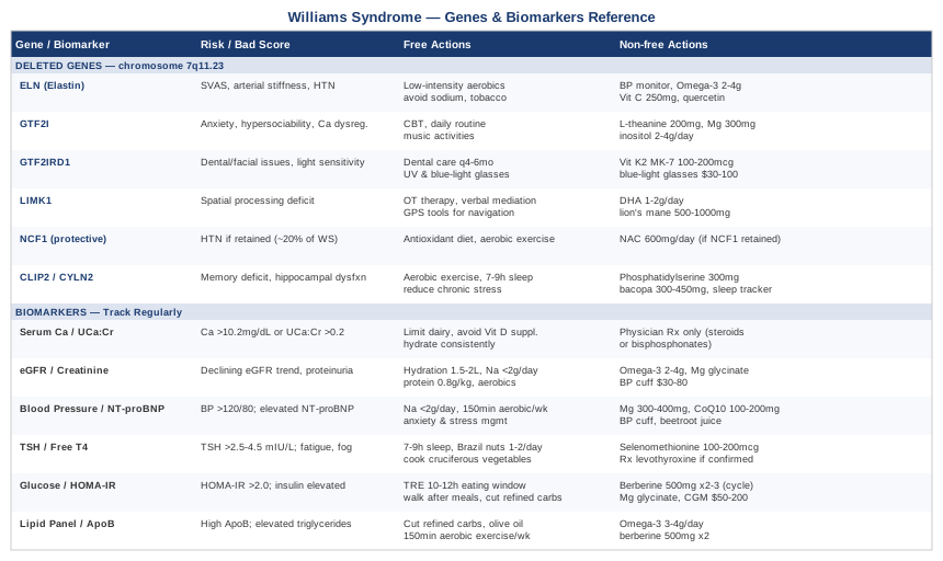
Understanding which genes are deleted in Williams syndrome — and what each one specifically controls — transforms how you interpret symptoms, why certain risks cluster together, and where monitoring and management should be prioritized. The deletion spans approximately 1.5–1.8 megabases on chromosome 7q11.23 and affects around 26–28 genes. According to the GeneReviews entry on Williams syndrome, six of these genes have the strongest evidence linking their loss to specific clinical consequences. Understanding each one gives meaning to the biomarker findings described above.
ELN (Elastin)
What the deletion causes. Elastin is the primary structural protein of arterial walls, giving blood vessels their ability to stretch and recoil with each heartbeat. Hemizygosity — one copy deleted — of ELN in Williams syndrome is the primary driver of supravalvular aortic stenosis (SVAS) and peripheral pulmonary artery stenosis. Without sufficient elastin production, arteries become stiffer, narrower, and more prone to progressive stenosis. This affects the aorta, pulmonary arteries, and peripheral vessels throughout the body, and explains directly why blood pressure and cardiovascular monitoring are so critical in WS throughout the lifespan.
If this gene is deleted — the plan without supplements. Since ELN hemizygosity permanently reduces elastin production, the management focus shifts entirely to protecting arterial health and managing consequences. This means rigorous blood pressure control, avoiding tobacco and high-sodium diets, maintaining a physically active lifestyle at appropriate intensity, and ensuring regular cardiology monitoring including echocardiography. Exercise should emphasize low-to-moderate intensity aerobic activity rather than high-intensity or heavy resistance training that creates acute blood pressure spikes dangerous in SVAS.
If this gene is deleted — the plan with supplements or equipment. No supplement restores elastin production. However, nutrients that support connective tissue and vascular health have indirect relevance. Vitamin C (250–500 mg per day, no cycling required, essential cofactor for collagen synthesis) provides mild support for arterial wall structural integrity. Flavonoids like quercetin (500–1000 mg per day, can be taken continuously, has anti-inflammatory and modest vascular-protective effects in human trials) contribute to endothelial support. Omega-3s at 2–4 g per day support arterial flexibility markers in clinical trials. A home blood pressure monitor is the most important equipment investment. Low-impact aerobic exercise tools — stationary bike, pool access, resistance bands for gentle strength maintenance — support vascular health without excessive hemodynamic stress.
GTF2I (General Transcription Factor IIi)
What the deletion causes. GTF2I encodes a transcription factor called TFII-I that regulates the expression of many other genes. It plays a central role in the distinctive social and cognitive profile of Williams syndrome — the characteristic hypersociability, strong verbal abilities, emotional expressivity, and the specific anxieties. Mouse models with GTF2I deletion reproduce key behavioral features including hypersociability and altered fear responses. GTF2I also appears to regulate calcium channel expression, providing a potential mechanistic link between the social phenotype and the calcium dysregulation seen in WS. This gene is currently one of the most actively studied in the WS deletion.
If this gene is deleted — the plan without supplements. The behavioral and cognitive features driven by GTF2I deletion are best addressed through structured, consistent environments that reduce ambiguity and provide social scaffolding. Anxiety — one of the most disabling features of WS in adults — benefits significantly from cognitive behavioral therapy adapted for intellectual disability, consistent daily routine, environmental predictability, and social support. Music-based activities exploit the preserved musical processing strength that appears partly linked to this gene region's influence on auditory-limbic circuitry.
If this gene is deleted — the plan with supplements or equipment. For anxiety related to GTF2I-driven alterations in brain circuitry, evidence-based options include L-theanine (200 mg per day, taken in the morning, can be used continuously, promotes calm alertness without sedation, well-tolerated with minimal side effects), magnesium glycinate (300–400 mg in the evening, continuous), and inositol (2–4 g per day, well-tolerated, has evidence in anxiety and OCD-spectrum presentations, can be used continuously without cycling). None of these replace structured psychological support but may support the nervous system context in which that support occurs.
GTF2IRD1 (GTF2I Repeat Domain Containing 1)
What the deletion causes. GTF2IRD1 is closely related to GTF2I and appears to influence craniofacial development, muscle tone, and some aspects of social behavior. Research in mouse models suggests GTF2IRD1 deletion contributes to the distinctive facial features of Williams syndrome and to altered social behavior independent of GTF2I. It also appears to play a role in retinal development and light sensitivity — relevant given that some individuals with WS report heightened sensitivity to bright environments. Dental abnormalities including enamel hypoplasia and crowding are also associated with this gene region.
If this gene is deleted — the plan without supplements. Craniofacial implications of GTF2IRD1 deletion include dental crowding, enamel hypoplasia, and bite abnormalities — all common in WS. Regular dental care every 4–6 months rather than annually, combined with early orthodontic assessment, is the most direct response. Light sensitivity can be managed practically through quality sunglasses with UV and blue light filtration and tinted lenses where indoor lighting creates discomfort.
If this gene is deleted — the plan with supplements or equipment. For enamel and dental health support: vitamin K2 in the MK-7 form (100–200 mcg per day, continuous, supports mineralization pathways) combined with adequate but monitored vitamin D supports dental mineral metabolism. High-quality blue-light-filtering glasses ($30–$100) and indoor lighting adjustments — warmer, lower-intensity light sources — are practical, low-cost tools for daily light sensitivity management.
LIMK1 (LIM Kinase 1)
What the deletion causes. LIMK1 encodes a kinase that regulates actin cytoskeleton dynamics in neurons — critical for dendritic spine formation, synaptic plasticity, and the physical structure of neural circuits involved in spatial processing. Hemizygosity of LIMK1 is a primary driver of the pronounced visual-spatial deficit characteristic of Williams syndrome: individuals with WS typically have strong language and face-processing abilities but marked impairment in tasks requiring spatial construction, such as drawing, navigating, and assembling objects. This specific dissociation — preserved language with severely impaired spatial cognition — is one of the most studied neuropsychological profiles in developmental neuroscience.
If this gene is deleted — the plan without supplements. Spatial processing challenges driven by LIMK1 hemizygosity benefit from repeated structured practice using compensatory strategies: verbal mediation of spatial tasks (talking through instructions step by step), spatial skill occupational therapy starting in childhood, and environments designed to reduce navigation demands. GPS and mapping tools effectively remove one of the most functionally limiting spatial deficits from daily life. Visual-spatial skill-building through occupational therapy in childhood shows meaningful benefit in adaptive function even without changing the underlying biology.
If this gene is deleted — the plan with supplements or equipment. LIMK1 drives actin polymerization in neurons; supporting overall synaptic health and neuroplasticity indirectly supports the function that remains. Evidence-based neuroplasticity support includes omega-3 DHA (1–2 g per day specifically as DHA, continuous, critical for synaptic membrane fluidity and structural integrity of dendritic membranes), lion's mane mushroom extract (500–1000 mg per day, continuous, stimulates nerve growth factor with human trial evidence for cognitive support, mild GI side effects possible), and consistent aerobic physical exercise, which remains the most robust known stimulus for BDNF-mediated synaptic plasticity broadly. These do not restore LIMK1 function but support the network resilience within which remaining circuits operate.
NCF1 (Neutrophil Cytosol Factor 1) — The Protective Deletion
What the deletion causes — and the surprising twist. NCF1 encodes p47-phox, a component of NADPH oxidase, an enzyme complex that generates reactive oxygen species. Higher NADPH oxidase activity increases oxidative stress in vascular tissue, contributing to endothelial damage and hypertension. In an unusual situation, when NCF1 is deleted as part of the Williams syndrome microdeletion, vascular oxidative stress is reduced — which appears to be protective against hypertension. Research has shown that WS individuals who retain NCF1 (due to a different deletion breakpoint, affecting approximately 20% of WS cases) have significantly higher rates of hypertension than those for whom NCF1 is also deleted.
Clinical implication and what to do with this information. Knowing whether NCF1 is deleted is clinically useful because it predicts hypertension risk with greater precision. This is determined through the specific molecular characterization of the deletion (chromosomal microarray), which should be part of the initial genetic workup and is increasingly standard. If NCF1 is retained, blood pressure monitoring should be more frequent and intervention thresholds should be set more conservatively. If NCF1 is deleted, hypertension risk is still present due to ELN-related arterial stiffness, but the baseline oxidative trajectory may be more favorable. For the approximately 20% of WS individuals who retain NCF1, antioxidant support becomes more relevant: N-acetylcysteine (600 mg per day, can be cycled with 4-week breaks after 3 months), and the same dietary and exercise strategies that reduce vascular oxidative stress in the general population.
CLIP2 / CYLN2 (Cytoplasmic Linker Protein 2)
What the deletion causes. CLIP2 encodes cytoplasmic linker protein 115, which is involved in microtubule dynamics, vesicle transport, and hippocampal development. The hippocampus is a region essential for memory consolidation, spatial navigation, and stress regulation. Mouse models with CLIP2 deletion show hippocampal abnormalities, learning and memory deficits, and altered stress responses. In Williams syndrome, CLIP2 hemizygosity contributes to the mild-to-moderate intellectual disability, memory challenges, and the exaggerated stress response that characterizes the condition's anxiety phenotype. It also appears to interact with the hippocampal mechanisms underlying the heightened music-emotional response that is distinctive to WS.
If this gene is deleted — the plan without supplements. Hippocampal health and memory function are most supported by consistent aerobic exercise (the most potent known stimulus of hippocampal neurogenesis), quality sleep (during which memory consolidation occurs), and low chronic stress exposure. Sleep quality in Williams syndrome deserves particular attention because sleep disturbances — including sleep apnea (more common in WS due to craniofacial anatomy) and non-restorative sleep — directly impair hippocampal consolidation. A sleep evaluation is appropriate if sleep is non-restorative or if snoring is noted.
If this gene is deleted — the plan with supplements or equipment. Phosphatidylserine (300 mg per day, continuous, has clinical trial evidence for memory and hippocampal-related cognitive function, generally well-tolerated), bacopa monnieri (300–450 mg of standardized extract per day, continuous use required for 8–12 weeks before full effect becomes apparent, take with food to minimize GI discomfort), and DHA as above. A sleep tracker (Oura Ring, Fitbit, or similar, $100–$350) provides objective data on sleep stages and helps identify non-restorative patterns worth pursuing clinically.
What Outlive Gets Right About Managing Williams Syndrome for the Long Term
Peter Attia's Outlive: The Science and Art of Longevity (2023) is not written for Williams syndrome, but its framework — what Attia calls Medicine 3.0, proactive and deeply biomarker-driven — maps directly onto what individuals and families managing WS actually need. Attia's central argument is that medicine intervenes too late, treating disease after it arrives rather than working to prevent it decades earlier. For a condition like WS, where cardiovascular disease, kidney failure, and metabolic dysfunction accumulate slowly over years, the Medicine 3.0 approach is not optional — it is the only approach that meaningfully improves long-term outcomes.
1. Start Tracking Cardiovascular Markers Now, Not When Symptoms Appear
Attia argues that most people are diagnosed with heart disease when it is already advanced — because standard thresholds for "normal" are calibrated to avoid over-treatment, not to optimize health. In WS, where ELN deletion creates inherent arterial vulnerability from birth, waiting for symptoms is particularly dangerous. Tracking ApoB, blood pressure trends, and NT-proBNP from early adulthood provides decades of early warning and early intervention opportunity.
2. ApoB Is the Number That Matters Most for Cardiovascular Risk
Attia, alongside Dayspring and Sniderman, has consistently emphasized that ApoB is the most predictive single cardiovascular risk marker, capturing actual atherogenic particle burden rather than estimating cholesterol mass. In WS, where stiff arteries are already a structural reality from the ELN deletion, minimizing the atherogenic particle count through diet, exercise, and where necessary medication meaningfully reduces additive cardiovascular risk. Request ApoB specifically — it is frequently not included in standard panels unless explicitly ordered.
3. Zone 2 Cardio Is the Most Important Exercise Investment
Attia's advocacy for Zone 2 training — low-intensity sustained aerobic effort at 60–70% of maximum heart rate, where you can speak in full sentences but feel effort — is well-matched to Williams syndrome. Zone 2 improves mitochondrial density, lowers resting heart rate, improves insulin sensitivity, and supports cardiovascular resilience without the dangerous blood pressure spikes of high-intensity training. For individuals with SVAS or peripheral artery stenosis, Zone 2 is also the safest intensity range. Thirty to forty-five minutes, four to five times per week, is the practical target.
4. Muscle Mass Protects Against Metabolic Disease
Attia frames muscle mass as the organ of longevity — its loss predicts mortality more reliably than most blood markers. For adults with WS who may be less physically active due to motor challenges or anxiety, preserving or building functional muscle mass through resistance training adapted to individual capacity is one of the highest-return health investments available. Even two sessions per week of resistance exercise meaningfully improves metabolic markers and reduces fall risk over time.
5. Sleep Is a Non-Negotiable Health Variable
Attia dedicates substantial material to sleep as a biological necessity. In WS, sleep disruption is common: anxiety delays sleep onset, and craniofacial anatomy increases sleep apnea risk. Poor sleep directly worsens insulin resistance, blood pressure, cognitive function, and emotional regulation — compounding every other WS vulnerability. Sleep apnea screening via polysomnography or home sleep testing ($100–$300 for home testing) is appropriate for any WS individual with non-restorative sleep, snoring, or daytime fatigue that is disproportionate to activity level.
6. Emotional Health Is Not Separate from Physical Health
Attia treats psychological health — particularly anxiety and depression — as major determinants of physical longevity, not a secondary concern. This is especially relevant in WS, where anxiety is pervasive, often severe, and poorly managed in routine care. Unmanaged chronic anxiety elevates cortisol, raises blood pressure, disrupts sleep, promotes insulin resistance, and accelerates arterial aging. The physical consequences of untreated WS anxiety are real and measurable in biomarkers. Psychological support is therefore not optional — it is a core medical intervention with cardiovascular and metabolic consequences.
7. Insulin Resistance Starts Decades Before Diabetes — Catch It Early
Attia is explicit that a normal fasting glucose is insufficient for metabolic assessment. HOMA-IR, CGM use, and fasting insulin are the tools that identify metabolic dysfunction at the stage where intervention is most effective. This is particularly relevant in WS, where reduced activity and anxiety-related eating patterns create metabolic risk that accumulates invisibly behind a "normal" fasting glucose result in routine blood work.
8. Blood Pressure Targets Should Be Tighter Than Standard Guidelines
Attia advocates for tighter blood pressure management than many clinical guidelines, particularly for individuals with identified cardiovascular risk. For WS, where arterial stiffness is structural from the ELN deletion, a target of under 120/80 mmHg — or lower with individual clinical guidance — is more protective than the commonly cited 130/80 threshold. Home monitoring enables this precision; office readings are too infrequent and often artificially elevated by white-coat effect.
9. Kidney Function Trends Matter More Than Single Values
Attia emphasizes longitudinal tracking — a single eGFR is far less informative than whether eGFR is stable, rising, or falling over three to five years. For WS, where structural renal abnormalities and cumulative calcium burden create inherent kidney risk, establishing a baseline eGFR in childhood and tracking it at consistent intervals provides decades of early warning for intervention that a snapshot result cannot.
10. The Goal Is Healthspan, Not Just Lifespan
The central message of Outlive — maintaining function, independence, and quality of life through the later decades — is precisely what a proactive biomarker approach to Williams syndrome enables. Managing calcium, blood pressure, kidney function, thyroid health, and metabolic markers is not about adding medical complexity to life. It is about reducing the probability that preventable complications steal function and independence in midlife and beyond.
Complementary Approaches With Meaningful Evidence
The following modalities have human clinical evidence relevant to specific challenges common in Williams syndrome: anxiety, blood pressure, cardiovascular health, and cognitive support. None replace medical care, but each offers a meaningful adjunctive contribution when used appropriately and realistically.
Music Therapy
People with Williams syndrome have a documented neurological affinity for music — heightened emotional response, unusually precise rhythm perception, and a social orientation toward musical interaction that is distinctive even compared to other developmental conditions. This is not a sentimental observation; it reflects altered auditory cortex organization and preserved musical processing circuits even where other cognitive domains are significantly impaired. Music therapy exploits this preserved channel for therapeutic benefit: regulating emotion, supporting communication, reducing anxiety, and building social connection through a modality that individuals with WS are naturally oriented toward.
The evidence for music therapy in WS is unusually strong for a complementary modality. Researchers at the Salk Institute, including Ursula Bellugi and Daniel Levitin, documented WS musical processing differences in neuroimaging studies showing greater amygdala and reward-circuit activation in response to music compared to typically developing individuals. Music therapy programs adapted for neurodevelopmental disabilities have demonstrated reductions in anxiety and improved social engagement in multiple controlled studies across conditions with overlapping profiles. The Williams Syndrome Association has incorporated music therapy into recommended support frameworks precisely because the neurological fit is so specific.
In practice, a music therapy program for Williams syndrome should be led by a board-certified music therapist (MT-BC credential), using receptive music listening, rhythmic entrainment exercises, and instrument-based interactive sessions. Sessions of 45–60 minutes, one to two times per week, are typical in structured programs. At home, structured listening to personally meaningful music — particularly before anxiety-provoking transitions or activities — can function as a self-regulation tool used daily without clinical cost, once the underlying approach has been introduced by a trained therapist.
Mindfulness Meditation and MBSR
Anxiety is one of the most impairing features of Williams syndrome in adolescents and adults, and one of the most undertreated. Mindfulness-Based Stress Reduction (MBSR) — the structured 8-week program originally developed by Jon Kabat-Zinn at the University of Massachusetts — has strong evidence for reducing generalized anxiety, improving emotional regulation, and lowering physiological markers of chronic stress including cortisol and blood pressure. In neurodevelopmental populations, adapted versions that use shorter sessions, more sensory anchors, and visual supports show meaningful engagement and measurable benefit.
A meta-analysis of mindfulness interventions in intellectual disability published in Mindfulness (Hwang and Kearney, 2013) found that adapted mindfulness approaches reduced anxiety and challenging behavior in participants with mild-to-moderate intellectual disability — the range that encompasses many adults with WS. More recent trials continue to support this direction, and adapted MBSR for neurodevelopmental conditions is a growing area of clinical development. The key adaptation for WS is simplification: shorter practices of 5–10 minutes rather than 45, sensory-grounded anchors including breath and tactile sensation, and visual prompts that reduce verbal demand on the participant.
To apply this realistically: begin with 5-minute guided breathing exercises using a free app at a consistent time each day, either morning or before typically anxious situations. Progress to 10–15 minute body scan practices over several weeks as comfort increases. For more structured benefit, an MBSR course adapted for neurodevelopmental needs — offered by some occupational therapy practices and disability support organizations — provides the full framework. A practical caution: some individuals with WS find open-monitoring mindfulness more anxiety-provoking than focused-attention practices, since unfocused attention can amplify rumination; start with breath-focused or music-guided options and adjust from there.
Biofeedback
Biofeedback trains individuals to consciously regulate physiological responses — heart rate, blood pressure, muscle tension, skin conductance — by providing real-time feedback on those signals. For Williams syndrome, where anxiety directly drives physiological dysregulation and elevated blood pressure, and where standard relaxation instructions may be insufficiently concrete for the learning profile, biofeedback's explicit feedback loop is well-suited. Heart rate variability (HRV) biofeedback in particular has evidence for reducing sympathetic nervous system activity, improving anxiety, and modestly lowering blood pressure by training a more flexible autonomic response.
Randomized trials published in journals including Applied Psychophysiology and Biofeedback have demonstrated meaningful HRV biofeedback benefits for anxiety reduction in adult samples. Studies in developmental populations with anxiety, including autism spectrum and ADHD, support generalization of these findings across neurodevelopmental profiles. No published trials in WS specifically exist at present, but the mechanism — reducing sympathetic overdrive through trained parasympathetic activation — is directly relevant to the anxiety and cardiovascular profile of Williams syndrome and makes this a reasonable adjunct.
Home HRV biofeedback devices such as the Inner Balance by HeartMath or the Muse headband ($100–$250) provide accessible starting points. A protocol of 10–20 minutes of HRV biofeedback training, five days per week for 8–12 weeks, is sufficient to establish the skill in most adults. For blood pressure specifically, resonance frequency breathing — typically around 6 breaths per minute — is the most evidence-supported biofeedback protocol and can be practiced without a device once the rhythm is learned, making the initial equipment investment a training tool rather than a permanent dependency.
Breathing-Based Therapies
Breathing directly modulates the autonomic nervous system — a fact that is physiologically fundamental and increasingly well-documented in clinical trials. For Williams syndrome, where chronic anxiety maintains elevated sympathetic tone and where cardiovascular health is a primary medical concern, controlled breathing practices offer a low-cost, always-available tool for both acute anxiety regulation and cumulative cardiovascular benefit that requires no equipment and no prescription.
Clinical trials of slow paced breathing at approximately 5–6 breaths per minute have demonstrated reductions in systolic blood pressure of 4–8 mmHg in hypertensive adults — comparable to first-line medications in mild hypertension. Device-assisted paced breathing (the FDA-cleared RESPeRATE device, approximately $300–$400) has the most controlled-trial evidence for blood pressure reduction through consistent daily use and may be appropriate for adults with WS hypertension as a supported adjunct. The underlying mechanism — increased baroreflex sensitivity and reduced sympathetic outflow — is directly applicable to the vascular physiology of WS.
A simple daily protocol: 5 minutes of 5–6 breath-per-minute breathing (inhale for 5 counts, exhale for 5 counts) practiced twice daily, morning and before sleep. This can be cued with a free app such as Breathwrk or Paced Breathing. Over 4–8 weeks of consistent practice, this builds baroreflex sensitivity and begins shifting the resting autonomic balance measurably. For acute anxiety — which in WS can escalate rapidly — an extended exhale pattern (inhale 4 counts, exhale 8 counts) activates the parasympathetic brake within 60–90 seconds and can be taught as a self-regulation skill even to individuals with moderate intellectual disability when introduced with appropriate support and visual cues.
Conclusion
Williams syndrome is a genuinely complex condition, but complexity is not the same as unmanageability. The genetic deletion on chromosome 7q11.23 creates real and specific vulnerabilities — in the arteries, kidneys, thyroid, metabolic system, and brain — but each of these vulnerabilities can be monitored, measured, and meaningfully supported when approached with the right tools and the right information.
Six biomarkers — serum calcium, kidney function, blood pressure, thyroid hormones, metabolic markers, and lipid particle count — provide the earliest and most actionable signals for the health risks that accumulate in WS across the lifespan. Six genes — ELN, GTF2I, GTF2IRD1, LIMK1, NCF1, and CLIP2 — explain why those risks exist and what each one specifically demands in terms of monitoring and support. Attia's longevity medicine framework applies directly, and complementary approaches from music therapy to controlled breathing add practical tools that align with the WS neurological profile. The next smart step may be as simple as requesting an ApoB and a calcium level at the next bloodwork visit, setting up a home blood pressure cuff, or asking a cardiologist whether the chromosomal microarray results specify the NCF1 deletion status. Better information consistently enables better decisions — and in a lifelong condition like Williams syndrome, the decisions made a decade before symptoms appear are the ones that protect function and independence when it matters most.
Cardiovascular: Blood Pressure Conditions Vascular Conditions
Mental Health: Anxiety Disorders Neurodevelopmental Conditions
Endocrine & Metabolic: Diabetes & Blood Sugar Thyroid Conditions
Autoimmune: Connective Tissue Conditions
Urological: Kidney Conditions