This article contains AI generated content.
X-Linked Hypophosphatemia - 5 Genes And 7 Biomarkers To Track
Living with X-linked hypophosphatemia (XLH) often means years of well-meaning but generic reassurance: keep taking your phosphate, come back in six months, your labs are "roughly stable." The condition is genetic, lifelong, and mechanically specific, yet the advice patients receive is frequently broad enough to fit any bone disorder. It rarely explains why a phosphate level moves the way it does, why alkaline phosphatase stays stubbornly high despite treatment, or why a sibling with the same gene variant has a milder course.
Generic guidance struggles with XLH because the condition sits at the intersection of bone biology, kidney physiology, and a small number of distinct genetic pathways that do not all behave the same way. A single instruction to "keep phosphate normal" glosses over the fact that the underlying gene actually changes the correct treatment: some forms of hypophosphatemic rickets respond to active vitamin D, and in at least one form, giving that same vitamin D is the wrong move entirely. Without that distinction, a well-intentioned plan can quietly work against itself.
This article takes a more detailed approach. It walks through the biomarkers that endocrinologists and nephrologists actually use to judge how the disease is behaving, what each one reveals, realistic costs, and what can genuinely be done - with and without medication - when a result comes back abnormal. It then adds a shorter section on the genetics behind XLH and related disorders, a set of ideas drawn from outside the endocrinology clinic, and a look at complementary approaches that have real supporting evidence for this specific condition.
None of this replaces a metabolic bone specialist, and burosumab, phosphate salts, and active vitamin D remain prescription decisions best made with one. But understanding what FGF23, tubular phosphate reabsorption, or urinary calcium actually measure - and what moves them - turns a lifelong diagnosis into something more concrete: a set of numbers to track, question, and act on with more precision than "see you in six months."
Summary
XLH is driven almost entirely by one hormone, fibroblast growth factor 23 (FGF23), running persistently too high relative to a person's phosphate level. Nearly every biomarker discussed below - serum phosphate, alkaline phosphatase, calcitriol, tubular phosphate handling - is really a downstream readout of that single problem, which is why tracking them together tells a clearer story than any one number alone. But the genetics underneath that hormone are not uniform: PHEX accounts for most cases, yet FGF23, DMP1, ENPP1, and SLC34A3 variants produce look-alike conditions that sometimes need the opposite treatment of classic XLH. Knowing which pathway is in play changes whether vitamin D helps or actively harms.
The sections ahead cover how to measure each of the seven key biomarkers, what a realistic non-drug plan looks like when a result is off, and what a medication or equipment-based plan adds - including dosing frequency and the side effects worth watching for. The genetics section explains, gene by gene, what tends to go wrong and how the body's biology is (or isn't) able to compensate. A summary of ideas from outside the endocrinology literature and a review of complementary approaches with actual evidence in bone and dental health round out the picture.
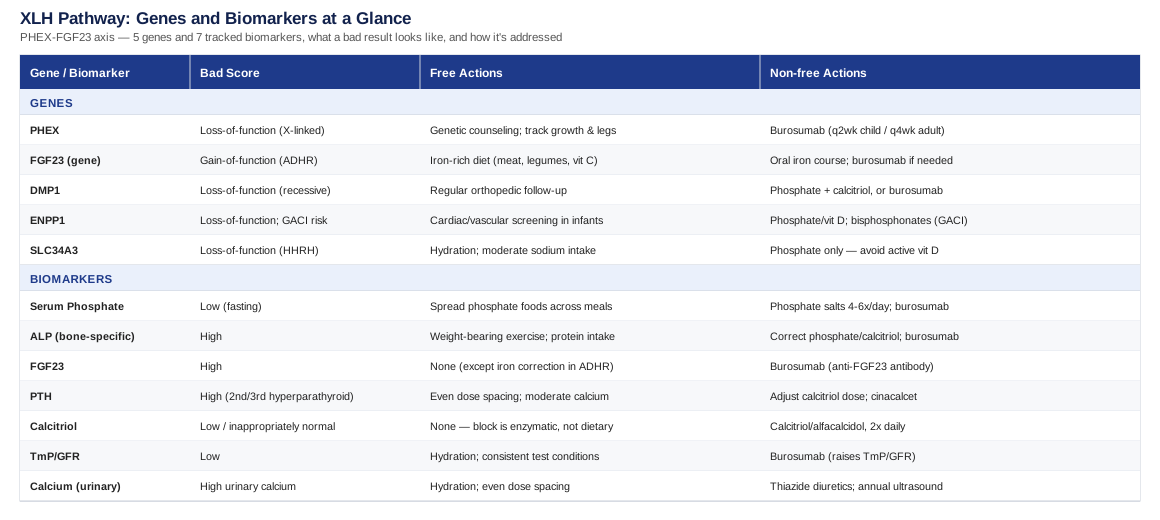
7 Biomarkers That Reveal How XLH Is Actually Behaving
Because XLH is caused by one overactive signal - FGF23 - rather than a single static defect, its severity moves over time, with growth spurts, puberty, pregnancy, and treatment changes. Blood and urine biomarkers are how clinicians and patients see that movement before it shows up as a fracture, a new dental abscess, or a child falling off their growth curve. The seven biomarkers below are the ones most consistently cited in the 2019 international consensus guideline on XLH management, and each one answers a slightly different question.
Serum Phosphate (Fasting)
Low serum phosphate is the defining biochemical feature of XLH: the kidneys, driven by excess FGF23, dump phosphate into the urine instead of reclaiming it. This single number is the most direct read on how much renal phosphate wasting is happening at any given time, and it is the value most treatment decisions are built around.
How to measure it
A fasting morning blood draw is standard, since phosphate has a natural diurnal rhythm and a non-fasting sample can look artificially reassuring. It is usually bundled into a basic or comprehensive metabolic panel, so the added cost is minimal - often free within a panel, or roughly 10 to 25 USD if ordered alone.If phosphate is low: the plan without supplements
Diet cannot correct XLH-driven phosphate wasting on its own, because the problem is renal loss, not dietary shortage. That said, spreading phosphate-containing foods - dairy, eggs, meat, fish, legumes - across smaller, more frequent meals can blunt the trough that shows up between doses of treatment, and staying consistent with meal timing makes lab trends easier to interpret. This is a supporting measure, not a fix, and it should be framed to patients that way.If phosphate is low: the plan with supplements or equipment
Conventional therapy uses oral phosphate salts, typically divided into four to six doses per day because phosphate is cleared quickly and single large doses cause gastrointestinal upset (diarrhea, cramping) without sustaining blood levels. It is almost always paired with active vitamin D (see below) to support absorption. The newer option, burosumab, is a subcutaneous anti-FGF23 antibody injected every two weeks in children or every four weeks in adults; it raises phosphate by blocking the hormone directly rather than replacing lost mineral. Reported side effects include injection-site reactions, headache, and restless leg sensations, and treatment requires phosphate monitoring to avoid overcorrection into hyperphosphatemia sustained efficacy data in children.Alkaline Phosphatase (ALP), Bone-Specific
ALP is produced by osteoblasts working to mineralize bone, and it rises when that mineralization is failing - which is exactly what happens in active rickets or osteomalacia. Where phosphate tells you about the kidney's behavior, ALP tells you about the skeleton's response to it, making the pair a useful before-and-after check on whether treatment is actually reaching the bone.
How to measure it
A standard chemistry panel includes total ALP for about 10 to 20 USD; a bone-specific isoenzyme assay, which filters out liver-derived ALP, is more precise and costs roughly 40 to 80 USD where available.If ALP is high: the plan without supplements
Appropriate, non-jarring weight-bearing activity supports normal bone remodeling once acute rickets is controlled, and adequate dietary protein gives osteoblasts the raw material to build with. Sensible, moderate sun exposure supports baseline vitamin D status, though in XLH this is a minor lever compared with the pathway problem described below.If ALP is high: the plan with supplements or equipment
Correcting the underlying phosphate and calcitriol deficits with conventional therapy or burosumab is what actually brings ALP down, typically over several months rather than weeks. Clinical trial data show burosumab normalizes ALP more consistently than phosphate and vitamin D alone. Labs are generally rechecked every three to six months; a plateauing or rising ALP despite treatment is a signal to review dosing with a specialist rather than assume it will resolve on its own.FGF23 (Intact Or C-Terminal Assay)
FGF23 is the hormone actually causing the disease - it suppresses renal phosphate reabsorption and blocks the kidney's ability to activate vitamin D. Measuring it directly is mostly used to confirm the diagnosis or screen relatives, since it is a specialized, less standardized assay and is affected by burosumab treatment itself.
How to measure it
This requires a send-out ELISA test, generally costing 100 to 300 USD and not part of routine panels. Ideally it is drawn before starting burosumab, since intact and C-terminal assays behave differently once anti-FGF23 therapy begins.If FGF23 is high: the plan without supplements
FGF23 overproduction in classic XLH is driven by the genetic loss of PHEX function, not by lifestyle, so diet and exercise do not meaningfully lower it. The one notable exception is in FGF23-related disease linked to iron status (discussed in the genetics section), where correcting iron deficiency can reduce FGF23 activity.If FGF23 is high: the plan with supplements or equipment
Burosumab is designed specifically to neutralize excess FGF23 signaling and is the only therapy that addresses this biomarker directly rather than downstream of it review of burosumab benefits versus conventional therapy in adults. Dosing starts low and is titrated to serum phosphate response rather than to the FGF23 level itself, since the assay is not standardized enough across labs to titrate on directly.Parathyroid Hormone (PTH)
PTH is not part of the primary XLH defect, but it is a common casualty of conventional treatment: repeated phosphate dosing can trigger reactive, and eventually autonomous, parathyroid overactivity known as secondary or tertiary hyperparathyroidism.
How to measure it
A routine intact PTH blood test costs approximately 30 to 60 USD and is typically checked alongside calcium every three to six months in treated patients.If PTH is high: the plan without supplements
Spacing phosphate doses evenly through the day, rather than in large infrequent boluses, reduces the swings that provoke parathyroid overactivity. Maintaining adequate but not excessive dietary calcium also helps avoid unnecessary parathyroid stimulation.If PTH is high: the plan with supplements or equipment
Calcitriol dosing is often adjusted to smaller, more frequent doses to blunt PTH stimulation, and in persistent tertiary hyperparathyroidism a calcimimetic such as cinacalcet may be used under specialist supervision. Hypercalcemia is the main side effect to watch when calcitriol doses are increased, so calcium should be rechecked alongside every PTH test.1,25-Dihydroxyvitamin D (Calcitriol)
This is the active form of vitamin D, and in classic XLH it is paradoxically low or inappropriately "normal" - low phosphate should normally push the kidney to make more of it, but excess FGF23 blocks the enzyme responsible for that conversion. This is one of the clearest examples of why generic "get more vitamin D" advice misses the actual mechanism.
How to measure it
This is a distinct, specialized assay from the common 25(OH)D test most people get, generally costing 80 to 150 USD and available mainly through reference labs.If calcitriol is low: the plan without supplements
Because the block is enzymatic rather than a supply problem, sun exposure and dietary vitamin D intake cannot fix this on their own - it is worth stating plainly to avoid false reassurance from otherwise-sensible general health advice.If calcitriol is low: the plan with supplements or equipment
Active vitamin D analogs - calcitriol or alfacalcidol, already in their hormonally active form - bypass the blocked enzyme and are typically dosed twice daily alongside phosphate. Calcium should be monitored regularly, since overcorrection risks hypercalcemia and hypercalciuria, which feeds into the kidney-related biomarkers below.Urinary Phosphate And Tubular Reabsorption Of Phosphate (TmP/GFR)
This calculation - combining a blood phosphate level with a timed urine collection - quantifies exactly how much phosphate the kidney is failing to reclaim, making it a more precise disease-activity marker than blood phosphate alone.
How to measure it
It requires a paired blood draw and a two-hour or 24-hour urine collection, calculated against a standard nomogram, generally costing 50 to 150 USD and usually ordered through a nephrology or endocrine clinic rather than a general lab.If TmP/GFR is low: the plan without supplements
Good hydration before and during the collection period improves the accuracy of the result, and repeating the test under similar conditions each time (same time of day, similar fluid intake) makes trend comparisons meaningful.If TmP/GFR is low: the plan with supplements or equipment
Burosumab has been shown to raise TmP/GFR by reducing FGF23's suppression of renal phosphate transporters, and this measure is often used at follow-up visits specifically to judge whether a dose adjustment is working.Calcium (Serum And 24-Hour Urinary)
Calcium is less about the disease itself and more about treatment safety: conventional phosphate and active vitamin D therapy can push calcium into the urine, raising the risk of nephrocalcinosis - calcium deposits in the kidney - over years of treatment.
How to measure it
Serum calcium is inexpensive, around 10 USD within a standard panel, while a 24-hour urinary calcium-to-creatinine ratio runs 40 to 70 USD. Periodic renal ultrasound, often once a year in treated patients, adds a structural check for early calcium deposits.If urinary calcium is high: the plan without supplements
Good hydration dilutes urinary calcium concentration, and evenly spacing phosphate and calcitriol doses avoids the sharp calcium peaks that concentrated dosing can cause.If urinary calcium is high: the plan with supplements or equipment
Thiazide diuretics are sometimes added specifically to reduce urinary calcium loss in patients on long-term conventional therapy, though they require monitoring for electrolyte shifts. Annual renal ultrasound functions as the "equipment" side of this plan, catching nephrocalcinosis early enough to adjust treatment before it progresses.Taken together, these seven values tell a more complete story than any single lab result - phosphate and TmP/GFR describe the kidney leak, ALP and calcitriol describe the skeletal and hormonal response, and PTH and calcium describe whether treatment itself is staying within a safe range. The genetics behind why this pathway misfires in the first place is where the next section picks up.
5 Genes Behind XLH And Look-Alike Phosphate-Wasting Disorders
Most of what shows up on the biomarker panel above traces back to one of five genes. Knowing which one is involved does more than satisfy curiosity - it can change which treatment is appropriate, and in one case, it flips the standard advice entirely.
PHEX
PHEX accounts for the large majority of XLH cases and is inherited in an X-linked dominant pattern, meaning one altered copy is enough to cause disease in both sexes, though severity varies. Loss of PHEX function indirectly drives up FGF23 production by osteocytes, setting off the renal phosphate wasting described throughout this article GeneReviews overview of X-linked hypophosphatemia.
If the gene is bad: the plan without supplements
The gene itself cannot be corrected through lifestyle, but genetic counseling and cascade testing of relatives catch the condition early, and tracking growth curves and leg alignment in children allows treatment to start before deformity progresses.If the gene is bad: the plan with supplements or equipment
Burosumab compensates for PHEX loss pharmacologically by neutralizing the FGF23 excess it causes, even though it does not touch the gene itself. Dosing is every two weeks in children and every four weeks in adults, with injection-site reactions and restless leg symptoms as the main reported side effects.FGF23
Gain-of-function variants in the FGF23 gene itself cause autosomal dominant hypophosphatemic rickets (ADHR), a related but distinct condition. Unusually, ADHR has a partly modifiable trigger: low iron status stabilizes the mutant FGF23 protein in its active form, and flares of the disease have been linked to iron deficiency, particularly around puberty or menstruation.
If the gene is bad: the plan without supplements
Maintaining iron sufficiency through diet - red meat, legumes, and vitamin C-rich foods to aid absorption - is a genuinely useful, evidence-linked lever specific to this gene, unlike PHEX-driven disease where diet has little direct effect on FGF23.If the gene is bad: the plan with supplements or equipment
When ferritin is low, oral iron supplementation is used, typically as a defined course until levels normalize rather than indefinitely, since iron overload carries its own risks. Common side effects include constipation and nausea, which is why cycling the course and rechecking ferritin rather than supplementing indefinitely is the standard approach. Burosumab remains an option when phosphate wasting persists despite iron correction.DMP1
DMP1 variants cause autosomal recessive hypophosphatemic rickets type 1, requiring one altered copy from each parent. DMP1 normally helps regulate osteocyte function and FGF23 production; its loss drives the same downstream FGF23 excess seen in PHEX-related disease, sometimes with more pronounced skeletal involvement overview of hypophosphatemic disorders in the FGF23 era.
If the gene is bad: the plan without supplements
Because skeletal disease can be more marked in this form, regular orthopedic follow-up alongside endocrine monitoring is worth prioritizing, even between lab checks.If the gene is bad: the plan with supplements or equipment
Treatment mirrors PHEX-related XLH - conventional phosphate and calcitriol, or burosumab where accessible - dosed and monitored the same way, since the downstream hormonal problem is identical even though the gene differs.ENPP1
ENPP1 loss-of-function causes autosomal recessive hypophosphatemic rickets type 2, but severe ENPP1 variants can also cause generalized arterial calcification of infancy (GACI), a distinct and more urgent vascular condition, making this gene's identification clinically important beyond bone health case report and review of ENPP1-related hypophosphatemic rickets.
If the gene is bad: the plan without supplements
Given the arterial calcification risk in severe cases, cardiology or vascular screening in infancy is a reasonable precaution when an ENPP1 variant is identified, separate from the bone-focused monitoring used elsewhere in this article.If the gene is bad: the plan with supplements or equipment
The rickets component is managed with phosphate and vitamin D therapy as in other forms; in infants with significant arterial calcification, bisphosphonates have been used under specialist care to limit ectopic calcification, with hypocalcemia and gastrointestinal irritation as notable side effects requiring close monitoring.SLC34A3
SLC34A3 encodes a kidney phosphate transporter, and its loss causes hereditary hypophosphatemic rickets with hypercalciuria (HHRH) - a genuinely different biochemical picture. Here, calcitriol is appropriately high, not low, because the kidney's vitamin D-activating enzyme is intact; the body responds normally to low phosphate, which unfortunately also raises calcium absorption and urinary calcium, increasing kidney stone risk.
If the gene is bad: the plan without supplements
Good hydration and moderate sodium intake help limit urinary calcium loss, and periodic renal ultrasound is worth discussing given the elevated stone risk specific to this form.If the gene is bad: the plan with supplements or equipment
This is the clearest case where standard XLH advice would backfire: active vitamin D should generally be avoided in HHRH, since calcitriol is already elevated, and adding more raises calcium and stone risk further. Treatment is phosphate supplementation alone, typically in three to five divided daily doses, with diarrhea and GI upset as the main side effects to manage through dose timing around meals.Understanding these five genes clarifies why two people carrying an "XLH-like" diagnosis can need opposite treatment plans - which is also where ideas from outside the endocrinology clinic can add a useful, complementary layer of thinking about lifelong disease management.
10 Ideas From Peter Attia's Outlive Worth Applying To A Lifelong Bone Condition
Peter Attia's book Outlive: The Science and Art of Longevity is not written about rare bone disease, but its central argument - shift from reactive to proactive medicine, and let objective data drive decisions rather than how a person feels in the moment - maps unusually well onto a condition like XLH, where irreversible skeletal and dental damage accumulates quietly between symptomatic flares. The ten ideas below are drawn from the book's framework and adapted specifically to the realities of tracking and managing XLH over a lifetime.
1. "Medicine 3.0" Means Acting Before The Damage Is Visible
Attia's central critique of conventional medicine is that it waits for disease to become symptomatic before intervening. In XLH, this translates directly: bowing, dental abscesses, and enthesopathy are late signs of a process that biomarkers like phosphate and ALP can flag years earlier.2. The "Marginal Decade" Reframes What Treatment Is For
Attia argues that most people's last decade of life is spent in decline, and that today's choices determine how functional that decade will be. For XLH, this reframes bone and joint protection in childhood and early adulthood as an investment in mobility decades later, not just symptom control now.3. Zone 2 Aerobic Training Fits Joint-Limited Conditions
Low-intensity, sustained aerobic exercise builds mitochondrial and metabolic capacity without the high-impact loading that can aggravate XLH-related joint and enthesis pain, making it a more appropriate cardio baseline than high-impact training.4. Strength Training Is Framed As Foundational, Not Optional
Attia calls resistance training one of the most powerful longevity interventions available. For XLH, this needs adaptation rather than avoidance - moderate, well-supervised resistance work supports bone loading and muscle support around vulnerable joints, ideally guided by a physical therapist familiar with the condition.5. VO2 Max As A Functional Capacity Target
Attia treats VO2 max as one of the strongest predictors of long-term function. For someone managing chronic phosphate wasting, tracking cardiorespiratory fitness alongside bone biomarkers gives a fuller picture of whether the body is coping well overall, not just biochemically.6. Stability Work Reduces Injury Risk
Attia emphasizes balance and stability training to prevent the falls that cause fractures later in life. This matters more, not less, in XLH, where bone quality is already compromised and a fall carries higher fracture risk than in the general population.7. Objective Data Beats Subjective Feeling
A recurring theme in the book is distrust of "I feel fine" as a health metric. This mirrors exactly the case for structured biomarker tracking in XLH - phosphate and FGF23 can be drifting unfavorably well before symptoms appear.8. Nutritional Flexibility Over Rigid Dogma
Attia is skeptical of one-size-fits-all diets. Applied to XLH, this supports including phosphate-rich, ordinary foods across the day rather than adopting restrictive eating patterns that offer no proven benefit for this specific condition.9. Emotional Health Is Inseparable From Physical Outcomes
The book dedicates significant space to mental health as a determinant of long-term physical outcomes. Chronic, lifelong conditions like XLH carry a real psychological load - repeated procedures, visible physical differences in childhood, ongoing monitoring - that deserves attention alongside the biochemistry.10. Build A Coordinated Team Rather Than See Specialists In Isolation
Attia argues patients should actively direct their own care team rather than passively receive fragmented advice. This lines up closely with the international consensus recommendation that XLH be managed by a coordinated multidisciplinary team anchored by a metabolic bone specialist, rather than juggling separate, uncoordinated referrals 2019 international consensus recommendations for XLH.These are general principles, not XLH-specific research, and none of them substitute for the disease-specific data covered earlier - but they offer a useful mental model for approaching a lifelong condition proactively rather than reactively, which connects naturally to the complementary approaches below.
Complementary Approaches With Real Supporting Evidence
Most complementary modalities have little to no XLH-specific research behind them, and it would be misleading to suggest otherwise. The four below are included because they have either direct evidence in XLH's dental complications or reasonably solid evidence in the specific bone and pain domains that overlap with this condition.
Cure Tooth Decay Approach (Ramiel Nagel)
Recurrent, spontaneous dental abscesses in teeth without cavities or trauma are one of the defining, and most distressing, features of XLH, caused by hypomineralized dentin that lets bacteria reach the pulp through microscopic clefts. Ramiel Nagel's Cure Tooth Decay approach centers on nutrient-dense eating to support dentin and enamel quality, and while it is not a peer-reviewed clinical protocol, the underlying idea - that dentin mineralization quality matters enormously in XLH - is strongly supported by clinical literature on XLH dental disease, which shows dentin defects, not hygiene, as the primary driver of these abscesses burosumab and dental abscesses in children with XLH. In practice, this means treating diet-focused approaches like Nagel's as a supportive layer - alongside, not instead of, the medical therapies shown to actually improve dentin mineralization - and coordinating any dietary changes with a dentist experienced in XLH rather than applying the book's protocol in isolation.Tai Chi
Tai chi is a slow, low-impact movement practice combining weight shifting, balance, and gentle isometric loading, which makes it relevant to XLH patients who need bone-supportive movement without the joint stress of higher-impact exercise. A systematic review and meta-analysis of randomized controlled trials found tai chi practice meaningfully attenuated bone mineral density loss compared with controls, primarily studied in postmenopausal populations rather than XLH specifically systematic review and meta-analysis of tai chi and bone mineral density. For someone with XLH, a realistic approach is starting with a beginner class two to three times weekly, done under guidance from a physical therapist familiar with the person's specific joint and bowing history, since individual deformities may require modified stances.Massage Therapy
Musculoskeletal pain from enthesopathy (where tendons and ligaments attach to bone) and joint stiffness is common in adults with XLH, and massage therapy has reasonably consistent evidence for reducing musculoskeletal pain and improving short-term function across various chronic pain conditions, though not XLH-specific trials. Regular sessions - often weekly for four to six weeks to assess benefit, then spaced out as needed - focused on affected joints and surrounding soft tissue is a low-risk way to manage discomfort alongside medical treatment. The main caution is avoiding deep, aggressive pressure directly over areas of active pseudofracture or recent orthopedic procedures.Biofeedback
Biofeedback trains conscious control over physiological signals like muscle tension and heart rate variability, and has reasonable evidence for reducing perceived intensity of chronic pain, which is relevant given how much of the adult XLH burden is ongoing musculoskeletal pain rather than acute rickets. A typical course involves six to ten sessions with a trained practitioner, sometimes supplemented with home devices for daily practice between sessions. It carries essentially no physical side effects, making it a reasonable adjunct for patients already managing multiple medications, though it should be viewed as a pain-management support rather than something that alters the underlying phosphate biology.Conclusion
XLH is ultimately a single hormonal problem - excess FGF23 - expressed through a small set of genes and made visible through seven trackable biomarkers, each answering a different question about how the disease and its treatment are behaving. Serum phosphate and TmP/GFR describe the underlying renal leak; alkaline phosphatase and calcitriol describe the skeletal and hormonal response to it; PTH and calcium describe whether treatment itself is staying within a safe range. Knowing which of the five genes is involved matters beyond curiosity, since at least one form - SLC34A3-related HHRH - calls for a treatment plan that is the direct opposite of standard XLH care.
The next practical step is not a lifestyle overhaul but a more precise conversation: bring this list of biomarkers to your next appointment, ask which ones have actually been checked recently, and ask whether genetic confirmation has been done if it hasn't. Track results over time rather than as isolated snapshots, and raise any complementary approach - dietary, movement-based, or otherwise - with the specialist coordinating your care before adding it. A lifelong condition managed with precise, current information is a fundamentally different experience than one managed with generic reassurance.