This article contains AI generated content.
· UpdatedBone Metastasis: 6 Key Genes And 6 Biomarkers To Track
Introduction
A bone metastasis diagnosis leaves most people trapped between two conversations that never quite connect: the imaging appointment that shows what is there, and the symptom conversation about pain management. What is almost never discussed is the biochemistry happening between those visits — the measurable signals in blood and urine, the gene expression patterns, the cellular machinery running beneath the surface. That gap is real, and it matters.
The challenge with standard monitoring is that total alkaline phosphatase or a quarterly bone scan can confirm that something is happening, but they rarely help you understand how fast, in which direction, or whether a given intervention is working. People end up reactive when the biology could have been tracked weeks earlier. It is not that doctors are withholding information — it is that most standard care protocols are not designed for this level of granularity.
This article takes a more practical and layered approach. Two parallel angles are worth developing together: specific blood and urine biomarkers that you can track longitudinally with your oncologist, and the gene-level pathways that explain why bone is such a common target for certain cancers. Neither of these replaces oncological treatment. But both give you a sharper picture and, critically, better questions to bring to your care team.
The first and most actionable angle covers six biomarkers — including bone resorption and formation markers — that can be monitored affordably and repeatedly. The second explores six key genes and molecular drivers behind bone metastasis biology, including what can be done when these are unfavorable. Beyond that, recent metabolic cancer research challenges some long-standing assumptions about how tumors grow, and four evidence-supported complementary approaches round out the picture. Better information does not promise better outcomes, but it almost always enables better decisions.
6 Biomarkers to Monitor for Bone Metastasis
Tracking bone health in the context of metastatic cancer means looking at both sides of bone metabolism: how much bone is being broken down and how much is being rebuilt. Adding a general tumor burden marker completes the picture. The six markers below are ranked by clinical utility, cost-accessibility, and depth of evidence in cancer-specific bone disease contexts.
Why Bone Turnover Markers Are Not Just for Osteoporosis
When cancer cells colonize bone, they corrupt the normal balance between osteoclasts (bone-dissolving cells) and osteoblasts (bone-building cells). In osteolytic metastasis — most common in breast, lung, and kidney cancer — osteoclasts dominate, releasing bone matrix growth factors that paradoxically feed further tumor growth. In osteoblastic metastasis — more common in prostate cancer — osteoblasts are pathologically overstimulated, producing disorganized, weak new bone. Both processes leave measurable byproducts in blood and urine, and tracking these over months reveals disease activity and treatment response faster and more cheaply than repeat imaging.
Multiple prospective studies have established that elevated bone resorption markers predict skeletal-related events — fractures, spinal cord compression, hypercalcemia — before they become clinical emergencies, offering a window to intervene.
Biomarker 1: CTX (Serum C-Terminal Telopeptide of Type I Collagen)
Why it matters: CTX is one of the most sensitive and specific bone resorption markers available in clinical practice. When osteoclasts degrade type I collagen — the primary structural protein of bone — they release C-terminal telopeptide fragments into the bloodstream. In bone metastasis, elevated CTX signals accelerated bone destruction, sometimes well before imaging shows progression. It is particularly relevant in osteolytic disease (breast, lung, thyroid cancers) where bone breakdown dominates.
What it reveals: High CTX correlates with increased skeletal-related event risk in solid tumor bone metastasis. It also responds rapidly to bone-protective treatment: effective bisphosphonate or denosumab therapy typically produces a 50–70% reduction in CTX within the first one to three months, making it one of the most useful markers for monitoring therapeutic response.
How to measure it: Serum β-CTX (CrossLaps) is a fasting morning blood draw — food intake, especially breakfast, can suppress CTX by up to 30%, making pre-breakfast collection essential for accurate results. Cost: approximately $60–150 USD through most clinical labs. Some oncology centers include it in bone health monitoring panels.
If the score is elevated — plan without supplements
Correct collection timing first: Before assuming the marker is truly elevated, confirm you drew blood fasting before 10am. A non-fasting sample can appear falsely high and lead to unnecessary alarm.
Resistance training: Weight-bearing and resistance exercise stimulates osteoblast activity and shifts the OPG/RANKL balance in a direction that reduces osteoclast activation. Three sessions per week, 30–45 minutes each, involving major muscle groups. Always confirm bone integrity with your oncologist before beginning impact exercise if lesion location is uncertain.
Reduce bone-depleting habits: Excess alcohol, high sodium intake, heavy caffeine use, and smoking all increase urinary calcium loss and are associated with higher bone resorption marker levels. Reducing these has measurable effects on CTX within weeks.
Sleep optimization: Cortisol — elevated chronically with poor sleep — drives bone resorption. Seven to nine hours of quality sleep nightly is a zero-cost intervention with solid mechanistic rationale. Frequency of retesting: every three months during active treatment; every six months in stable monitoring.
If the score is elevated — plan with supplements or equipment
Vitamin D3 + K2: Vitamin D deficiency impairs calcium absorption and is consistently associated with higher bone resorption rates. Dose: 2000–5000 IU D3 daily, paired with K2 in the MK-7 form at 90–200 mcg/day, taken with a fat-containing meal for absorption. K2 directs calcium into bone rather than soft tissue — the combination matters. Side effects: rare at these doses; monitor serum 25-OH vitamin D every 6 months. Use caution with K2 if on warfarin — discuss with your physician.
Magnesium (glycinate or malate form): Works synergistically with D3 and calcium metabolism. Dose: 200–400 mg/day. Common side effect: loose stools at higher doses — start at 150 mg and titrate. No cycling required.
Medical options: For significantly elevated CTX confirmed on repeat testing, your oncologist may initiate or adjust bisphosphonate therapy (zoledronic acid 4 mg IV every three to four weeks, or every 12 weeks in stable disease) or denosumab (Xgeva, 120 mg subcutaneous every four weeks). Both are evidence-based and have direct, measurable effects on CTX.
Biomarker 2: P1NP (Procollagen Type 1 N-Terminal Propeptide)
Why it matters: P1NP is the gold standard bone formation marker — the counterpart to CTX. The International Osteoporosis Foundation recommends CTX and P1NP as the reference pair for monitoring bone turnover. Together they tell a more complete story: whether the disruption is primarily destructive, primarily formative, or both. This context changes everything about interpretation and intervention.
What it reveals: P1NP is released when osteoblasts synthesize new type I collagen, the first step in bone matrix formation. In osteoblastic bone metastasis — characteristic of advanced prostate cancer — P1NP is often markedly elevated, reflecting pathological bone formation. Tracking P1NP alongside CTX over time shows whether the bone is primarily being destroyed, rebuilt chaotically, or maintaining some equilibrium under treatment.
How to measure it: Serum P1NP via standard blood draw. Fasting is not strictly required, but morning samples improve consistency. Cost: approximately $75–200 USD. Widely available through clinical labs; some oncology programs include it in bone health panels.
If the score is abnormal — plan without supplements
Weight-bearing exercise: Osteoblasts are mechanosensitive — physical load is their primary biological signal to form bone. Walking, resistance training, and low-impact weight-bearing activities (three to five sessions per week, at least 20–30 minutes) stimulate P1NP production in a meaningful and measurable way.
Adequate dietary protein: Bone matrix is primarily collagen, which requires sufficient amino acid supply. Target 1.2–1.6 g of protein per kg of body weight per day from whole food sources — fish, eggs, legumes, and lean meat all qualify. This supports osteoblast function and collagen synthesis without expensive interventions.
Sunlight and vitamin D synthesis: Fifteen to twenty minutes of direct midday sun exposure on arms and legs (when practical and safe) stimulates endogenous vitamin D production, which regulates bone metabolism signaling at a foundational level.
If the score is abnormal — plan with supplements or equipment
Vitamin D3 + K2: Same protocol as CTX — D3 2000–5000 IU daily with K2 MK-7 90–200 mcg. K2 activates osteocalcin, a protein specifically responsible for directing calcium into bone matrix rather than arteries.
Hydrolyzed collagen peptides: Five to ten grams of hydrolyzed collagen daily taken with vitamin C may support bone matrix formation. The evidence base comes primarily from osteoporosis studies but the mechanism applies. No cycling required; minimal side effects.
Medical options: In specific cases, bone anabolic agents like teriparatide (a PTH analogue) may be considered to stimulate osteoblast activity. Note that teriparatide is contraindicated in some metastatic contexts — this is a discussion for your oncologist. Retesting: every three months during active monitoring.
Biomarker 3: BALP (Bone-Specific Alkaline Phosphatase)
Why it matters: Total alkaline phosphatase appears on most standard chemistry panels, but it reflects enzyme activity from multiple organs including the liver and intestine. Bone-specific alkaline phosphatase (BALP) is produced exclusively by osteoblasts, giving a clean, unambiguous signal of bone formation activity. This specificity makes it particularly valuable in prostate cancer bone metastasis, where osteoblastic activity is the defining pathological process, and in monitoring treatment response over time.
What it reveals: Elevated BALP indicates osteoblast overactivation — which in a cancer context usually reflects pathological bone formation driven by metastatic disease. When BALP rises alongside clinical progression markers, it signals increasing disease activity. When it normalizes under treatment, it confirms a therapeutic response more precisely than total ALP alone would allow.
How to measure it: BALP requires a specialized isoenzyme assay — not the same as the total ALP on a standard chemistry panel. Cost: approximately $100–250 USD. Not universally available through all labs; request it specifically, or use specialty reference labs such as Mayo Clinic Laboratories or ARUP. Some academic oncology centers include it in prostate cancer bone metastasis panels.
If the score is elevated — plan without supplements
Confirm that total ALP is also elevated and rule out liver as the primary source — this clinical distinction changes the interpretation entirely. A GGT test run alongside ALP helps confirm liver vs. bone origin before acting on BALP elevation.
Weight-bearing physical activity three to five times per week helps modulate osteoblast signaling over time and supports overall bone homeostasis. Reduce alcohol intake, which impairs vitamin D metabolism and undermines bone mineral density.
If the score is elevated — plan with supplements or equipment
Vitamin D3 adequacy is foundational: Target 25-OH vitamin D levels of 40–60 ng/mL. Supplement D3 as needed (2000–5000 IU daily) to reach and maintain that range. Suboptimal vitamin D disrupts bone metabolism regulation at a fundamental level.
Boron: A trace mineral at three to six milligrams per day that supports vitamin D metabolism and may modulate bone formation markers. Evidence comes primarily from osteoporosis research; data in metastatic cancer contexts are limited. Low risk at these doses.
Medical options: If BALP elevation is confirmed as bone-derived and tied to metastatic progression, zoledronic acid and denosumab have well-established evidence for reducing skeletal-related events and influencing bone remodeling marker levels. Reassessment of systemic treatment is typically the primary response.
Biomarker 4: NTX (N-Terminal Telopeptide of Type I Collagen)
Why it matters: NTX is a bone resorption marker with a particularly strong track record in cancer bone metastasis research. Landmark studies by Lipton, Coleman, and colleagues established that elevated urinary NTX is one of the most powerful available predictors of skeletal-related events, disease progression, and reduced survival in solid tumor bone metastasis — independently of other clinical factors. It was among the first markers to be prospectively validated for this specific purpose in cancer patients.
What it reveals: High urinary NTX (above approximately 64 nmol BCE/mmol creatinine in validated assays) in cancer patients correlates with significantly elevated fracture risk, bone pain progression, and shorter survival compared to those with lower levels. Importantly, it responds to bisphosphonate and denosumab therapy in a measurable way — often dropping 50–70% within the first three months of effective bone-targeted treatment, making it a responsive treatment monitoring tool.
How to measure it: Urinary NTX uses the second morning urine void for best standardization — not the first morning collection. Serum NTX is also available but the urine version has historically stronger validation in cancer contexts. Cost: approximately $50–100 USD through most clinical labs.
If the score is elevated — plan without supplements
Proper collection protocol matters before any intervention: the second morning urine void, not first thing, gives the most consistent results. Confirm collection timing before interpreting an elevated result.
Resistance training and regular physical activity modulate bone resorption markers over time and are the most evidence-supported free interventions available. Begin with physician clearance if bone lesions are present.
Smoking cessation has direct measurable effects on bone resorption markers within months — nicotine directly accelerates osteoclast activity and increases bone turnover.
If the score is elevated — plan with supplements or equipment
Calcium from food or supplement: Adequate total calcium intake (1000–1200 mg/day combined from food and supplement) reduces the physiological pressure to resorb calcium from bone. Prefer food-first sources — dairy, fortified plant milks, bone broth, leafy greens. If supplementing, use calcium citrate in divided doses of no more than 500 mg per dose for absorption efficiency.
D3 + K2 + Magnesium triad: Same protocol as CTX — these three nutrients form the foundational bone metabolism support stack and address the most common nutritional deficiencies driving elevated resorption.
Medical options: Zoledronic acid and denosumab have the most direct and robust evidence for lowering NTX in cancer patients with bone metastasis. Your oncologist can guide dosing frequency based on disease stability.
Biomarker 5: DKK-1 (Dickkopf-1)
Why it matters: DKK-1 is a secreted protein that inhibits the WNT signaling pathway — a critical pathway that drives osteoblast differentiation and normal bone formation. When tumor cells produce high levels of DKK-1, they actively suppress osteoblast function, creating a bone microenvironment biased toward osteolytic destruction. DKK-1 has been most extensively studied in multiple myeloma bone disease, where it is a key pathological driver, and is increasingly recognized in solid tumors including advanced breast cancer.
What it reveals: Elevated serum DKK-1 may signal tumor-driven suppression of bone formation, even in cases where standard bone formation markers appear within normal range. It is also being studied as a therapeutic target — monoclonal antibodies against DKK-1 are in clinical trials for myeloma and are showing early promise. As a diagnostic signal, elevated DKK-1 may represent early disease activity before imaging changes become apparent.
How to measure it: DKK-1 is not yet a routine clinical test at most institutions. It is primarily available through research labs, academic medical centers with specialized bone disease programs, and certain advanced diagnostic panels. Cost: $150–400 USD where available, but access is genuinely limited at this time. Ask your oncologist whether your center measures it, especially in the context of multiple myeloma or advanced breast cancer with bone involvement.
If the score is elevated — plan without supplements
Weight-bearing exercise activates WNT pathway signaling in osteoblasts, which may partially counteract DKK-1's suppressive effects — though direct clinical evidence in bone metastasis patients is limited and emerging.
An anti-inflammatory, polyphenol-rich dietary pattern (berries, leafy greens, olive oil, turmeric) reduces the chronic inflammatory cytokines — particularly IL-6 and TNF-α — that amplify DKK-1-mediated WNT suppression in the bone microenvironment.
If the score is elevated — plan with supplements or equipment
Curcumin with piperine: Curcuminoids have shown DKK-1 modulating and WNT-pathway-supportive effects in preclinical models. Dose: 500–1000 mg curcumin daily with 5–20 mg piperine (black pepper extract) for absorption. Take with food. Evidence in humans is early-stage; GI sensitivity at higher doses is the main side effect.
EGCG (green tea polyphenols): EGCG has shown WNT-activating and DKK-1-modulating effects in bone cell research. Dose: 400–800 mg EGCG daily; avoid on an empty stomach (GI irritation) and separate from iron supplementation (EGCG inhibits non-heme iron absorption). Decaffeinate if caffeine sensitive.
Medical options: DKK-1-targeting antibodies remain in clinical trials. If you are eligible for myeloma or solid tumor bone disease trials, this pathway is worth discussing with a research oncologist.
Biomarker 6: LDH (Lactate Dehydrogenase)
Why it matters: LDH is not bone-specific, but it is one of the most accessible and prognostically meaningful markers in metastatic oncology. Elevated LDH reflects increased cellular turnover, anaerobic glycolysis (the Warburg effect, discussed further in the book summary below), and tissue necrosis — all of which accompany aggressive tumor behavior. In multiple metastatic cancer types, LDH above the upper limit of normal is incorporated into staging criteria and consistently correlates with poorer overall prognosis.
What it reveals: In bone metastasis, significantly elevated LDH suggests aggressive systemic disease extending well beyond local bone involvement. It is also responsive to systemic treatment — falling LDH over weeks to months of effective therapy is a meaningful positive signal. Conversely, rising LDH during treatment is an early warning of resistance or disease progression.
How to measure it: LDH is part of standard comprehensive metabolic panels or ordered as a standalone. It is among the most affordable oncology biomarkers: approximately $15–50 USD at virtually any clinical lab. Interpret results in context — hemolysis from blood handling and liver dysfunction can falsely elevate LDH independently of tumor activity.
If the score is elevated — plan without supplements
Aerobic exercise: Moderate-intensity aerobic activity — walking, cycling, swimming — at 150–200 minutes per week improves mitochondrial function, reduces reliance on anaerobic glycolysis in normal tissues, and has been associated with reductions in circulating inflammatory markers. The effect on LDH in metastatic cancer is indirect, but the metabolic rationale is solid.
Reduce refined carbohydrate and sugar load: High glycolytic demand from high-glycemic, processed foods supplies the glucose substrate that tumor cells preferentially use. A lower-glycemic, whole-food pattern (vegetables, legumes, lean protein, healthy fats) reduces substrate availability for tumor Warburg metabolism.
Prioritize sleep and stress management: Chronic cortisol elevation from stress and poor sleep worsens the inflammatory and pro-tumor microenvironment. Seven to nine hours of quality sleep per night is not optional in this context.
If the score is elevated — plan with supplements or equipment
CoQ10 (ubiquinol form): Elevated LDH often signals a shift away from efficient oxidative phosphorylation toward anaerobic glycolysis. CoQ10 supports mitochondrial electron transport chain function and may support the metabolic shift back toward aerobic efficiency. Dose: 200–400 mg/day ubiquinol form, taken with food containing fat. Minimal side effects; may mildly lower blood pressure. No cycling required.
Alpha-lipoic acid: A mitochondria-targeted antioxidant with some evidence in cancer metabolism contexts. Dose: 300–600 mg/day. Separate from thyroid medication by at least two hours. Evidence in metastatic cancer specifically is early-stage.
Medical focus: Significantly elevated LDH is primarily a trigger for oncology reassessment — it signals a need to evaluate systemic therapy rather than target LDH directly with supplements. No supplement independently normalizes LDH in the setting of active metastatic cancer.
Moving from what is measurable in the lab to what is driving the process at a genetic level gives a clearer picture of why these biomarkers behave the way they do and where the most targeted interventions may be possible.
The Genetic Machinery Behind Bone Metastasis
Understanding the genes driving bone metastasis does not require a genome sequencing appointment — though tumor molecular profiling is increasingly available and useful. What it requires is understanding which biological pathways are most active and what each one means for the clinical picture. These are primarily somatic changes (occurring in tumor cells) rather than germline variants, though some overlap with inherited susceptibility exists. Tumor tissue analysis, liquid biopsy, and gene expression profiling can reveal which of these pathways are most active.
Gene 1: RANKL (TNFSF11) — The Master Switch for Bone Destruction
What it does: RANKL (Receptor Activator of Nuclear Factor Kappa-B Ligand) is the single most important cytokine in bone metastasis biology. It binds to RANK receptors on osteoclast precursors, driving their differentiation into mature, bone-dissolving osteoclasts. In normal bone, RANKL is balanced by osteoprotegerin (OPG), a decoy receptor that neutralizes excess RANKL. In bone metastasis, tumor cells directly produce RANKL and stimulate surrounding stromal cells to produce more of it — tipping the OPG/RANKL balance catastrophically toward osteoclast activation and bone destruction. The bone-derived TGF-β released during resorption then stimulates further tumor RANKL production, completing a destructive vicious cycle.
What it affects: Osteoclast activation rate, rate of bone destruction, skeletal-related event risk, calcium release from bone, tumor growth cycle in the bone niche.
If the gene is overexpressed — plan without supplements
Resistance training directly shifts the OPG/RANKL balance in favor of OPG production in bone stromal cells — effectively a biomechanical brake on osteoclast activation. Three to five sessions per week of resistance or weight-bearing exercise, cleared by your oncologist based on bone integrity at the lesion sites.
Adequate calcium intake prevents PTH-driven RANKL upregulation: calcium deficiency causes PTH to rise, which directly stimulates osteoblasts to increase RANKL expression. Prioritize food-based calcium sources.
If the gene is overexpressed — plan with supplements or equipment
Vitamin D3 + K2: Adequate vitamin D (target 40–60 ng/mL serum) downregulates osteoclast activity and modulates RANKL signaling at the receptor level. D3 2000–5000 IU daily with K2 MK-7 90–200 mcg daily, taken with a fat-containing meal.
Medical options — the most direct intervention available: Denosumab (Xgeva) is a fully human monoclonal antibody that directly binds and neutralizes RANKL. It is the most mechanistically precise pharmacological intervention targeting this pathway and is standard of care in bone metastasis from solid tumors. Bisphosphonates (zoledronic acid) reduce osteoclast activity downstream. Both require prescription and oncology supervision.
Gene 2: CXCR4 — The Bone-Seeking GPS
What it does: CXCR4 is a chemokine receptor that functions as a molecular GPS guiding tumor cells to bone marrow. Its ligand, CXCL12 (SDF-1), is abundantly produced by bone marrow stromal cells. Cancer cells — particularly breast cancer, prostate cancer, and multiple myeloma — that overexpress CXCR4 are chemically attracted to CXCL12-rich bone marrow, where they establish the metastatic niche. CXCR4 overexpression correlates with greater bone metastasis frequency and worse clinical outcomes across multiple tumor types. Importantly, CXCR4 is often epigenetically regulated: hypomethylation of its promoter region leads to overexpression — making it theoretically responsive to epigenetic lifestyle interventions.
What it affects: Tumor homing to bone, seeding efficiency of metastatic cells in bone marrow, extent of bone marrow involvement, progression speed.
If the gene is overexpressed — plan without supplements
Methyl-donor rich dietary pattern: folate (from leafy greens, legumes), betaine (from beets, quinoa), and choline (from eggs, liver) are key building blocks for DNA methylation. Supporting healthy methylation machinery may help maintain appropriate promoter methylation of genes like CXCR4.
Moderate aerobic exercise has shown effects on immune cell and tumor cell trafficking in preclinical models. While direct human evidence in bone metastasis is limited, exercise consistently reduces systemic inflammation that amplifies metastatic spread.
If the gene is overexpressed — plan with supplements or equipment
Methylated B vitamins (methylfolate + methylcobalamin): For those with MTHFR variants (which impair methylation, as highlighted in functional medicine contexts by Gary Brecka and Ali Torkamani), methylated B vitamins support the methylation cycle that regulates epigenetic gene silencing. Methylfolate 400–800 mcg + methylcobalamin B12 500–1000 mcg daily. Minimal side effects; foundational support for epigenetic regulation.
EGCG: Green tea polyphenols, particularly EGCG, have shown CXCR4 downregulation effects in cancer cells in preclinical research. Dose: 400–800 mg EGCG daily. Early-stage human evidence.
Medical options: Plerixafor (AMD3100) is an FDA-approved CXCR4 antagonist (currently used for stem cell mobilization) that is under investigation in cancer metastasis clinical trials. Discuss with an academic oncologist if relevant.
Gene 3: PTHLH (PTHrP) — The Calcium Signal Tumor Cells Hijack
What it does: The PTHLH gene encodes parathyroid hormone-related protein (PTHrP). In normal tissues, PTHrP is produced locally in small amounts for paracrine signaling. When breast cancer cells invade bone, they often dramatically upregulate PTHrP, which binds PTH receptors on osteoblasts and drives RANKL expression — activating osteoclasts and accelerating osteolysis. The bone-derived TGF-β released feeds back to increase tumor PTHrP further: another self-amplifying loop. PTHrP is also directly responsible for humoral hypercalcemia of malignancy — one of the most dangerous metabolic emergencies in oncology, occurring when circulating PTHrP reaches systemic levels.
What it affects: Bone resorption rate, hypercalcemia risk, RANKL-driven osteoclast activation, tumor proliferation signaling in the bone niche.
If the gene is overexpressed — plan without supplements
Maintain adequate hydration — this is a critical safety priority when PTHrP is active, since calcium is lost through urine and dehydration worsens hypercalcemia risk. Aim for two to three liters of water daily unless contraindicated by cardiac or renal conditions.
A moderately alkaline, plant-forward diet reduces the acid load that promotes calcium mobilization from bone — a modest but real effect.
If the gene is overexpressed — plan with supplements or equipment
Vitamin D3 requires careful management here: When PTHrP is active and hypercalcemia is a risk, supplemental vitamin D can worsen calcium levels — a critical safety exception to the usual D3 recommendation. Vitamin D supplementation in this context must be guided by your oncologist with regular calcium monitoring.
Medical options: Denosumab and bisphosphonates address the downstream consequence of PTHrP by blocking osteoclast activation regardless of the stimulus. For PTHrP-driven hypercalcemia itself, bisphosphonates IV (pamidronate, zoledronic acid) and cinacalcet are the standard interventions. All require oncological supervision.
Gene 4: VEGFA — Fueling the Metastatic Niche
What it does: VEGF-A (vascular endothelial growth factor A) is the primary driver of tumor angiogenesis — the formation of new blood vessels that supply tumors with oxygen and nutrients. In bone metastasis, VEGFA plays a dual role: supplying the metabolic needs of the metastatic colony and directly stimulating osteoclast differentiation (RANK is expressed on both endothelial cells and osteoclast precursors that respond to VEGF signals). High VEGFA expression in primary tumors correlates with greater bone metastatic potential in breast, lung, and prostate cancers.
What it affects: Vascular density in the bone metastatic niche, osteoclast activation, lesion growth rate, tumor-bone interface remodeling.
If the gene is overexpressed — plan without supplements
Regular moderate-intensity aerobic exercise paradoxically reduces pathological tumor angiogenesis while improving normal tissue vascular efficiency — supported by multiple preclinical and emerging human studies. Target 150–200 minutes per week of moderate-intensity cardio (walking, cycling, swimming).
Anti-inflammatory dietary patterns — Mediterranean diet emphasis on olive oil, fish, leafy greens, reduced processed foods — reduce the chronic inflammatory signals (IL-6, TNF-α, NF-κB activation) that upregulate VEGFA expression in tumor microenvironments.
If the gene is overexpressed — plan with supplements or equipment
Omega-3 fatty acids (EPA + DHA): High-dose fish oil has demonstrated anti-angiogenic effects and may modestly reduce VEGF-driven signaling in clinical and preclinical research. Dose: 3–4 g EPA+DHA combined daily, taken with meals to minimize GI side effects. If on anticoagulants, discuss dose safety with your physician before exceeding 2 g. No strict cycling required.
Medical options: Bevacizumab (Avastin) is a VEGF-targeting monoclonal antibody used in some metastatic regimens (ovarian, colorectal, and others). Tyrosine kinase inhibitors targeting VEGF receptors (cabozantinib, sunitinib) are also used in certain solid tumors. These are prescription medications under oncologist direction.
Gene 5: RUNX2 — The Bone Transcription Factor Gone Rogue
What it does: RUNX2 is the master transcription factor for osteoblast differentiation — essential for normal skeletal development. In cancer, RUNX2 is aberrantly expressed in tumor cells themselves (particularly breast, prostate, and thyroid cancers), driving a gene expression program that gives tumor cells bone-like invasive properties. Aberrant RUNX2 in cancer cells upregulates MMP-9 and MMP-13 (matrix metalloproteinases that digest bone matrix), VEGFA, and osteopontin — essentially equipping tumor cells with a molecular toolkit for invading and remodeling bone tissue.
What it affects: Matrix degradation, tumor invasiveness into bone, bone remodeling patterns, downstream VEGFA and MMP expression.
If the gene is overexpressed — plan without supplements
Weight-bearing exercise stimulates normal RUNX2 signaling in resident osteoblasts, maintaining healthy transcription factor competition within the bone niche and partially occupying the downstream signaling space that aberrant RUNX2 in tumor cells seeks to exploit.
Systemic inflammation reduction — through sleep, stress management, and anti-inflammatory diet — lowers the cytokine levels (particularly TGF-β and TNF-α) that amplify RUNX2 expression in cancer cells within bone.
If the gene is overexpressed — plan with supplements or equipment
Curcumin with piperine: Has shown RUNX2-inhibitory effects in breast and prostate cancer cell lines in preclinical models. Dose: 500–1000 mg curcumin daily with 5–20 mg piperine. Evidence is preclinical; randomized human trial data are not yet available.
Medical options: Zoledronic acid has demonstrated RUNX2-modifying effects in bone metastasis models beyond its standard anti-osteoclast mechanism — one reason it remains a first-line bone-targeted agent even in contexts beyond pure osteolytic disease.
Gene 6: CDH1 (E-Cadherin) — The Lost Gatekeeper
What it does: CDH1 encodes E-cadherin, a cell adhesion protein and key tumor suppressor that keeps epithelial cells bound together and resists invasion. When CDH1 is silenced — most commonly through epigenetic methylation of its promoter, not permanent gene mutation — cancer cells lose contact inhibition and undergo epithelial-to-mesenchymal transition (EMT). EMT-transformed cells become migratory, invasive, and capable of seeding distant sites including bone marrow. CDH1 silencing is one of the earliest and most consequential steps in the metastatic cascade.
Critically, because CDH1 silencing is usually epigenetically driven (a methylation-based switch), it is theoretically modifiable — which makes this gene particularly relevant for dietary and lifestyle interventions that support healthy DNA methylation patterns.
If the gene is silenced — plan without supplements
Cruciferous vegetables (broccoli, kale, Brussels sprouts, cabbage, arugula) contain sulforaphane and indole-3-carbinol — compounds that modulate DNMT (DNA methyltransferase) enzyme activity and support normal epigenetic regulation. Consuming these daily — cooked or raw, with mustard seeds added to cooked broccoli to improve sulforaphane yield — provides meaningful epigenetic support.
Minimize alcohol: acetaldehyde, a metabolite of ethanol metabolism, directly disrupts DNA methylation fidelity and may worsen epigenetic silencing of tumor suppressors including CDH1.
If the gene is silenced — plan with supplements or equipment
Sulforaphane (broccoli sprout extract): 30–60 mg/day standardized sulforaphane from broccoli sprout extract. Confirm the product contains myrosinase enzyme for efficient glucoraphanin-to-sulforaphane conversion. Take with food. A five-days-on/two-days-off pattern is sometimes used for cycling; minimal side effects, occasional GI adjustment in the first week.
Methylfolate + Methylcobalamin B12: Adequate methyl group supply — critical for maintaining healthy DNA methylation across the genome — supports proper epigenetic regulation of tumor suppressor promoters. For those with MTHFR variants (which impair methylation capacity, as identified in functional genomics approaches), methylated B vitamins are particularly important. Methylfolate 400–800 mcg + methylcobalamin 500–1000 mcg daily. Minimal side effects; foundational for epigenetic methylation support.
Medical options: DNMT inhibitors (decitabine, azacitidine) are established in hematological malignancies and have epigenetic demethylation mechanisms. Their role in solid tumor epigenetic reprogramming is investigational. Discuss with an academic oncologist if molecular profiling confirms CDH1 silencing.
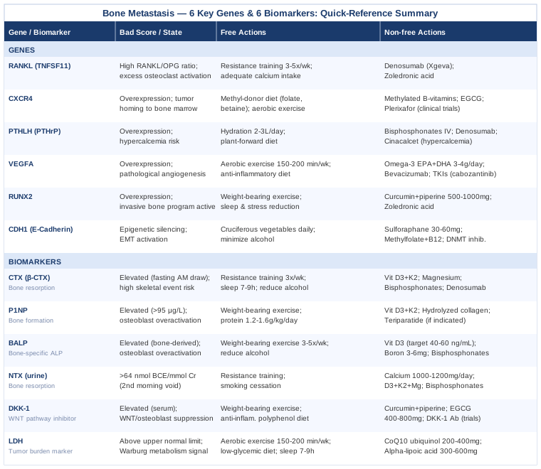
The table above provides a quick-reference summary of every gene and biomarker covered, with clear action pathways for both accessible and medical-grade interventions. From here, it is worth stepping back and looking at the broader metabolic framework that some of the most impactful recent cancer research has proposed — one that reframes how bone metastasis biology is understood at a systems level.
What "The Cancer Code" by Dr. Jason Fung Gets Right About Tumor Metabolism
Published in 2020 and citing extensive clinical and basic science literature, The Cancer Code by Dr. Jason Fung (nephrologist and fasting researcher) makes a compelling case that cancer is not purely a genetic disease — it is also, fundamentally, a metabolic one. His framework draws on Otto Warburg's century-old observation, updated with modern mechanistic understanding, and has direct implications for how bone metastasis develops and might be slowed. Below are ten of the most impactful insights from the book, each with specific relevance to bone metastasis.
1. Cancer Cells Run on a Primitive Fuel System
The Warburg effect describes how cancer cells preferentially ferment glucose into lactate even in the presence of adequate oxygen — a metabolically inefficient strategy that normal cells abandon after birth. This primitive fuel preference is not a random mutation. It reflects a return to an ancient survival program that prioritizes growth over efficiency. For bone metastasis, this means that high circulating glucose and insulin create an optimal fuel-rich environment for tumor cells colonizing bone marrow.
2. Insulin and IGF-1 Are Tumor Growth Accelerators
Fung's central thesis is that chronic hyperinsulinemia — driven by high-sugar and refined-carbohydrate diets, insulin resistance, and obesity — activates IGF-1 (insulin-like growth factor 1), one of the most potent tumor growth signals in oncology. IGF-1 directly activates PI3K/Akt/mTOR pathways in tumor cells, drives RANKL expression in bone metastasis, and suppresses apoptosis. Reducing insulin load through diet and fasting is, in Fung's framework, one of the most accessible interventions available to most cancer patients.
3. Intermittent Fasting Activates Autophagy — the Body's Cleanup Program
During fasting periods (typically 16–24 hours), cellular autophagy — the process of digesting and recycling damaged cellular components — is strongly upregulated. Autophagy can selectively degrade damaged mitochondria (mitophagy) and, in some research contexts, has shown effects on tumor cell survival. Fung argues that regular metabolic cycling between fed and fasted states creates a hostile environment for cells relying on Warburg-type metabolism. A 16:8 intermittent fasting approach (16 hours fasting, 8-hour eating window) is the most accessible entry point; check with your oncologist regarding timing relative to treatments.
4. mTOR Is the Growth Signal That Fasting Suppresses
mTOR (mechanistic target of rapamycin) is a central nutrient-sensing hub. When activated by amino acids, insulin, and growth factors, it drives cell growth and inhibits autophagy. Chronically elevated mTOR activity is associated with faster tumor progression. Fasting, time-restricted eating, and reduced protein intake during certain windows all suppress mTOR activity. In oncology, mTOR inhibitors (everolimus, temsirolimus) are already standard treatment in some metastatic cancers — Fung's argument is that dietary approaches offer a partial analogue.
5. Obesity Worsens Bone Metastasis Risk Through Adipokine Signaling
Adipose tissue — particularly visceral fat — produces leptin, adiponectin, and inflammatory cytokines that modulate bone marrow microenvironment. High leptin (associated with obesity) promotes tumor proliferation and invasion. Low adiponectin (also associated with obesity) removes a key tumor-suppressive signal. Bone marrow contains significant adipose tissue that directly interacts with metastatic cells. Maintaining healthy body composition — through exercise and dietary quality — is not merely cosmetic.
6. Time-Restricted Eating Aligns Metabolism With Circadian Biology
Tumor cells, unlike normal cells, appear to lose circadian rhythm regulation — they metabolize and divide at abnormal hours. Keeping one's own metabolic activity tightly time-restricted (eating within an 8–10 hour window aligned with daytime) supports circadian gene expression in normal tissues and creates metabolic pressure on dysrhythmic tumor cells. This is an emerging area of chronobiology applied to oncology.
7. Ketogenic Dietary Patterns May Create a Selective Disadvantage for Tumor Cells
Because most cancer cells have impaired mitochondria and cannot efficiently metabolize ketones, a low-carbohydrate, high-fat ketogenic diet reduces the primary fuel source tumor cells use (glucose) while providing normal cells with an alternative energy substrate. Human trial data in cancer are limited and mixed, but preclinical evidence is substantial. In bone metastasis specifically, several ongoing trials are examining ketogenic dietary patterns as adjuncts to systemic therapy. This approach requires careful nutritional planning and oncologist coordination to avoid unintended weight loss.
8. Chronic Stress Elevates Cortisol and Feeds Tumor Biology
Cortisol — the primary stress hormone — has profound immunosuppressive and pro-tumor effects at chronically elevated levels. It suppresses natural killer cell activity (the immune cells most important for killing circulating tumor cells), raises blood glucose (fueling Warburg metabolism), increases RANKL expression in bone (directly accelerating osteoclast activation), and promotes angiogenesis through VEGF upregulation. Stress management is not a soft adjunct in this context — it is a mechanistically relevant intervention.
9. Caloric Restriction Mimetics Offer Drug-Free mTOR Inhibition
Several compounds mimic the molecular effects of caloric restriction without requiring food restriction: rapamycin (prescription mTOR inhibitor), metformin (biguanide that activates AMPK and inhibits mTOR), resveratrol, and berberine (both available OTC). Of these, metformin has the strongest and most extensive human evidence — multiple retrospective studies associate its use in diabetic cancer patients with improved outcomes, and prospective trials are underway. Discuss metformin with your oncologist — it is low-cost, widely available, and has a well-established safety profile.
10. Cancer Is a Disease of the Microenvironment — Not Just the Cell
Fung's final and perhaps most paradigm-shifting point is that focusing exclusively on killing cancer cells misses the environment that enabled them to grow and spread in the first place. The bone marrow niche — with its inflammatory cytokines, growth factors, adipokines, and metabolic substrates — is as much a target as the tumor cell itself. Interventions that normalize the microenvironment: exercise, dietary quality, sleep, fasting, stress reduction, and bone-targeted agents, all have mechanistic roles in making the niche less hospitable to metastatic cells. This frames lifestyle medicine not as alternative therapy but as microenvironment-targeting therapy.
These insights connect directly to every biomarker and gene discussed in earlier sections — and explain why so many of the recommended interventions converge on the same foundational behaviors: exercise, dietary quality, sleep, and stress reduction.
Complementary Approaches With Meaningful Evidence for Bone Metastasis
The following modalities are selected specifically for their evidence base in cancer patients — particularly for pain management, quality of life, and biological markers relevant to bone metastasis. None are presented as alternatives to oncological treatment; all are adjuncts with specific human clinical evidence worth knowing.
Mindfulness-Based Stress Reduction (MBSR)
MBSR is an eight-week, standardized program that combines body scan meditation, mindfulness sitting practice, gentle movement, and group inquiry. Its relevance to bone metastasis is multilayered: chronic stress activates the HPA axis and sympathetic nervous system in ways that elevate cortisol, suppress NK cell activity, increase RANKL-driving cytokines, and promote the pro-tumor bone microenvironment. MBSR directly targets this stress-biological cascade.
A well-cited randomized trial by Carlson et al. (PMID 13129986) demonstrated that MBSR in breast cancer survivors produced significant reductions in cortisol and improvements in immune markers. A Cochrane-informed body of evidence consistently supports MBSR for cancer-related distress, fatigue, and quality of life, with effects on anxiety and depression that are robust across tumor types and stages.
In practice: most oncology centers now offer MBSR programs or can refer to community-based programs. Online MBSR programs (including Dr. Jon Kabat-Zinn's original curriculum adapted for digital delivery) are also available. The eight-week format with 45-minute daily practice is the validated protocol; even adapted ten-minute daily mindfulness practice shows measurable benefit on stress biomarkers over eight weeks. Begin with whatever commitment is sustainable.
Yoga
Yoga's relevance to bone metastasis operates on two levels: the mechanical effect of gentle weight-bearing postures on bone microarchitecture and the anti-inflammatory, stress-reducing effects of the mind-body practice. In cancer patients with bone involvement, standard yoga must be significantly modified to avoid impact poses, inversions, or positions that stress at-risk lesion sites — but modified yoga remains meaningful.
The YOCAS (Yoga for Cancer Survivors) randomized controlled trial (PMID 24280826) demonstrated significant improvements in sleep quality, fatigue, and quality of life in cancer survivors undergoing treatment, with a structured twice-weekly restorative yoga protocol. In the context of bone health specifically, a pilot trial in breast cancer survivors found that twelve weeks of yoga improved bone mineral density markers compared to controls — an effect likely mediated by combination of mechanical loading and cortisol reduction.
For realistic application in bone metastasis: seek an oncology-certified yoga instructor (RCYT with oncology specialization or C-IAYT certified) who can adapt postures to your specific lesion locations and current functional status. Restorative yoga — using props, minimal load, and breath focus — is typically the safest entry point. Twice per week for 45–60 minutes is the most-studied protocol.
Massage Therapy
Massage therapy in the oncology setting — specifically oncology massage, which differs significantly from standard Swedish or deep tissue massage — has meaningful evidence for pain management, anxiety reduction, and quality of life in metastatic cancer patients. Relevant to bone metastasis: pain is frequently the most disabling symptom, and pharmacological pain management is often incomplete. Oncology massage offers a non-pharmacological adjunct.
A randomized controlled trial by Kutner et al. published in Annals of Internal Medicine (PMID 18838724) found that simple touch and massage both reduced pain and mood disturbance in patients with advanced cancer, with massage showing stronger and more sustained effects on pain scores. The evidence base for oncology massage for cancer pain includes multiple systematic reviews supporting its use as an integrative adjunct.
In practice: oncology massage must be performed by a therapist trained specifically in cancer patient care — standard deep tissue massage is contraindicated near bone lesion sites, over radiation fields, and in areas of lymphedema. The protocol typically involves light-to-moderate pressure, avoiding lesion sites, working with medical clearance, and focusing on peripheral areas of the body for generalized relaxation and pain modulation. Weekly 30–60 minute sessions are the most commonly studied format.
Music Therapy
Music therapy — distinct from simply listening to music — involves trained therapists using live music, guided imagery with music, lyric analysis, or music-assisted relaxation specifically designed for clinical therapeutic goals. In oncology, it has the strongest evidence for acute pain relief, reduction of procedure-related anxiety, and improved mood in patients with advanced cancer.
A Cochrane systematic review of music interventions in people with cancer (PMID 27092593) analyzing 52 trials and 3,731 patients found that music interventions produced moderate-quality evidence for reductions in anxiety, pain, fatigue, and improvements in quality of life compared to standard care. The pain-reducing effect is particularly relevant for bone metastasis patients who experience chronic breakthrough pain.
For practical application: board-certified music therapists (MT-BC credential) specialize in oncology settings and can provide individual or group sessions. Many major cancer centers now offer music therapy as part of integrative oncology programs. For those without access to a music therapist, guided music-based relaxation protocols (available through validated apps or oncology-focused online programs) offer a meaningful approximation. Sessions of 20–45 minutes, one to three times per week, showed benefit in reviewed trials.
Conclusion
Bone metastasis is a condition where the difference between reactive and proactive monitoring can translate directly into better symptom control, more informed treatment decisions, and fewer surprises. The six biomarkers covered here — CTX, P1NP, BALP, NTX, DKK-1, and LDH — give you a longitudinal window into bone biology that imaging alone cannot provide. The six genes — RANKL, CXCR4, PTHLH, VEGFA, RUNX2, and CDH1 — explain the molecular machinery underneath those biomarker numbers and point toward both pharmacological and lifestyle interventions with real mechanistic rationale.
The clearest next step is a conversation with your oncologist about which of these biomarkers are already being tracked, which are worth adding to your monitoring protocol, and how the results connect to your current treatment plan. Tracking trends over time — not single-point snapshots — is where these markers deliver their most useful signal. Bring your biomarker results, ask about patterns, and use this information to make more targeted and confident decisions alongside your medical team.
Cancer & Oncology Endocrine & Metabolic
Musculoskeletal: Bone Conditions
Cancer & Oncology: Breast Cancer Lung Cancer Prostate Cancer Bone Cancer