This article contains AI generated content.
· UpdatedCervical Myelopathy: 6 Genes and 7 Biomarkers to Track
If you've been told you have cervical myelopathy — or you're watching early signs of it in a parent, a partner, or yourself — you've probably noticed that most of the advice out there stops at the same place: "see a spine specialist," "avoid neck strain," "consider surgery if it progresses." None of that is wrong. But it also doesn't explain why one person's spinal cord tolerates decades of degenerative change with barely a symptom, while another develops hand clumsiness and gait changes in their forties.
That gap between generic advice and your actual situation is where genetics and biomarkers become useful. Cervical myelopathy isn't one uniform process — it's the downstream result of mechanical compression, bone and ligament biology, inflammation, and how well an individual spinal cord tolerates chronic pressure. Some of that is fixed by anatomy. A meaningful part of it is measurable, and in some cases, modifiable.
This article won't pretend there's a supplement stack that reverses spinal cord compression — there isn't, and anyone who claims otherwise is not being straight with you. What it will do is walk through the specific genes tied to ossification and disc degeneration, the blood and imaging biomarkers that track disease activity and surgical readiness, a genuinely interesting line of neuroscience research on nerve recovery, and a short list of complementary approaches with real evidence behind them for neck-related conditions.
None of this replaces a spine surgeon or neurologist. What it can do is help you ask better questions at your next appointment, understand what your labs and scans are actually telling you, and make more deliberate decisions about the parts of this condition you do have some influence over.
Summary
Cervical myelopathy develops at the intersection of biology you inherited and biology you can track. On the genetic side, variants in collagen genes like COL6A1 and COL11A2, bone-signaling genes like BMP2/BMP4 and RUNX2, the vitamin D receptor gene, and a pyrophosphate-regulating gene called ENPP1 all shape how likely ligament and disc tissue is to calcify or degenerate around the cervical spinal cord. None of them guarantee myelopathy — but they shift the odds, and in ENPP1's case, they may even point toward a specific, correctable metabolic deficiency.
On the biomarker side, blood tests for neurofilament light chain, GFAP, and interleukin-6 are starting to show real promise for tracking spinal cord injury severity and predicting recovery after surgery, while more familiar tools — vitamin D and phosphate panels, hs-CRP, and above all MRI signal changes and nerve conduction studies — remain the most clinically actionable data you can get today. Below, you'll find what each of these markers actually measures, realistic cost ranges, and what a sensible response looks like when a result comes back unfavorable — with and without supplements. There's also a look at cutting-edge nerve-recovery research and a short, evidence-checked list of complementary approaches that are actually appropriate for a spinal cord condition (and one that generally is not).
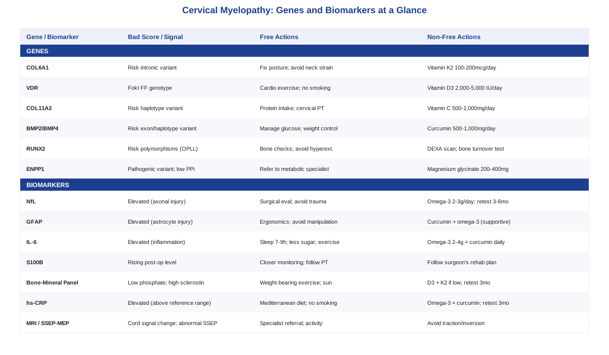
7 Biomarkers Worth Tracking in Cervical Myelopathy
Unlike cardiometabolic conditions, cervical myelopathy doesn't yet have a mature, direct-to-consumer biomarker culture built around it — there's no equivalent of ordering your own ApoB the way Peter Attia or Thomas Dayspring would recommend for cardiovascular risk. But that's changing quickly, and a handful of markers already have enough evidence to be worth understanding, whether you order them yourself, ask your neurologist about them, or simply learn to read your existing scans and labs more critically. The list below moves from newer blood-based research markers to the older, more established imaging and electrophysiology tools that still do most of the clinical heavy lifting.
1. Serum Neurofilament Light Chain (NfL)
NfL is a structural protein released into the bloodstream when axons — the long fibers that carry signals up and down the spinal cord — are damaged. It's not specific to cervical myelopathy; it rises in multiple sclerosis, ALS, and traumatic brain injury too. But in a 2025 prospective study of patients with degenerative cervical myelopathy, presurgical serum NfL, combined with interleukin-6 and BDNF, distinguished myelopathy patients from controls with good accuracy, and higher presurgical NfL correlated with how much grip strength patients regained after decompression surgery (serum protein biomarkers for degenerative cervical myelopathy, J Neurosurg Spine 2025). That makes it one of the more promising "how bad is the damage, and how well will I recover" markers currently in development.
How to Measure It
NfL is measured with an ultrasensitive assay (Simoa), which is still mostly confined to research hospitals, neurology referral centers, and a small number of specialty labs. Where available, a single blood draw runs roughly $100–$300 out of pocket. It is not yet a standard order at a regular commercial lab, so the realistic path is asking a neurologist or spine surgeon involved in your care whether it's part of a research protocol you could join.If the Marker Is Off: The Plan Without Supplements or Equipment
An elevated NfL in the context of myelopathy is a signal of active axonal injury, not a lifestyle problem to fix with diet. The most evidence-backed response is timely evaluation for decompression surgery if you haven't had it, strict avoidance of activities that risk neck trauma or repetitive hyperextension (contact sports, high-speed cycling without neck support, aggressive yoga inversions), and structured physical therapy focused on cervical stabilization rather than end-range stretching.If the Marker Is Off: The Plan With Supplements or Equipment
There is no supplement shown to lower NfL in myelopathy specifically. Omega-3 fatty acids (roughly 2–3g combined EPA/DHA daily) have general anti-neuroinflammatory evidence in other neurological conditions and are reasonable as a supportive measure, taken with food to reduce GI upset; people on anticoagulants or antiplatelet medication should check with their physician first, since high-dose fish oil modestly increases bleeding risk. Retesting every 3–6 months makes sense if you're being monitored for surgical timing; there is no cycling protocol, since this isn't a hormonal or enzymatic marker that responds to intermittent dosing.2. GFAP (Glial Fibrillary Acidic Protein)
GFAP is released by astrocytes — the support cells of the spinal cord — when they're injured or become chronically reactive. Most of the direct evidence comes from chronic spinal cord injury rather than degenerative myelopathy specifically: in patients with chronic cord injury and tethered cord, CSF GFAP (along with phosphorylated neurofilament heavy chain) was elevated and tracked with ongoing neurological deterioration (CSF GFAP and pNF-H in chronic spinal cord injury, Acta Neurochirurgica 2020). It's a reasonable extrapolation to myelopathy given the shared mechanism of chronic cord compression, but it hasn't been validated in cervical myelopathy cohorts the way NfL has, so treat it as a supporting marker rather than a decisive one.
How to Measure It
Blood-based GFAP uses the same Simoa-type platform as NfL and has the same limited availability — mostly research centers and a few specialty neurology labs, at a similar $100–$300 range. A CSF version requires a lumbar puncture, which is more invasive and costlier ($300–$1,000+ depending on setting), and is not something to pursue outside of a specific clinical or research reason.If the Marker Is Off: The Plan Without Supplements or Equipment
As with NfL, the priority is limiting further mechanical stress on the cord: ergonomic correction if you spend long hours at a screen (monitor at eye level, avoiding prolonged forward neck flexion), avoiding chiropractic neck manipulation or high-velocity neck movements until a specialist has ruled out significant compression, and keeping up with scheduled specialist follow-up rather than "watching and waiting" indefinitely on your own.If the Marker Is Off: The Plan With Supplements or Equipment
Anti-inflammatory nutrition (curcumin 500–1,000mg/day with a black pepper extract for absorption, omega-3s as above) is a reasonable, low-risk supportive addition, but should not be framed as something that reverses astrocyte injury. Curcumin can cause GI upset at higher doses and should be used cautiously alongside anticoagulants or in people with gallstones. There's no equipment intervention with direct evidence here.3. Interleukin-6 (IL-6)
IL-6 is a pro-inflammatory cytokine, and it was one of the three markers (alongside NfL and BDNF) that improved diagnostic accuracy for degenerative cervical myelopathy in the same 2025 study referenced above (J Neurosurg Spine, 2025). Chronic low-grade inflammation is also mechanistically plausible as a contributor to both disc degeneration and the secondary injury cascade that happens once the cord is compressed, which is discussed in a broader review of degenerative cervical myelopathy pathobiology (Degenerative Cervical Myelopathy: Insights into Its Pathobiology, J Clin Med 2021).
How to Measure It
Unlike NfL and GFAP, IL-6 is a standard ELISA test available at most major commercial labs (LabCorp, Quest, and equivalents outside the US), typically costing $50–$150 and orderable directly through many direct-to-consumer lab services without a specialist referral.If the Marker Is Off: The Plan Without Supplements or Equipment
Because IL-6 is a general inflammation marker, the usual levers apply: consistent sleep (7–9 hours), reducing ultra-processed food and added sugar, maintaining a healthy body weight, regular moderate-intensity exercise (walking, swimming, and stationary cycling are lower-risk for the neck than activities involving impact or extension), and addressing any untreated dental or gum disease, which is an underrated chronic inflammation source.If the Marker Is Off: The Plan With Supplements or Equipment
Omega-3s (2–4g/day EPA+DHA) and curcumin (500–1,000mg/day) both have reasonable evidence for lowering inflammatory markers generally. Retest at 3 months before adjusting dose further. Side effects are mild but real: fish oil at higher doses can cause loose stools and increases bleeding risk in combination with blood thinners; curcumin can interact with several medications metabolized by the liver, so check with a pharmacist if you're on regular prescriptions. No cycling is needed — these are meant to be taken consistently, not in on/off blocks.4. S100B
S100B is released by astrocytes under stress and has been studied specifically as a perioperative monitoring tool in cervical spondylotic myelopathy. In a study of 51 surgical CSM patients, serial S100B measurements didn't predict outcomes in straightforward cases, but they did track postoperative neurological deterioration in patients whose surgical course was complicated (Marquardt et al., Acta Neurochirurgica 2009). In other words, this is less a marker you'd track proactively and more one your surgical team might use around the time of your operation if recovery isn't going as expected.
How to Measure It
S100B is a blood test, generally only used in the perioperative/hospital setting rather than ordered independently. Cost (typically $50–$200) is usually bundled into surgical monitoring rather than billed as a standalone consumer test.If the Marker Is Off: The Plan Without Supplements or Equipment
This is a context where the "plan" is coordination, not lifestyle change: if your surgical team flags elevated or rising S100B after decompression surgery, the appropriate response is closer neurological monitoring and following their recovery protocol, including graded physical therapy milestones rather than pushing activity levels on your own timeline.If the Marker Is Off: The Plan With Supplements or Equipment
No supplement or device has evidence for modifying S100B directly. The most useful "equipment" in this window is whatever your surgeon prescribes for early mobilization and rehabilitation — this is not a marker to self-manage.5. Bone-Mineral Metabolism Panel (Vitamin D, Phosphate, PTH, Sclerostin)
For people with — or at risk of — ossification of the posterior longitudinal ligament (OPLL), a major cause of cervical myelopathy, a 2023 study of 107 cervical OPLL patients found that younger age, lower serum phosphate, and higher sclerostin levels predicted faster ossification progression, more so than vitamin D status alone (predictive biomarkers of ossification progression in cervical OPLL, European Spine Journal 2023). This is a genuinely useful, if underused, panel for anyone already diagnosed with OPLL or with a strong family history of it.
How to Measure It
25-hydroxyvitamin D, phosphate, and PTH are routine, inexpensive tests available at any commercial lab, usually $50–$150 combined. Sclerostin is less commonly offered and typically requires a specialty or research lab, running $100–$200 when available.If the Marker Is Off: The Plan Without Supplements or Equipment
If phosphate is low, addressing the underlying cause matters more than a fix at home — this can reflect kidney handling of phosphate, certain medications, or (rarely) an ENPP1-related condition discussed below, and warrants a conversation with your physician rather than dietary phosphate loading, since intact regulation of phosphate is complex. General bone health measures — weight-bearing exercise, adequate protein intake, and sensible sun exposure — are reasonable regardless of the specific numbers.If the Marker Is Off: The Plan With Supplements or Equipment
If vitamin D is low (below roughly 30 ng/mL), a common approach is 2,000–5,000 IU of D3 daily paired with 100mcg of vitamin K2 (MK-7) to support proper calcium utilization, retesting 25(OH)D after 3 months and adjusting toward a 40–60 ng/mL target. Avoid sustained doses above 10,000 IU/day without medical supervision, since prolonged over-supplementation can cause hypercalcemia; people on warfarin should discuss K2 with their prescriber, since it can interfere with anticoagulation. Sclerostin-lowering treatments (such as romosozumab) are prescription drugs used for osteoporosis, not a self-directed supplement — mentioned here only so you recognize the term if a specialist raises it, not as something to pursue independently.6. High-Sensitivity CRP (hs-CRP)
hs-CRP is a standard, non-specific inflammation marker. In the context of myelopathy, it's mainly used diagnostically — to help rule out inflammatory or infectious causes of spinal cord dysfunction that can mimic or coexist with degenerative myelopathy, as outlined in the NIH's overview of cervical myelopathy workup (Cervical Myelopathy, StatPearls/NCBI Bookshelf). It's not a strong predictor of surgical outcome on its own, but it's cheap, widely available, and a reasonable general health marker to keep an eye on.
How to Measure It
hs-CRP is one of the most accessible tests on this list — $10–$30 at nearly any lab, frequently bundled into routine or direct-to-consumer panels (Quest, LabCorp, Function Health, and similar services).If the Marker Is Off: The Plan Without Supplements or Equipment
A Mediterranean-style eating pattern, consistent sleep, smoking cessation, regular low-impact exercise, and treating any underlying infection or autoimmune condition are the primary, well-evidenced ways to bring chronic hs-CRP elevation down.If the Marker Is Off: The Plan With Supplements or Equipment
Omega-3s and curcumin (dosing as above) are reasonable additions with modest evidence for lowering CRP. Retest every 3 months. Avoid stacking multiple anti-inflammatory supplements at high doses simultaneously, particularly if you're already on prescription anti-inflammatories or blood thinners, due to additive bleeding or GI risk.7. MRI Signal Change and Electrophysiological Testing (SSEP/MEP)
This is, honestly, the marker category that matters most in clinical practice today. T2-weighted MRI hyperintensity within the spinal cord, along with the cord compression ratio (how much the cord is flattened at the point of maximal compression), remains the primary tool for staging cervical myelopathy severity and predicting how well someone will respond to surgery. Somatosensory and motor evoked potentials (SSEP/MEP) add a functional read on how well signals are actually conducting through the compressed segment, which is especially useful when clinical exam findings are ambiguous.
How to Measure It
A cervical spine MRI typically costs $500–$3,000 out of pocket in the US (far less or fully covered depending on insurance or country), and is ordered by a neurologist, spine surgeon, or primary care physician when myelopathy is suspected. SSEP/MEP testing runs roughly $300–$1,500 and is generally ordered by a neurologist or as part of a surgical workup.If the Marker Is Off: The Plan Without Supplements or Equipment
If imaging shows significant cord compression or signal change, the most important "plan" is a timely conversation with a spine specialist about decompression surgery — this is the point where lifestyle measures are supportive, not primary. Short of surgery, activity modification (avoiding contact sports, roller coasters, and any activity involving sudden neck extension or high-impact loading), posture correction, and a structured, specialist-guided cervical stabilization program can help avoid unnecessary additional strain while a treatment decision is made.If the Marker Is Off: The Plan With Supplements or Equipment
No supplement changes cord compression visible on MRI. Be cautious with equipment marketed for neck relief: home cervical traction devices and inversion tables are frequently promoted for neck pain, but in the presence of documented cord compression they can be actively harmful and are generally discouraged without explicit clearance from your treating physician. A properly fitted ergonomic setup (monitor height, supportive pillow for neutral neck alignment during sleep) is a lower-risk, genuinely useful adjustment. Repeat imaging is typically done every 6–12 months for mild, stable findings under watchful waiting, or sooner if new symptoms appear.Biomarkers tell you what's happening inside the cord and surrounding tissue right now. Genetics tell you something different — why some people are more prone to the ossification and disc changes that lead there in the first place. That's worth understanding too, even though the science here is earlier-stage and comes with more caveats.
What Your Genes May Reveal About Cervical Myelopathy Risk
Genetic research on cervical myelopathy has focused almost entirely on ossification of the posterior longitudinal ligament (OPLL) and, to a lesser extent, cervical disc degeneration — both major mechanical drivers of cord compression. Most of the strongest studies come from Japanese and Chinese cohorts, since OPLL is markedly more common in East Asian populations, so some findings may not generalize evenly across ethnic backgrounds. This is exactly the kind of nuance that researchers like Ali Torkamani emphasize in personalized genomics — a risk variant's effect size and even its direction can shift depending on the population it was studied in, which is why single-SNP results should inform your questions, not your certainty. Gary Brecka's approach of pairing genetic risk with actionable nutrient and metabolic status is also a reasonable mental model here: a gene is a predisposition, not a sentence, and several of the pathways below are influenced by nutrients you can actually measure and adjust.
1. COL6A1
COL6A1, on chromosome 21, was the first gene strongly linked to OPLL, identified through a sibling-pair linkage study that found a specific intronic variant significantly overrepresented in affected individuals (Koga et al., American Journal of Human Genetics 2003). A more recent study found that a related COL6A1 variant increases gene expression and pushes ligament cells toward an osteogenic (bone-forming) state, offering a plausible mechanism for how this variant drives ossification (Mol Med Rep 2020).
If the Gene Is Unfavorable: The Plan Without Supplements
Since the mechanism involves excess osteogenic signaling in ligament tissue, minimizing repetitive mechanical strain on the cervical spine is sensible: correcting forward-head posture from prolonged screen use, avoiding repetitive high-load neck flexion/extension activities, and maintaining a healthy body weight to reduce overall mechanical and metabolic stress.If the Gene Is Unfavorable: The Plan With Supplements or Equipment
Vitamin K2 (MK-7, 100–200mcg/day) is sometimes proposed to help direct calcium toward bone and away from soft-tissue calcification, though direct evidence in OPLL specifically is lacking — treat this as a plausible, low-risk option rather than a proven one. It should be avoided or discussed with a physician first if you're on warfarin. No specific equipment is indicated; an ergonomic standing desk or monitor riser to reduce sustained neck flexion is a reasonable, evidence-neutral addition.2. VDR (Vitamin D Receptor)
A specific variant of the vitamin D receptor gene, the FokI "FF" genotype, was identified as an independent risk factor for OPLL in a multi-hospital case-control study, alongside atherosclerosis-related metabolic factors (Spine, 2008). This ties OPLL risk not just to bone metabolism but to broader cardiometabolic health.
If the Gene Is Unfavorable: The Plan Without Supplements
Because this variant's effect appears tied to cardiovascular and metabolic risk factors, the most defensible response is addressing those directly: regular aerobic exercise, a diet that limits refined carbohydrates and supports healthy lipids, smoking cessation, and routine blood pressure monitoring.If the Gene Is Unfavorable: The Plan With Supplements or Equipment
If your 25-hydroxyvitamin D level is actually low, D3 supplementation (2,000–5,000 IU/day, ideally alongside K2) to bring levels into the 40–60 ng/mL range is reasonable, with retesting every 3 months. Sustained high-dose supplementation above 10,000 IU/day without monitoring risks hypercalcemia and should be avoided. This variant is about receptor sensitivity, not just circulating vitamin D, so normalizing your level is a sound but not guaranteed fix — routine cardiovascular risk management matters just as much.3. COL11A2
An intronic variant in COL11A2, which affects mRNA splicing, was found to be more common in people without OPLL, suggesting a possible protective association — and by extension, that the more common allele may confer relative susceptibility (Journal of Bone and Mineral Research 2001). A related study found the associated haplotype was significantly more frequent specifically in male OPLL patients, suggesting a sex-specific effect (follow-up cohort study). This is earlier-stage, genetic-association evidence rather than a well-established causal pathway, so hold it loosely.
If the Gene Is Unfavorable: The Plan Without Supplements
General connective-tissue-supportive habits apply: adequate dietary protein, avoiding smoking (which impairs collagen cross-linking), and structured physical therapy to build cervical stabilizing muscle support rather than relying on passive stretching.If the Gene Is Unfavorable: The Plan With Supplements or Equipment
Vitamin C (a required cofactor for collagen synthesis, 500–1,000mg/day if dietary intake is low) and adequate copper and zinc intake support normal collagen turnover generally, though there's no direct evidence this offsets COL11A2-related OPLL risk specifically. No equipment is indicated here; this is one of the genes where the honest answer is "monitor, don't over-intervene."4. BMP2 and BMP4
Bone morphogenetic proteins drive osteoblast activity, and variants in both BMP2 and BMP4 have been linked to OPLL susceptibility and severity. A BMP2 exon variant was associated with OPLL susceptibility in a Chinese Han male cohort (Chinese Medical Journal 2008), and a specific BMP4 haplotype was associated with both OPLL risk and greater disease severity, more than doubling the odds in one study (Journal of Orthopaedic Research 2012).
If the Gene Is Unfavorable: The Plan Without Supplements
Since BMP signaling intersects with metabolic bone activity, managing blood glucose and metabolic syndrome (both independently associated with OPLL in the broader literature) is a sensible, evidence-aligned target: regular exercise, weight management, and limiting added sugar.If the Gene Is Unfavorable: The Plan With Supplements or Equipment
Curcumin (500–1,000mg/day with piperine) has preclinical evidence for modulating BMP and related osteogenic signaling pathways, though this hasn't been tested in OPLL patients directly — treat it as a low-risk, biologically plausible option rather than a demonstrated fix. Avoid it if you have gallstones or are on anticoagulants without medical guidance. No cycling is required; consistent daily use is typical.5. RUNX2
RUNX2 is a master transcription factor for osteoblast differentiation. Two RUNX2 polymorphisms were statistically associated with both OPLL and ossification of the ligamentum flavum in a Chinese Han cohort (Clinical Orthopaedics and Related Research 2010), reinforcing that OPLL is, at its core, a bone-formation-gone-wrong process in the wrong tissue.
If the Gene Is Unfavorable: The Plan Without Supplements
Because RUNX2 governs systemic osteoblast activity rather than a local, modifiable process, the most useful non-supplement step is monitoring: periodic bone health assessment and physical therapy that avoids repetitive cervical hyperextension, which may mechanically stimulate ossification at ligament attachment points.If the Gene Is Unfavorable: The Plan With Supplements or Equipment
There's no supplement shown to modulate RUNX2 activity specifically. If you're being monitored for OPLL, periodic DEXA scanning or bone turnover marker testing (alongside the vitamin D/phosphate panel above) is a more useful "equipment" investment than any supplement stack.6. ENPP1
This is arguably the most actionable gene on the list, because it connects to a measurable, correctable metabolic pathway rather than just a statistical risk association. ENPP1 regulates plasma inorganic pyrophosphate, a natural inhibitor of tissue mineralization. Loss-of-function ENPP1 mutations have been documented in patients with both hypophosphatemic rickets and OPLL (Bone, 2011), and a 2026 study found that 14% of OPLL patients carried pathogenic ENPP1 variants with correspondingly low plasma pyrophosphate — with an ENPP1 enzyme replacement therapy showing promise in reducing spinal ossification in an animal model (Journal of Bone and Mineral Research 2026).
If the Gene Is Unfavorable: The Plan Without Supplements
If you have a confirmed ENPP1 variant or unexplained low plasma pyrophosphate, the most useful non-supplement step is a referral to a metabolic bone specialist or geneticist, since this pathway can sometimes reflect a broader, manageable phosphate-handling condition and, increasingly, a potential future treatment target as enzyme therapy research matures.If the Gene Is Unfavorable: The Plan With Supplements or Equipment
Magnesium plays a natural role in inhibiting soft-tissue mineralization, and magnesium glycinate (200–400mg/day) is a reasonable, low-risk addition if dietary intake is low — though it has not been directly tested for OPLL prevention, and people with impaired kidney function should have their dose supervised, since magnesium is renally cleared. ENPP1 enzyme replacement therapy itself remains investigational as of this 2026 mouse-model research and is not yet available for human use outside of clinical trials — worth knowing so you can recognize the term without expecting it as a near-term option.Genes and biomarkers describe the biology of compression and calcification. But cervical myelopathy also raises a separate question that's gotten far more research attention recently than most people realize: once the spinal cord has been compressed and then decompressed, how much function can actually come back, and what accelerates that?
The Nerve-Recovery Research Reshaping How Doctors Think About Spinal Cord Rehab
A 2025 Nature study — discussed in detail on the Huberman Lab podcast episode featuring UT Dallas neuroscientist Dr. Michael Kilgard (How to Rewire Your Brain & Learn Faster, Huberman Lab) — is one of the more genuinely important developments in spinal cord rehabilitation in years. Kilgard's group tested closed-loop vagus nerve stimulation (VNS) paired with intensive, gamified physical therapy in people with chronic, incomplete cervical spinal cord injury — a population whose functional recovery windows are traditionally assumed to be mostly closed. In a double-blinded, sham-controlled trial, 19 participants who received active VNS timed precisely to successful movements during 12 weeks of therapy showed significant improvements in arm and hand strength and daily function (Closed-loop vagus nerve stimulation aids recovery from spinal cord injury, Nature 2025). This matters for cervical myelopathy specifically because post-decompression recovery follows a similar logic: the nervous system's capacity to rewire around old damage doesn't vanish just because the injury is chronic. Here are the ten most useful things to take from this line of research.
1. The nervous system stays plastic far longer than assumed
The chronic spinal cord injury patients in this study were, on average, years past their injury — a population conventional rehab timelines often treat as having plateaued. Meaningful functional gains were still possible, reinforcing that "chronic" doesn't mean "fixed."2. Timing of neuromodulator release matters enormously
The underlying neuroscience holds that neuromodulators like acetylcholine and norepinephrine need to be released within roughly a two-second window of a successful movement to reinforce the neural circuit responsible for it. This is the actual mechanism VNS is exploiting — it's artificially delivering that reinforcement signal at precisely the right moment.3. Focused, effortful practice outperforms passive repetition
Rehab exercises done with genuine attention and effort appear to drive more rewiring than the same movements performed passively or on autopilot. For anyone in post-surgical cervical myelopathy rehab, this argues for quality of engagement during physical therapy, not just logging reps.4. Success signals, not effort alone, drive the strongest reinforcement
In the study design, stimulation was paired specifically with successful movements rather than all attempts. This suggests that structuring rehab around achievable, gradually progressing tasks — where success is frequent — may reinforce recovery circuits more effectively than repeatedly attempting movements that fail.5. Sleep is part of the recovery mechanism, not separate from it
Consolidation of newly rewired motor circuits is understood to depend heavily on sleep. Skimping on sleep during an active rehab period, whether post-surgical or during a structured therapy program, likely blunts the very plasticity the therapy is trying to build.6. Real-world, variable movement builds more durable circuits than rigid drills
The broader research this podcast draws on suggests that practicing movements in varied, naturalistic conditions builds more robust and generalizable motor recovery than repeating identical, isolated drills — relevant to how physical therapists sequence hand and arm exercises after cervical decompression.7. Reflection reinforces learning even after the practice session ends
Briefly reflecting on what worked and didn't during a rehab session appears to extend the plasticity benefit beyond the active practice window itself — a low-cost addition to any home exercise program.8. Vagus nerve stimulation is a targeted amplifier, not a blanket treatment
Closed-loop VNS doesn't create new function from nothing — it amplifies whatever the nervous system is doing at the exact moment of stimulation. That's why the pairing with successful, task-specific movement was essential to the trial's design and results.9. This approach is still investigational and access is limited
The VNS device used in this research is implanted and was tested in a controlled trial — it is not an over-the-counter or readily accessible treatment today. It's worth knowing about and asking a specialist whether trial enrollment is an option, not something to pursue as a DIY approach.10. The same principles apply even without access to the technology
Even without VNS, the underlying rehab principles — focused effort, success-oriented task design, adequate sleep, and reflection — are things any physical therapy program for post-decompression cervical myelopathy recovery can incorporate today, at no cost.Alongside the biology of nerve recovery, a number of lower-tech, well-studied approaches can support how you manage the day-to-day experience of neck-related pain and stiffness — with one important caveat about which techniques are appropriate given spinal cord involvement.
Complementary Approaches Worth Considering
A quick safety note before the list: spinal manipulation and chiropractic adjustment are commonly recommended for general neck pain, but cervical myelopathy is a different situation — the spinal cord itself is compressed, not just surrounding soft tissue. Case reports have documented myelopathy and other neurological complications following cervical manipulation (acute presentation of cervical myelopathy following manipulation therapy, radiculomedullary complications of cervical spinal manipulation), even though a separate case series found manipulation was generally tolerated in patients with incidental cord encroachment but no significant myelopathy signs (Journal of Manipulative and Physiological Therapeutics 2006). Given that mixed picture, it's excluded from the recommendations below, and anyone with diagnosed or suspected cervical myelopathy should treat high-velocity neck manipulation as something to avoid unless explicitly cleared by the treating spine specialist.
Tai Chi
Tai chi is a slow, low-impact movement practice combining gentle postural shifts, weight transfer, and breath control, which makes it inherently gentler on a compromised cervical spine than higher-impact exercise options. For someone managing myelopathy-related stiffness or balance concerns, its emphasis on controlled, deliberate movement is also directly useful for the gait and coordination issues myelopathy can cause. A randomized controlled trial of 114 participants with chronic nonspecific neck pain found tai chi more effective than a wait-list control, performing comparably to conventional neck exercises (Journal of Pain, 2016). In practice, this means starting with a beginner class or instructor experienced with neurological or orthopedic conditions, avoiding any postures involving deep neck extension, and treating it as a supplement to prescribed physical therapy rather than a replacement for it.Qigong
Qigong shares much of tai chi's gentle, low-load movement philosophy but places more emphasis on breath and slow, repetitive arm and torso movements, which can be adapted even for those with more limited neck mobility. A systematic review found that four of five included studies showed a significant effect of qigong on neck pain and disability, though it wasn't shown to outperform standard exercise therapy (Complementary Therapies in Clinical Practice, 2019). A realistic approach is a short daily practice (10–15 minutes) focused on shoulder and upper-back mobility rather than neck-specific rotation, ideally learned initially from an instructor who can adapt movements around any restrictions your surgeon or physical therapist has flagged.Massage Therapy
Massage therapy addresses the muscular guarding and secondary myofascial tension that often accompanies chronic neck conditions, even though it doesn't affect the underlying cord compression itself. A systematic review and meta-analysis of 15 randomized trials (1,062 participants) found moderate evidence that massage improves neck pain compared to inactive controls, though evidence for improving functional disability was weaker (Evidence-Based Complementary and Alternative Medicine, 2014). For myelopathy specifically, this means seeking a therapist experienced with spinal conditions, avoiding deep or forceful cervical manipulation-style techniques during the massage itself, and focusing sessions on the shoulders and upper back rather than aggressive direct neck work.Biofeedback
Biofeedback trains awareness and control over muscle tension using real-time sensor feedback, which can help reduce the compensatory muscle guarding that often develops around a painful or stiff neck. A systematic review and meta-analysis of 15 trials (990 participants) found a moderate effect on short-term disability from neck pain, a smaller effect at intermediate follow-up, and no significant effect on pain intensity or work ability (Musculoskeletal Science and Practice, 2021). This is best approached as a several-week structured program with a trained provider (physical therapist or psychologist offering EMG biofeedback), rather than an at-home gadget purchase, given the modest and short-lived effect sizes reported.Mindfulness Meditation
Mindfulness-based approaches target the pain-amplifying and stress-related components of living with a chronic spinal condition, which can meaningfully affect quality of life even when they don't touch the structural problem directly. It's worth being upfront that the strongest randomized evidence for mindfulness meditation comes from general chronic pain populations rather than neck-specific or myelopathy-specific trials — one well-cited RCT found meaningful reductions in pain-related measures for chronic pain patients broadly (Randomized Controlled Trial, 2014). A realistic starting point is an 8-week structured MBSR (mindfulness-based stress reduction) program, which requires no equipment beyond a quiet space and roughly 20–30 minutes daily, used as a way to manage the psychological load of a chronic diagnosis alongside — not instead of — medical management.Taken together, none of these four strategies is a substitute for an actual diagnosis and a relationship with a spine specialist or neurologist who can order the right imaging and decide whether and when surgery is warranted. What they offer is a way to understand your own case with more precision, and to know which levers are genuinely yours to pull.
Conclusion
Cervical myelopathy sits at the intersection of inherited biology and measurable disease activity — genes like COL6A1, VDR, and ENPP1 help explain why ossification and disc degeneration happen faster in some people than others, while biomarkers ranging from serum NfL and IL-6 to the still-central tools of MRI and electrophysiology tell you where your specific case actually stands right now. Neither replaces the other, and neither replaces surgical evaluation when imaging and symptoms point that way — but together they turn a vague diagnosis into something you can actually track, question, and act on with your care team.
If you take one concrete step after reading this, make it a simple one: pull your last cervical MRI report and vitamin D/phosphate panel (or request them if you don't have recent ones), and bring specific questions from this article to your next appointment with a spine specialist or neurologist. That single conversation, grounded in your actual numbers rather than general advice, is where better decisions start.
Musculoskeletal: Bone Conditions Spine Conditions
Neurological: Nerve Conditions