This article contains AI generated content.
· UpdatedDiastrophic Dysplasia Genes and Biomarkers — 3 Genes and 6 Biomarkers to Track
Introduction
Living with diastrophic dysplasia — or caring for someone who does — means navigating a condition that most clinicians know only in outline. The genetics are well-characterized in academic literature, but the practical, actionable guidance for optimizing bone and joint health, tracking meaningful markers, and making informed decisions about supplementation and monitoring rarely comes together in one place. That gap has real consequences over time, because the choices made across years of management compound in ways that matter.
What gives this condition an unusual degree of molecular clarity is its primary cause: mutations in a single gene, SLC26A2, disrupt the transport of sulfate into cartilage cells, resulting in proteoglycans that are inadequately sulfated and a cartilage matrix that is structurally deficient from early development. That specificity is actually useful. It points toward precise targets — sulfate metabolism, cartilage turnover, inflammatory load, bone remodeling — rather than the vague terrain of generic musculoskeletal care.
Generic advice such as "stay active" or "eat anti-inflammatory foods" is not harmful, but it was not designed for this condition. Diastrophic dysplasia creates specific patterns of joint stress, differential bone development, and tissue vulnerability that call for targeted thinking. Managing inflammation in a joint that bears unusual mechanical load requires a different framework than managing it in a typical patient.
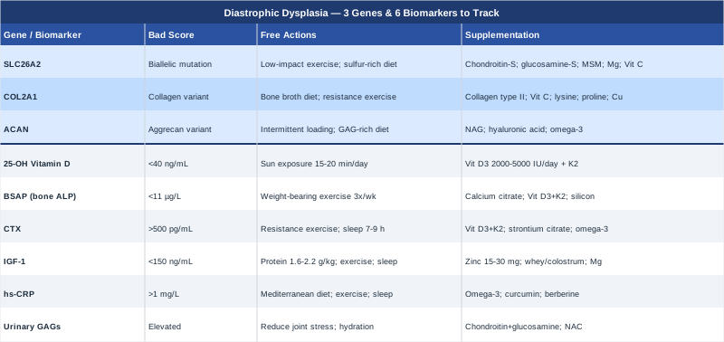
This article explores that framework from two directions. The primary section examines the three most relevant genes — what each does at a molecular level, what goes wrong when it is disrupted, and what evidence-informed steps can partially compensate for those disruptions, with and without supplementation. A second section identifies six biomarkers worth tracking consistently, explaining what each reveals, how it is measured, what optimal ranges look like, and how to act on abnormal readings. Combined, these two lenses offer a more precise path than standard management alone — not a cure, but a more informed way forward.
Summary
This article covers: the 3 key genes involved in diastrophic dysplasia (starting with SLC26A2, the core driver), practical compensatory plans for each gene variant both with and without supplements, and 6 biomarkers worth tracking regularly — including ones that can guide bone health, joint inflammation, and metabolic function in real clinical time. Beyond genetics and biomarkers, you will find an evidence-based review of complementary approaches with real study citations, a distillation of the most relevant research insights in the field, and practical protocols grounded in what the science actually supports. Whether you are newly diagnosed, years into management, or supporting a family member, this article is designed to help you make better-informed decisions at every step.
---
3 Key Genes in Diastrophic Dysplasia — What They Reveal and What You Can Do
Diastrophic dysplasia is among the most genetically well-defined skeletal dysplasias. Its molecular origins are centered on sulfate metabolism and cartilage matrix synthesis. Understanding the three genes described below provides a foundation not only for understanding the condition but for making decisions that can partially compensate for the molecular deficit.
Gene 1: SLC26A2 (DTDST) — The Core Driver
SLC26A2, also known as the diastrophic dysplasia sulfate transporter gene (DTDST), encodes a transmembrane protein responsible for transporting inorganic sulfate into chondrocytes — the cells that build cartilage. Sulfate is required to sulfate glycosaminoglycan chains on proteoglycan molecules, particularly aggrecan, which gives cartilage its compressive resilience and structural integrity. When SLC26A2 is mutated, chondrocytes cannot import sufficient sulfate, proteoglycans remain undersulfated, and the cartilage matrix formed during fetal development and throughout growth is mechanically inferior.
Diastrophic dysplasia is caused by biallelic loss-of-function mutations in this gene — meaning both copies are affected. The severity of the phenotype depends heavily on the residual transporter activity: some mutation combinations leave barely any functional transporter (as in the more severe achondrogenesis type IB), while others preserve partial function (as in the milder multiple epiphyseal dysplasia type 4). Diastrophic dysplasia occupies the middle range of this spectrum. The Finnish founder mutation (a splice site change in intron 1, IVS1+2T→C) is highly prevalent in Finland and has been identified in molecular epidemiological work as a key determinant of disease in that population. Other well-characterized mutations include R279W, A715V, and C653S, each with defined transporter activity reductions. The MedlinePlus genetics page for SLC26A2 provides a useful clinical overview of this gene's function and disease associations.
If the SLC26A2 gene is mutated — the plan without supplements
Since SLC26A2 mutations cannot be corrected without gene-level intervention (gene therapy for this condition is in early research stages and not yet available clinically), the compensatory approach focuses on reducing stress on an already-compromised cartilage system and optimizing the biological environment around it.
- Protect joint cartilage mechanically: Low-impact exercise — swimming, cycling, aqua therapy — preserves joint range of motion and muscle strength without adding compressive load to vulnerable joints. Daily 20–30 minute sessions, 5 days per week, are a practical target. Avoid high-impact activities and repetitive loading. - Dietary sulfur intake: Sulfur-containing foods (eggs, cruciferous vegetables, alliums like garlic and onion, and organ meats) provide dietary precursors to inorganic sulfate through metabolism. While this does not bypass the transporter defect, maintaining adequate systemic sulfate availability may reduce the severity of the deficit. Aim for 2–3 servings of sulfur-rich vegetables daily. - Orthopedic surveillance: Cervical spine imaging every 2–3 years, scoliosis monitoring from childhood, and annual joint assessment are not optional — cervical kyphosis in particular can lead to spinal cord compression if unmonitored. - Minimize inflammatory load: A whole-foods, low ultra-processed diet reduces systemic inflammation that accelerates cartilage degradation in already-stressed joints. The Mediterranean dietary pattern is the best-studied for musculoskeletal protection.
If the SLC26A2 gene is mutated — the plan with supplements
The rationale for supplementation centers on providing bioavailable sulfate substrate through alternative routes and supporting the downstream cartilage matrix as much as possible. Evidence for these supplements in the specific context of SLC26A2 mutations is largely mechanistic and extrapolated from general chondroprotective research, not yet from controlled trials in diastrophic dysplasia patients specifically.
- Chondroitin sulfate: 800–1200 mg daily, providing pre-sulfated glycosaminoglycan chains that can be incorporated into cartilage matrix without requiring cellular sulfation. Take consistently; effects are slow-building over 3–6 months. Widely available and well-tolerated. Side effects: occasional mild gastrointestinal discomfort. No known cycling requirement — continuous use is standard. - Glucosamine sulfate: 1500 mg daily, as a sulfate donor and glycosaminoglycan precursor. Use the sulfate form specifically (not hydrochloride) to maximize sulfate delivery. Begin with 6-week trials to assess individual response. Side effects: mild GI symptoms; caution if shellfish allergy (plant-derived versions exist). Take continuously or cycle 3 months on, 1 month off if cost is a factor. - Methylsulfonylmethane (MSM): 1000–3000 mg daily in divided doses. MSM is an organosulfur compound that serves as a systemic sulfur donor. Some human trials in osteoarthritis show reduced joint pain and improved function. Side effects: occasional headache or GI symptoms at higher doses; these typically resolve. No firm cycling protocol — continuous low-dose use is practical. - Magnesium (glycinate or malate form): 300–400 mg daily. Magnesium supports bone mineral density and has anti-inflammatory properties. The glycinate form is better tolerated gastrointestinally. Take in the evening. No cycling required. - Vitamin C: 500–1000 mg daily, as a cofactor for collagen synthesis and antioxidant protection of cartilage tissue. Time-release formulations may improve tolerability. Avoid megadosing (>2000 mg/day) due to risk of kidney stones.
Gene 2: COL2A1 — The Collagen Scaffold
COL2A1 encodes type II collagen, the primary structural protein of articular cartilage and intervertebral discs. While mutations in COL2A1 are the direct cause of a separate group of skeletal dysplasias (including spondyloepiphyseal dysplasia and Stickler syndrome), this gene is relevant to diastrophic dysplasia in a different way. When the cartilage matrix is already abnormal due to undersulfated proteoglycans from SLC26A2 mutations, the integrity of the collagen scaffold becomes particularly important. Variants in COL2A1 that reduce collagen II quantity or alter its structure can significantly amplify joint vulnerability in a genetic background already compromised by sulfation deficiency.
In clinical genetic panels run for skeletal dysplasias, COL2A1 variants of uncertain significance are frequently returned alongside the primary SLC26A2 pathogenic variants. Understanding whether such variants impair collagen synthesis — and acting to support that synthesis pathway — is a practical and underused strategy. Collagen II forms the fibrillar meshwork that holds proteoglycans in place within cartilage; when both the proteoglycan component (via SLC26A2) and the collagen component (via COL2A1) are suboptimal, joint degeneration accelerates markedly.
If the COL2A1 variant is relevant — the plan without supplements
- Collagen-supportive diet: Bone broth, slow-cooked connective tissue, fish skin, and egg whites provide hydroxyproline-rich peptides that stimulate endogenous collagen synthesis through fibroblast and chondrocyte signaling. Aim for inclusion in 4–5 meals per week. - Minimize collagen-degrading exposures: Smoking accelerates collagen breakdown; UV overexposure damages dermal and potentially articular collagen. These should be consistently avoided. - Load management: Gradual, controlled mechanical loading through resistance exercise (adapted for joint limitations) stimulates COL2A1 expression in chondrocytes. This is the best non-supplement intervention for collagen turnover. 2–3 sessions per week of resistance exercise adapted for mobility constraints is a reasonable protocol.
If the COL2A1 variant is relevant — the plan with supplements
- Hydrolyzed collagen type II (undenatured or hydrolyzed): 10–40 mg of undenatured type II collagen (UC-II) or 10–15 g of hydrolyzed collagen daily. Taken 30 minutes before meals for UC-II (immune tolerance mechanism) or with food for hydrolyzed collagen. Human trials in knee osteoarthritis show meaningful reductions in joint pain and stiffness. Side effects: minimal; occasional mild digestive discomfort. No cycling required — continuous use is appropriate. - Vitamin C: As above (500–1000 mg daily) — essential cofactor for the hydroxylation of proline and lysine residues in collagen synthesis. Non-negotiable if supporting COL2A1-dependent collagen production. - Lysine and proline (amino acid supplementation): 500–1000 mg of each, if dietary intake is inadequate. These are the two primary rate-limiting amino acids for collagen synthesis. Side effects: very low at these doses. Can be taken continuously. - Copper: 1–2 mg daily (as copper bisglycinate). Copper is essential for lysyl oxidase, the enzyme that crosslinks collagen fibers. Deficiency impairs collagen structural integrity. Note: balance with zinc intake (high-dose zinc supplementation competes with copper absorption). Side effects minimal at physiological doses.
Gene 3: ACAN (Aggrecan) — The Primary Proteoglycan Substrate
ACAN encodes aggrecan, the large proteoglycan that dominates the cartilage extracellular matrix. Aggrecan is the molecule that requires sulfation — specifically, its chondroitin sulfate and keratan sulfate chains must be sulfated post-translationally for the molecule to function. When SLC26A2 is mutated, the aggrecan produced by ACAN-expressing chondrocytes is undersulfated and functionally deficient, regardless of how much aggrecan protein is produced.
ACAN variants themselves are known causes of short stature and accelerated bone age (autosomal dominant ACAN mutations) and are associated with familial idiopathic short stature, advanced bone age, and early-onset osteoarthritis. In the context of diastrophic dysplasia, ACAN variants that reduce aggrecan production (reduced substrate) or alter its structure (modified sulfation sites) compound the impact of SLC26A2 dysfunction. Research has consistently shown that the functional output of aggrecan — measured by cartilage compressive stiffness — is disproportionately sensitive to combined disruptions in both sulfation capacity and aggrecan quantity or quality.
From a systems perspective, ACAN and SLC26A2 function in the same molecular pathway: ACAN provides the proteoglycan, SLC26A2 enables its sulfation, and the final sulfated aggrecan determines cartilage mechanical properties. Disruptions at either node degrade the output of the entire system.
If the ACAN variant is relevant — the plan without supplements
- Prioritize cartilage compressive resilience: Intermittent joint loading (alternating compression and decompression, as in swimming or cycling) is more protective of aggrecan-deficient cartilage than sustained static loading. Avoid prolonged standing or sitting in fixed positions. - Diet rich in proteoglycan precursors: N-acetylglucosamine and galactose (found in dairy, legumes, and specific vegetables) provide the backbone sugars for glycosaminoglycan chain synthesis. While not a substitute for functional aggrecan, ensuring substrate availability is a low-cost approach. - Monitor growth velocity in children: Compound ACAN-SLC26A2 dysfunction may impact longitudinal bone growth more severely than either mutation alone. Regular height and bone age assessment in pediatric patients with both variants is clinically indicated.
If the ACAN variant is relevant — the plan with supplements
- N-acetylglucosamine (NAG): 500–1000 mg daily. NAG serves as a precursor to glycosaminoglycan chains and may partially compensate for reduced aggrecan output by providing additional building material. Better tolerated than standard glucosamine in some patients. Side effects: minimal. Take continuously. - Hyaluronic acid (oral): 80–200 mg daily of low-molecular-weight hyaluronic acid. Oral hyaluronic acid has shown in several human trials to reduce joint stiffness and support synovial fluid quality, which is critically important when cartilage matrix is deficient. Side effects: minimal. Can be taken continuously. - Omega-3 fatty acids (EPA/DHA): 2–3 g daily from fish oil or algae-based sources. EPA and DHA reduce the activity of matrix metalloproteinases (MMPs) that degrade aggrecan in inflamed joints. Human trials consistently show reductions in joint pain with this dose range. Cycle: continuous use appropriate; monitor for anticoagulant interactions if on blood thinners.
---
With the genetic picture mapped, the next practical question is: which blood and urine tests can actually track what is happening in real time? Biomarkers provide ongoing feedback that genetic information alone cannot — they tell you not just about predisposition but about current status.
6 Biomarkers Worth Tracking in Diastrophic Dysplasia
These six biomarkers were selected for their direct relevance to the molecular and clinical vulnerabilities of diastrophic dysplasia: bone turnover, sulfate metabolism, inflammation, growth signaling, and vitamin status. Together, they provide a practical monitoring panel that any clinician familiar with the condition should be able to order.
Biomarker 1: 25-Hydroxyvitamin D
Why it matters
Vitamin D is not simply a bone vitamin — it is a steroid hormone precursor that regulates hundreds of genes involved in calcium absorption, bone mineralization, immune modulation, and chondrocyte differentiation. Individuals with limited mobility or indoor lifestyles (common in skeletal dysplasias where outdoor physical activity is restricted) are at elevated risk for vitamin D deficiency, which accelerates bone loss, reduces muscle strength, and amplifies joint inflammation. In the context of diastrophic dysplasia, adequate vitamin D is essential for maintaining what bone density and joint integrity remains possible.
How to measure it
A serum 25-hydroxyvitamin D [25(OH)D] test is standard, widely available, and inexpensive ($30–80 USD without insurance). Optimal range for musculoskeletal health: 40–60 ng/mL (100–150 nmol/L). Below 30 ng/mL is deficient; below 20 ng/mL is severely deficient. Test twice yearly: once in late winter (nadir) and once in late summer (peak).
If the score is low — the plan without supplements
Maximize direct sun exposure during peak UVB hours (10 am–2 pm), targeting 15–20 minutes of arm and leg exposure 4–5 days per week. Dietary sources (fatty fish, egg yolks, fortified foods) make a modest contribution but rarely normalize levels without sun or supplementation.
If the score is low — the plan with supplements
Vitamin D3: 2000–5000 IU daily for maintenance; up to 10,000 IU daily for 8–12 weeks to correct deficiency (under clinical supervision). Always combine with Vitamin K2 (MK-7): 100–200 mcg daily — K2 directs calcium into bones and away from soft tissue calcification, which is particularly important in diastrophic dysplasia given the risk of ectopic calcification (ear calcifications are a known feature). Retest after 3 months of supplementation. Side effects: rare at doses below 10,000 IU/day; toxicity typically requires sustained very high doses. No cycling required; continuous supplementation appropriate for most patients.
Biomarker 2: Bone-Specific Alkaline Phosphatase (BSAP)
Why it matters
Bone-specific alkaline phosphatase (BSAP) is the most specific circulating marker of osteoblast activity — it reflects how actively new bone is being formed. In skeletal dysplasias, bone formation dynamics are often abnormal, and tracking BSAP provides real-time information about whether bone-building activity is adequate, insufficient, or excessively elevated (which can indicate pathological remodeling or compensatory hyperactivity in response to mechanical stress).
How to measure it
Serum BSAP test (bone-specific ALP isoform); cost: $60–120 USD. More specific than total alkaline phosphatase, which includes liver and intestinal isoforms. Normal adult range: 11–30 µg/L (varies slightly by laboratory and sex). Request this specifically rather than the generic ALP panel. Test annually as part of a bone health panel.
If the score is low — the plan without supplements
Low BSAP reflects insufficient osteoblast activity and often signals nutritional inadequacy or sedentary mechanical environment. Weight-bearing or resistance activities (adapted for mobility limitations) are the most potent stimulus for osteoblast activation — even 20–30 minutes of resistance-based activity 3 times per week produces measurable improvements in bone formation markers within 8–12 weeks.
If the score is low — the plan with supplements
- Calcium (calcium citrate form): 500–1000 mg daily (split doses for absorption). Calcium citrate absorbs well without requiring stomach acid. Side effects: constipation (mitigate with magnesium). Continuous use appropriate. - Vitamin D3 + K2: As above — essential for calcium to reach bone tissue. - Silicon (orthosilicic acid form): 10–25 mg daily. Silicon stimulates osteoblast differentiation and BSAP activity. Human studies in women with osteoporosis show increased BSAP with silicon supplementation. Side effects: very low. Take continuously.
Biomarker 3: C-Telopeptide (CTX) — Bone Resorption Marker
Why it matters
CTX (C-terminal telopeptide of type I collagen) is the most validated marker of bone resorption — it rises when osteoclasts are breaking down bone matrix faster than it is being rebuilt. Thomas Dayspring and other leading lipid/metabolic experts have emphasized that bone resorption markers like CTX, interpreted alongside BSAP, give a dynamic picture of bone turnover balance. Elevated CTX in a patient with reduced BSAP is a red flag for progressive net bone loss, which carries serious implications in diastrophic dysplasia where spinal and joint integrity are already compromised.
How to measure it
Serum CTX (morning fasting sample for best accuracy); cost: $60–100 USD. Optimal range: below 500 pg/mL in adults; higher values indicate elevated resorption. Must be drawn in the morning and fasting (CTX has a strong circadian and food-dependent variation). Test annually alongside BSAP for a paired bone turnover assessment.
If the score is elevated — the plan without supplements
Elevated CTX responds strongly to mechanical loading — resistance exercise consistently reduces bone resorption within 6–8 weeks. Sleep optimization is also critical: growth hormone (which suppresses osteoclast activity) is primarily released during deep sleep, so 7–9 hours of quality sleep directly affects CTX levels. Reduce caffeine and alcohol intake, both of which elevate CTX.
If the score is elevated — the plan with supplements
- Vitamin D3 + K2: As above — vitamin K2 specifically inhibits osteocalcin carboxylation in osteoclasts, reducing resorption activity. - Strontium citrate (non-prescription form): 340–680 mg daily in the evening, taken separately from calcium. Strontium reduces osteoclast activity and has been studied for bone preservation. Side effects: avoid prescription strontium ranelate (cardiac risk); the citrate form is far safer. Cycle: 6 months on, 1 month off as a general caution for long-term use. - Omega-3 fatty acids: As above (2–3 g EPA/DHA daily) — reduces inflammatory signaling that drives osteoclast activation.
Biomarker 4: IGF-1 (Insulin-like Growth Factor 1)
Why it matters
IGF-1 is the primary mediator of growth hormone's anabolic effects on bone and cartilage. It stimulates chondrocyte proliferation, osteoblast activity, and proteoglycan synthesis — all of which are already compromised in diastrophic dysplasia. Suboptimal IGF-1 levels (common in sedentary individuals, those with poor sleep, or poor protein intake) amplify the intrinsic biological deficit. Peter Attia has extensively emphasized IGF-1 as a critical longevity and musculoskeletal health biomarker, noting that both extremes (very low and very high) carry risk, but that low levels in adults consistently associate with accelerated bone and muscle loss.
How to measure it
Serum IGF-1 (fasting not strictly required, but consistent timing improves tracking); cost: $60–120 USD. Optimal range for adults: 150–300 ng/mL. Values below 100 ng/mL suggest significant growth hormone axis insufficiency. Test annually, or more frequently if interventions are being trialed.
If the score is low — the plan without supplements
IGF-1 responds substantially to: high-quality protein intake (1.6–2.2 g/kg body weight per day), resistance exercise (particularly compound movements), deep sleep (growth hormone peaks in slow-wave sleep), and reduction of insulin resistance. These lifestyle factors can raise IGF-1 by 30–60% in low-normal individuals within 3–4 months.
If the score is low — the plan with supplements
- Zinc: 15–30 mg daily (as zinc picolinate or bisglycinate). Zinc deficiency suppresses growth hormone release and IGF-1 production. Balanced with copper (add 1–2 mg copper if taking >25 mg zinc). Side effects: nausea at high doses; take with food. - Colostrum or whey protein concentrate: 20–30 g daily. Both contain bioactive peptides that modestly stimulate IGF-1 production. Whey is well-studied for lean mass preservation; colostrum contains growth factors. Side effects: minimal if dairy is tolerated. - Magnesium: As above — magnesium deficiency impairs growth hormone secretion and thus IGF-1 levels.
Biomarker 5: High-Sensitivity CRP (hs-CRP)
Why it matters
High-sensitivity C-reactive protein (hs-CRP) is the most practical general marker of systemic inflammation. In diastrophic dysplasia, joint stress and abnormal biomechanical loading create chronic low-grade inflammation that accelerates cartilage degradation and worsens pain. Chronically elevated hs-CRP (above 3 mg/L) is also an independent cardiovascular risk marker — a concern for long-term health planning. Peter Attia considers hs-CRP one of the most useful inexpensive tests precisely because it integrates so many inputs: diet, sleep, gut health, visceral fat, and infection load.
How to measure it
Serum hs-CRP; cost: $15–40 USD. Optimal: below 1 mg/L. Borderline: 1–3 mg/L. Elevated: above 3 mg/L. Note: acute illness or recent injury will temporarily spike hs-CRP and should be excluded before interpreting the result as a chronic elevation. Test twice yearly.
If the score is elevated — the plan without supplements
Mediterranean-pattern diet, elimination of ultra-processed foods and refined sugars, sleep optimization (7–9 hours), and regular moderate exercise reduce hs-CRP by 20–40% in most individuals within 8–12 weeks. Visceral fat reduction is the most potent single driver of chronic hs-CRP elevation.
If the score is elevated — the plan with supplements
- Omega-3 fatty acids (EPA/DHA): 2–3 g daily — well-established anti-inflammatory effect. A 2003 meta-analysis in BMJ confirmed significant reductions in inflammatory markers with omega-3 supplementation. Continuous use appropriate. - Curcumin (phospholipid-complexed or liposomal form): 500–1000 mg daily. Standard curcumin has poor bioavailability; choose formulations that address this (Meriva, Longvida, or liposomal). Human trials show hs-CRP reductions comparable to low-dose NSAIDs in chronic inflammatory conditions. Side effects: mild GI symptoms; avoid high doses if on blood thinners. - Berberine: 500 mg twice daily with meals. Berberine activates AMPK and has documented anti-inflammatory effects. Cycle: 2–3 months on, 1 month off to prevent tolerance. Side effects: GI discomfort at initiation (start with 250 mg twice daily and titrate up).
Biomarker 6: Urinary Glycosaminoglycans (GAGs)
Why it matters
Urinary glycosaminoglycans (GAGs) — including chondroitin sulfate, dermatan sulfate, and heparan sulfate fragments — reflect cartilage and proteoglycan turnover. In conditions involving abnormal proteoglycan metabolism, urinary GAG excretion can be elevated as undersulfated or dysfunctional proteoglycans are catabolized more rapidly. This marker is less commonly ordered in routine clinical practice than the others in this list, but it is one of the few tests with direct relevance to the SLC26A2-dependent pathway. Specialized metabolic genetics laboratories offer quantitative urinary GAG profiling.
How to measure it
24-hour urine collection or random urine normalized to creatinine; typically ordered through metabolic genetics or pediatric biochemistry laboratories. Cost: $100–250 USD, often covered when ordered by a specialist for a known genetic diagnosis. Reference ranges depend on age (GAG excretion is higher in children). Elevated GAGs in the context of diastrophic dysplasia suggest accelerated proteoglycan catabolism — a potential target for intervention.
If the score is elevated — the plan without supplements
Reduce mechanical joint stress (as described above) to limit cartilage degradation. Anti-inflammatory dietary measures (above) reduce protease activity that breaks down proteoglycans. Adequate hydration ensures efficient renal clearance of GAG fragments and reduces their reabsorption.
If the score is elevated — the plan with supplements
- Chondroitin sulfate + glucosamine sulfate: As described in the SLC26A2 section — providing pre-sulfated GAG substrate may reduce the catabolic demand on endogenous proteoglycans. - N-acetylcysteine (NAC): 600–1200 mg daily. NAC is a glutathione precursor with direct free-radical scavenging properties in cartilage tissue. It reduces matrix metalloproteinase activity that degrades proteoglycans. Side effects: occasional GI discomfort; rare: unpleasant sulfur smell. Cycle: continuous or 3 months on, 1 month off.
---
What Leading Researchers Know: Key Insights From the Science
No single book or podcast addresses diastrophic dysplasia with the depth it deserves, but the molecular cartilage biology research from groups led by Andrea Superti-Furga (University of Lausanne) and colleagues has produced the most clinically relevant insights. Below are the ten most impactful findings from this body of work, framed for practical application.
1. Residual SLC26A2 transporter activity predicts clinical severity
Not all SLC26A2 mutations are equal. Mutations that preserve even 5–10% of sulfate transport activity produce substantially milder phenotypes than complete loss-of-function. Knowing the specific mutation and its functional consequence is more informative than the diagnosis alone.
2. Dietary and supplemental sulfate can partially compensate for transporter deficiency
Animal model studies have demonstrated that increased dietary sulfate intake can partially rescue the proteoglycan sulfation defect when transporter function is reduced but not abolished. This is the mechanistic basis for the chondroitin sulfate and glucosamine sulfate approach described above.
3. The cartilage matrix defect is present from early fetal development
Proteoglycan sulfation is essential during the earliest stages of chondrocyte differentiation in the embryo. This means the architectural consequences of the mutation are established before birth. Post-natal interventions address progression and preservation, not reversal of the original developmental deficit.
4. Cervical kyphosis is the most life-threatening skeletal manifestation
Progressive cervical kyphosis can compress the spinal cord and has caused sudden neurological deterioration in undisclosed cases. Surveillance imaging of the cervical spine every 2–3 years from early childhood is not cautionary boilerplate — it is a clinical imperative.
5. Inflammation dramatically accelerates cartilage loss in deficient matrix
Undersulfated cartilage is more vulnerable to inflammatory protease attack than normal cartilage. This means elevated hs-CRP is not a coincidental finding — it is a direct accelerant of disease progression in this condition.
6. Proteoglycan sulfation status affects chondrocyte survival, not just matrix quality
Chronically undersulfated chondrocytes show increased apoptosis rates in cell culture models. This means the cartilage cell population is itself at risk over time — another reason that reducing inflammatory load and providing substrate support matters.
7. Gene therapy is being explored but is not clinically available
Several research groups are investigating lentiviral and adeno-associated viral vectors for SLC26A2 correction in chondrocytes. Results in cellular models have been promising, but translation to clinical use remains years away at minimum.
8. Scoliosis is progressive and often requires surgical intervention
Spinal deformity in diastrophic dysplasia is not static. The progressive nature of scoliosis in this condition is distinct from idiopathic scoliosis, and bracing alone is frequently insufficient. Early surgical consultation with a surgeon experienced in skeletal dysplasias is advisable when curves exceed 40 degrees.
9. Ear calcifications are a near-pathognomonic feature but are not purely cosmetic
The cauliflower ear deformity in diastrophic dysplasia results from episodic auricular cartilage inflammation and subsequent calcification. These episodes can be painful and may affect hearing through external canal involvement. Anti-inflammatory management during acute episodes is standard of care.
10. Multi-disciplinary care teams produce substantially better outcomes
The MedlinePlus overview of diastrophic dysplasia and international skeletal dysplasia registries consistently show that patients managed by teams including orthopedic surgery, physical medicine, genetics, respiratory medicine, and nutrition have significantly fewer complications than those followed by a single provider.
---
Complementary Approaches With Meaningful Evidence
The following three modalities were selected because they have evidence — mostly from related musculoskeletal or chronic pain conditions — that is clinically plausible for diastrophic dysplasia. None offers disease modification; each offers symptom management and quality-of-life support.
Low-Level Laser Therapy (LLLT) / Photobiomodulation
Low-level laser therapy (LLLT), also called photobiomodulation, involves applying specific wavelengths of near-infrared or red light (typically 630–950 nm) to tissue at non-thermal intensities. The mechanism relevant to diastrophic dysplasia: photobiomodulation reduces oxidative stress and inflammatory cytokine production in chondrocytes, stimulates mitochondrial function in cartilage cells, and has demonstrated modest but consistent reductions in joint pain and stiffness in human trials of knee osteoarthritis — a condition sharing the feature of degraded articular cartilage matrix.
A 2009 systematic review and meta-analysis in The Lancet (PMID 19913903, available on PubMed) found significant short-term pain relief with LLLT in chronic neck pain compared to sham treatment. For knee osteoarthritis, multiple controlled trials have shown improvements in pain, function, and inflammatory markers. The evidence is strongest for musculoskeletal pain conditions; direct evidence in diastrophic dysplasia does not yet exist.
Practically: professional-grade LLLT devices (650–850 nm, 50–500 mW output) are available through physiotherapy and sports medicine clinics. Home devices of lower power exist but require longer treatment times. A typical protocol is 5–10 minutes per joint area, 3 times per week for 4 weeks, then as needed for maintenance. Avoid direct application over active growth plates in pediatric patients, and ensure any practitioner is aware of the underlying diagnosis and the specific joint vulnerabilities involved.
Massage Therapy
Manual massage therapy is relevant to diastrophic dysplasia not as a cartilage-modifying intervention but as a tool for managing the muscle tension, joint stiffness, and pain that accumulate around joints under abnormal biomechanical stress. In this condition, compensatory muscle patterns — muscles working harder to stabilize hypermobile or stiff joints — create secondary myofascial pain that compounds the primary articular symptoms.
A systematic review of massage therapy for musculoskeletal pain conditions (PMID 24753618 on PubMed) found consistent evidence for short-term improvements in pain, disability, and quality of life. Myofascial release techniques specifically targeting paraspinal and hip musculature are often the most relevant for patients with diastrophic dysplasia, given the typical pattern of spinal involvement.
Practically: sessions of 45–60 minutes with a practitioner experienced in working with clients with skeletal conditions, every 2–4 weeks for maintenance. It is essential that the therapist be briefed on the cervical instability risk and avoid high-velocity or deep pressure techniques near the cervical spine. Self-massage tools (foam rollers, massage guns on low intensity) can supplement professional sessions for accessible daily use on the lower extremities and paraspinal muscles.
Mindfulness-Based Stress Reduction (MBSR)
Mindfulness-based stress reduction (MBSR) is an 8-week structured program combining meditation, body scan, and gentle movement to build non-reactive awareness of pain and stress. Its relevance to diastrophic dysplasia is primarily through its well-documented effects on chronic pain perception: MBSR does not reduce structural joint damage, but it consistently reduces the suffering and disability associated with chronic pain — which is pervasive in this condition across the lifespan.
A landmark RCT comparing MBSR to standard care for chronic low back pain (PMID 27002445 on PubMed) showed that MBSR produced significant improvements in functional disability and pain bothersomeness at 26 and 52 weeks. Secondary evidence from musculoskeletal pain populations broadly supports similar effects. The mechanisms include altered descending pain modulation, reduced cortisol (which elevates inflammatory markers), and improved sleep quality.
MBSR programs are available in hospital-based formats, outpatient settings, and online. The standard 8-week format requires 30–45 minutes of daily practice. For patients with limited mobility, seated and lying meditation practices replace movement-based components. The key is consistency over weeks, not intensity within sessions. Combining MBSR with physical therapy creates an evidence-based chronic pain management package that addresses both peripheral joint pain and central pain sensitization.
---
Conclusion
Diastrophic dysplasia is a condition defined by molecular precision: a single gene, a single functional deficit, and a cascade of consequences that flow from that deficit throughout the skeleton over a lifetime. That precision is not only a clinical reality — it is an opportunity. When you know where the bottleneck is, you can make targeted rather than generic decisions about supplementation, monitoring, and lifestyle.
The three genes described here — SLC26A2, COL2A1, and ACAN — define the molecular architecture of the problem. The six biomarkers provide ongoing feedback about how that architecture is holding up in real time. Neither replaces clinical judgment or specialist oversight, but both offer something that standard care often does not: a framework for making better-informed decisions at each stage of management.
The practical next step is not to implement everything here at once. Start with the most accessible: have your 25-hydroxyvitamin D and hs-CRP tested, optimize sleep and protein intake, and introduce one or two well-tolerated supplements from the list above. From there, build toward a fuller biomarker panel reviewed annually with your treating team. Bring this information to your orthopedist, geneticist, or physiatrist — not to replace their guidance, but to make the conversations more specific, and the decisions better grounded in your individual biology.
Musculoskeletal: Bone Conditions Joint Conditions Spine Conditions
Autoimmune: Connective Tissue Conditions