This article contains AI generated content.
· UpdatedDiffuse Idiopathic Skeletal Hyperostosis Genes and Biomarkers — 6 Genes And 7 Biomarkers To Track
Introduction
Diffuse Idiopathic Skeletal Hyperostosis — DISH — is one of those conditions that tends to arrive quietly. Most people first hear the name when a radiologist mentions "flowing calcifications" on a spine X-ray taken for something else entirely. By then, the process has often been underway for years, slowly turning ligaments and tendons around the vertebrae into bony bridges. The result is stiffness, dull back pain, and sometimes difficulty swallowing when the cervical spine is involved. It affects an estimated 10 to 25 percent of adults over fifty, with men and people carrying excess weight around the middle disproportionately represented.
Standard medical advice for DISH tends to stop at symptom management: non-steroidal anti-inflammatory drugs, physical therapy for range of motion, weight loss. That advice is not wrong, but it is incomplete. It does not address the underlying question of why some people calcify so aggressively while others with similar lifestyles do not. It does not ask whether certain blood markers are signaling a metabolic environment that actively promotes ossification, or whether specific genetic variants are quietly tilting the odds. Without that layer of information, interventions remain generic.
The evidence increasingly suggests that DISH is not a random consequence of aging. It has deep roots in insulin metabolism, bone-signaling pathways, and the chemistry of mineralization regulation. Several genetic variants affect how efficiently the body neutralizes signals that drive ectopic bone formation. Several blood biomarkers can reveal whether those mechanisms are running hot right now — and crucially, most of them respond to targeted lifestyle and nutritional interventions.
This article covers two main angles. First, seven measurable biomarkers that reflect the core metabolic drivers of DISH — the ones most likely to reveal something actionable in your next lab panel. Second, six genes whose variants have been linked to abnormal ossification tendencies — with practical plans for each. Neither angle offers a cure. But together they offer something more useful than generic advice: a precise picture of your individual risk landscape and a starting point for decisions that actually match your biology.
7 Biomarkers That Reveal What Is Driving Your DISH
Blood tests cannot show you the calcium deposits forming in spinal ligaments, but they can show you the metabolic conditions that make those deposits more likely. The seven biomarkers below were selected because each one sits at a mechanistic junction point in the pathophysiology of DISH. They are also all measurable, most are affordable, and every single one responds to intervention.
Biomarker 1 — Fasting Insulin and HOMA-IR
Of all the metabolic drivers of DISH, chronic elevated insulin is the most consistently implicated. Insulin receptors are present on osteoblasts, and sustained hyperinsulinemia promotes bone formation not just in the right places but also at entheses — the attachment points of ligaments and tendons. This is thought to be a key mechanism behind the characteristically distributed calcifications seen in DISH. Population studies repeatedly show a disproportionate overlap between DISH and insulin resistance, independent of body weight.
How to measure it: Request fasting insulin (after at least 10 hours of fasting) and fasting glucose together. HOMA-IR is then calculated as (fasting insulin in μIU/mL × fasting glucose in mg/dL) ÷ 405. Cost: $30 to $60 for both values in most labs. Many standard metabolic panels omit fasting insulin, so it often needs to be specifically requested.
Target: Fasting insulin below 7 μIU/mL. HOMA-IR below 1.5 (optimal under 1.0). A HOMA-IR above 2.5 represents meaningful insulin resistance and, in the context of DISH, a significant metabolic risk signal.
If the score is bad — the plan without supplements: Time-restricted eating with an 8-to-10-hour eating window (16:8 or 14:10 protocols) is one of the most reliable tools for lowering fasting insulin without medication or supplements. Pair this with a significant reduction in refined carbohydrates and liquid sugars. Post-meal walks of 10 to 15 minutes after each main meal blunt the glucose and insulin response substantially, as demonstrated in multiple randomized trials. Resistance training three times per week improves skeletal muscle insulin sensitivity within four to six weeks. These changes collectively lower HOMA-IR by 20 to 40 percent in most people within three months.
If the score is bad — the plan with supplements or equipment: Berberine at 500 mg three times daily with meals is among the most studied plant-derived insulin sensitizers. Meta-analyses including over 2000 participants show reductions in fasting insulin and HOMA-IR comparable to low-dose metformin. Cycle at three months on, one month off, to avoid adaptation. Magnesium glycinate or malate at 300 to 400 mg before bed improves insulin receptor signaling in magnesium-depleted individuals and also supports sleep. Myo-inositol at 2 g twice daily has shown benefit particularly in women with insulin-resistance-related conditions, with few side effects. A continuous glucose monitor (CGM) worn for 14 days is a powerful non-supplement tool that makes insulin spikes visible, enabling highly targeted dietary adjustments without guesswork.
Biomarker 2 — IGF-1 (Insulin-Like Growth Factor 1)
IGF-1 is a potent anabolic signal produced primarily by the liver in response to growth hormone. In bone biology, IGF-1 directly activates osteoblast proliferation and increases collagen synthesis at entheses. Several studies comparing DISH patients to age-matched controls have found elevated IGF-1 in the DISH group, suggesting that the growth hormone–IGF-1 axis may be chronically overactivated in susceptible individuals. Elevated fasting insulin also stimulates hepatic IGF-1 production, creating a compounding loop.
How to measure it: A standard serum IGF-1 test, fasting or non-fasting, is sufficient. Cost: $60 to $120. Reference ranges are age-adjusted; the concern is not an IGF-1 in the lower third of the normal range but one consistently in the upper third or above it.
Target: Middle of the age-adjusted reference range. For adults in their 40s and 50s, this typically means 100 to 200 ng/mL. Chronically elevated IGF-1 above 250 to 300 ng/mL in this age group warrants attention.
If the score is bad — the plan without supplements: The most reliable way to reduce chronically elevated IGF-1 without medication is to moderate total protein intake, particularly from animal dairy. Casein and whey in milk are among the most potent stimulators of IGF-1. This does not mean eliminating protein — adequate protein is essential — but reducing dairy-derived protein to one serving per day or less, while relying more on plant-based and lean animal proteins, consistently lowers IGF-1 by 10 to 20 percent in intervention studies. Multi-day modified fasting protocols (the 5:2 method or periodic 24-hour fasts) significantly suppress GH and IGF-1 signaling. Prioritizing seven to eight hours of quality sleep matters: paradoxically, chronic sleep deprivation disrupts the pulsatile release of growth hormone in ways that dysregulate IGF-1 over time.
If the score is bad — the plan with supplements or equipment: There are no well-supported direct IGF-1 supplements to lower an elevated level. The most effective indirect approach is correcting insulin resistance (which drives hepatic IGF-1 production), using the berberine and myo-inositol protocol above. Evening melatonin at 0.5 to 3 mg, taken 30 minutes before sleep, may support more regular sleep architecture and indirectly normalize GH pulsatility over time. Avoid high-dose leucine supplements and protein powders emphasizing muscle hypertrophy if IGF-1 is already elevated.
Biomarker 3 — HbA1c and Fasting Glucose
DISH overlaps substantially with type 2 diabetes and prediabetes in epidemiological data. The mechanism is partially independent of insulin: chronic hyperglycemia promotes the formation of advanced glycation end-products (AGEs), which cross-link collagen fibers and stiffen connective tissue. AGEs also directly stimulate calcification in entheseal tissue. HbA1c reflects average glucose over three months and is a more reliable signal than a single fasting glucose reading.
How to measure it: HbA1c is included in most standard metabolic panels and costs $20 to $40. Fasting glucose is typically under $15 as a standalone test. Both together give the most complete picture.
Target: HbA1c below 5.4 percent (optimal), below 5.7 percent (acceptable). Fasting glucose below 85 mg/dL (optimal). Values in the prediabetic range (HbA1c 5.7 to 6.4 percent, fasting glucose 100 to 125 mg/dL) represent a meaningful risk signal in a DISH patient.
If the score is bad — the plan without supplements: A low-glycemic or low-carbohydrate diet remains the most effective non-pharmacological intervention for HbA1c reduction. Mediterranean dietary patterns also show consistent HbA1c-lowering effects in randomized trials. The glycemic load of any meal can be cut significantly by eating vegetables and protein before carbohydrates within the same meal — a simple sequencing strategy shown to reduce post-meal glucose by up to 37 percent. Resistance training three to four times per week substantially increases glucose uptake in skeletal muscle, the body's primary glucose disposal site.
If the score is bad — the plan with supplements or equipment: Berberine (500 mg three times daily, cycles of three months with a one-month break) is the most evidence-backed option for both fasting glucose and HbA1c outside of prescription medication. Alpha-lipoic acid at 600 mg per day improves insulin-mediated glucose uptake and has an additional antioxidant effect on AGE formation, which is specifically relevant to DISH. Chromium picolinate at 200 to 400 mcg per day may improve insulin sensitivity modestly, particularly in people with chromium depletion. A CGM remains the most powerful feedback device available for dietary optimization in this area.
Biomarker 4 — Serum Uric Acid
Hyperuricemia is found in DISH patients at higher rates than in the general population. Uric acid is not merely a gout marker — at elevated levels, it activates the NLRP3 inflammasome, promotes oxidative stress, drives vascular and soft-tissue calcification, and impairs insulin signaling. Fructose metabolism is a primary driver of uric acid production, which is one of several reasons why high fructose corn syrup and sugar-sweetened beverages are implicated in both metabolic syndrome and DISH progression.
How to measure it: A serum uric acid test is included in some basic metabolic panels and can be ordered standalone for under $20. Cost: $15 to $30.
Target: Below 5.0 mg/dL for optimal metabolic protection. In the context of active DISH or concurrent joint calcification, targeting below 4.5 mg/dL is reasonable.
If the score is bad — the plan without supplements: The most impactful dietary change is eliminating fructose from liquid sources: no sugar-sweetened beverages, no fruit juice, minimal added sugars. Reducing organ meats (liver, kidney) and shellfish (anchovies, sardines, mussels) matters in high-intake individuals. Beer specifically raises uric acid through both alcohol and purine content and should be minimized. Adequate hydration — at least 2.5 liters of water per day — increases renal uric acid clearance. Moderate aerobic exercise five days per week supports uric acid excretion.
If the score is bad — the plan with supplements or equipment: Tart cherry extract at 480 mg per day (or 240 mL of tart cherry juice) has shown consistent uric acid-lowering effects in randomized controlled trials, with reductions of 0.2 to 0.5 mg/dL over four to six weeks. Quercetin at 500 mg per day inhibits xanthine oxidase (the enzyme that produces uric acid) and has supporting human evidence. Vitamin C at 500 mg per day lowers serum uric acid by approximately 0.5 mg/dL through increased renal excretion, as demonstrated in a large randomized trial published in Arthritis & Rheumatism (Juraschek et al., 2011). These can be combined safely. Cycling is optional for quercetin and vitamin C; tart cherry extract can be taken continuously.
Biomarker 5 — High-Sensitivity CRP (hsCRP)
Chronic low-grade inflammation plays an underappreciated role in DISH. Inflammatory cytokines — particularly IL-6, TNF-alpha, and IL-17 — have been shown to upregulate bone morphogenetic protein (BMP) signaling and promote osteoblast activity at entheses. High-sensitivity CRP is the most widely available and affordable proxy for systemic inflammatory load. While DISH is traditionally classified as non-inflammatory (distinguishing it from ankylosing spondylitis), emerging research suggests that subclinical inflammation contributes to its progression.
How to measure it: High-sensitivity CRP is distinct from standard CRP and must be specifically requested. Cost: $20 to $45. Some labs include it in premium cardiovascular panels alongside ApoB and Lp(a).
Target: Below 0.5 mg/L (excellent). Below 1.0 mg/L (good). Values above 2.0 mg/L represent a significant inflammatory signal warranting investigation for triggers.
If the score is bad — the plan without supplements: An anti-inflammatory dietary pattern (Mediterranean or autoimmune protocol variants) is the most effective dietary lever. Eliminating ultra-processed foods, trans fats, and excess omega-6 vegetable oils (canola, soy, corn) is foundational. Improving sleep to seven to eight hours reduces inflammatory cytokine production markedly. Stress management, specifically chronic psychological stress reduction, is frequently overlooked but is a meaningful driver of IL-6 and CRP through HPA axis activation. Moderate cardio exercise at 60 to 70 percent of max heart rate, four to five times per week, consistently lowers CRP over twelve weeks.
If the score is bad — the plan with supplements or equipment: Omega-3 fatty acids at 2 to 4 grams of combined EPA and DHA per day lower hsCRP through multiple mechanisms (reduction of eicosanoid synthesis, resolution of inflammation via resolvins and protectins). Meta-analyses of randomized trials confirm clinically meaningful CRP reductions at this dose range. Purchase triglyceride-form fish oil (higher bioavailability than ethyl ester form), take with the largest meal, and store in the refrigerator to prevent oxidation. Curcumin with piperine at 500 mg twice daily has shown significant CRP reduction in several randomized trials; piperine is essential for bioavailability (increases absorption by 2000 percent). Boswellia extract (AKBA form) at 100 to 200 mg per day inhibits the 5-LOX inflammatory pathway and is particularly relevant for connective tissue inflammation. Cycle curcumin at two months on, two weeks off.
Biomarker 6 — Undercarboxylated Osteocalcin and Vitamin K2 Status
This is arguably the most underappreciated biomarker in DISH. Matrix Gla Protein (MGP) is the most potent known inhibitor of soft-tissue and ectopic calcification in the body. For MGP to do its job, it must be carboxylated — a process that requires vitamin K2 as a cofactor. Without adequate K2 activity, MGP circulates in an uncarboxylated, inactive form, and calcification in soft tissues — including spinal ligaments — proceeds unchecked. Osteocalcin undergoes the same carboxylation process; measuring undercarboxylated osteocalcin (ucOC) provides an indirect marker of K2 status in bone tissue.
How to measure it: The most specific test is desphospho-uncarboxylated MGP (dp-ucMGP), available through specialty labs such as VitaK (Netherlands) or some European reference labs. Cost: $80 to $200. A more accessible proxy in the United States is undercarboxylated osteocalcin, available through Quest or LabCorp with a physician order, for $80 to $150. Some functional medicine practitioners use it routinely.
Target: Low undercarboxylated osteocalcin. The exact optimal range varies by assay, but the goal is to maximize the ratio of carboxylated to undercarboxylated osteocalcin. High dp-ucMGP (above 600 pmol/L in most assays) is associated with vascular and soft-tissue calcification risk.
If the score is bad — the plan without supplements: Increase dietary vitamin K2 through food. Natto (fermented soybeans) is the richest food source of MK-7, the most bioavailable form, with a single 100-gram serving providing 800 to 1000 mcg. Hard aged cheeses (gouda, brie, munster) provide MK-4 to MK-9 forms. Egg yolks from pasture-raised hens, liver, and grass-fed butter contain meaningful MK-4. Building a dietary pattern that includes one or more of these daily can materially improve K2 status over three to six months.
If the score is bad — the plan with supplements or equipment: MK-7 (vitamin K2 as menaquinone-7) at 180 to 400 mcg per day with a fat-containing meal is the most studied supplemental form. MK-7 has a half-life of 72 hours (versus four hours for MK-4), meaning once-daily dosing is effective. Research published in Thrombosis and Haemostasis and reviewed in multiple cardiovascular calcification trials shows that MK-7 supplementation significantly lowers dp-ucMGP within 12 weeks. Critical safety note: Vitamin K2 supplements interact with warfarin (and related anticoagulants) — do not combine without medical supervision. For patients not on anticoagulants, K2 supplementation at these doses is considered safe. MK-4 at 1500 mcg three times daily (4500 mcg total) is used in Japanese clinical practice for osteoporosis and also supports MGP carboxylation, but the higher dose required makes MK-7 more practical.
Biomarker 7 — 25-OH Vitamin D
Vitamin D's role in DISH is more complex than the simple "supplement more D" narrative. Vitamin D is a calcium-regulating hormone. Adequate D is essential for normal bone mineralization and immune regulation. But very high vitamin D levels, in the absence of adequate vitamin K2 to direct calcium into bone and prevent ectopic deposits, may promote soft-tissue calcification. In DISH patients, the D-K2 relationship is particularly important to track and optimize together.
How to measure it: A standard 25-hydroxyvitamin D blood test. Cost: $30 to $60. Widely available through any lab or as an at-home finger-prick test from companies such as Everlywell.
Target: 40 to 60 ng/mL (100 to 150 nmol/L). Below 30 ng/mL represents deficiency; above 80 ng/mL without confirmed adequate K2 status is a caution zone specifically for DISH patients.
If the score is bad — the plan without supplements: Daily midday sun exposure (15 to 30 minutes of arms and legs) raises vitamin D levels reliably in fair-skinned individuals, more gradually in darker skin tones. Fatty fish (salmon, mackerel, sardines) and egg yolks are the best dietary sources. Avoid excess sunscreen during the short window of intentional vitamin D synthesis, then apply for extended outdoor time.
If the score is bad — the plan with supplements or equipment: Vitamin D3 at 2000 to 5000 IU daily should always be paired with vitamin K2 (MK-7, 180 to 200 mcg) in a DISH context — this combination ensures that any calcium mobilized by D is directed toward bone rather than soft tissue. Magnesium glycinate or malate at 300 to 400 mg per day is a necessary cofactor for vitamin D activation in the liver and kidneys; many people supplementing D without magnesium see blunted responses. Recheck 25-OH vitamin D levels every three months while adjusting dose. Avoid "megadosing" above 10,000 IU daily without medical supervision.
6 Genes Linked to DISH and What They Mean for Your Biology
Genetics do not determine fate in DISH, but they do raise or lower the activation energy required for ectopic ossification. Several genes influence how efficiently the body regulates pyrophosphate (a natural calcification inhibitor), how robustly it signals bone formation, and how it maintains the balance between bone deposition and resorption. Knowing your variants allows you to prioritize the most relevant interventions and understand why some strategies may matter more for you than for someone else.
Gene 1 — ENPP1 (Ectonucleotide Pyrophosphatase/Phosphodiesterase 1)
ENPP1 produces pyrophosphate (PPi) in the extracellular space. PPi is one of the body's primary brakes on mineralization — it physically inhibits calcium phosphate crystal formation in soft tissues. The K121Q variant (rs1044498) reduces ENPP1 enzyme activity, lowering extracellular PPi and effectively removing a critical anti-calcification signal.
If the gene is bad — the plan without supplements: Since this gene impairs the production of the PPi brake on calcification, lifestyle choices that would otherwise add more mineralization stimulus become more consequential. Avoid prolonged sedentary postures that place sustained mechanical load on spinal ligaments. Prioritize low-impact movement (swimming, walking, cycling) over high-impact loading that stresses entheses. Maintain low calcium intake from supplements specifically (dietary calcium from whole foods is far less likely to cause issues than calcium carbonate supplements). Minimize dietary phosphate from ultra-processed foods (phosphate additives are widespread in packaged foods and significantly raise serum phosphate, driving calcification when PPi is low).
If the score is bad — the plan with supplements or equipment: Magnesium competes with calcium at mineralization sites and can partially compensate for reduced PPi. Magnesium citrate or glycinate at 300 to 400 mg per day, long-term, is a low-risk strategy. Vitamin K2 (MK-7, 200 mcg/day) is particularly important for ENPP1 variant carriers because it activates the alternative anti-calcification pathway via MGP. Some research on related calcification disorders (like generalized arterial calcification of infancy, caused by ENPP1 loss-of-function mutations) suggests that dietary pyrophosphate sources — specifically whole foods rich in inositol hexaphosphate (IP6), such as brown rice, legumes, and seeds — may modestly support extracellular PPi. Evidence in DISH specifically is limited, but the mechanism is relevant.
Gene 2 — ANKH (Progressive Ankylosis Protein)
The ANKH gene encodes a transmembrane protein that transports PPi from inside cells to the extracellular space. Loss-of-function variants in ANKH reduce this transport, lowering extracellular PPi in the same downstream manner as ENPP1 variants. ANKH variants have been linked to both diffuse skeletal hyperostosis and calcium pyrophosphate deposition (CPPD), suggesting a shared pyrophosphate-deficiency mechanism. Multiple gain-of-function variants paradoxically also cause joint disease, illustrating the tight regulation required in this pathway.
If the gene is bad — the plan without supplements: The approach mirrors the ENPP1 strategy: minimize additional calcification load by controlling dietary calcium supplementation (aim for dietary calcium from whole foods, not supplements, unless a physician recommends otherwise), stay well-hydrated to support renal mineral clearance, and limit foods high in phosphate additives. Periodic monitoring of serum calcium and phosphate (included in a basic metabolic panel) is appropriate for ANKH variant carriers to confirm mineral levels remain within normal range.
If the gene is bad — the plan with supplements or equipment: Magnesium glycinate (300 to 400 mg/day) and MK-7 (180 to 200 mcg/day) are the most rationally supported supplements for reducing soft-tissue calcification risk in individuals with impaired PPi pathways. There is early-stage interest in dietary etidronate (a bisphosphonate) as a pharmaceutical option for severe calcification conditions related to PPi dysregulation, but this is not a current standard of care for DISH and requires specialist supervision.
Gene 3 — TNFRSF11B (Osteoprotegerin Gene)
TNFRSF11B encodes osteoprotegerin (OPG), a decoy receptor that inhibits RANKL from activating osteoclasts. The OPG/RANKL balance is the central regulator of bone turnover. In DISH, the concern is not simply excessive bone destruction but excessive net formation — variants that reduce OPG expression tilt the balance toward more net bone deposition. OPG also plays a direct role in vascular and soft-tissue calcification: OPG-knockout mice develop severe arterial calcification, and low serum OPG is associated with both vascular and spinal calcification in human studies.
If the gene is bad — the plan without supplements: Weight-bearing exercise (resistance training, walking) stimulates OPG production by osteoblasts and shifts the OPG/RANKL ratio favorably. Reduce chronic inflammatory load through dietary means (this is important because TNF-alpha, itself, suppresses OPG expression). Avoiding smoking is particularly relevant here, as smoking consistently lowers serum OPG. Estrogen (in women) and testosterone (in men) both upregulate OPG — sex hormone optimization through lifestyle (adequate sleep, healthy body composition, resistance training) is supportive, though this is not a direct pharmacological strategy.
If the gene is bad — the plan with supplements or equipment: Vitamin K2 (MK-7, 200 mcg/day) supports OPG-independent anti-calcification through MGP activation and is worth prioritizing for this gene variant. Vitamin D3 at appropriate levels (40 to 60 ng/mL) supports bone remodeling balance. Strontium ranelate has shown OPG-stimulating effects in vitro but is not widely available or recommended for routine use. The omega-3 fatty acid protocol (2 to 4 g EPA+DHA/day) reduces TNF-alpha, which indirectly preserves OPG expression — a reasonable indirect strategy.
Gene 4 — COL11A2 (Collagen Type XI Alpha-2)
COL11A2 encodes a structural collagen present in cartilage, spinal discs, and ligaments. Variants in COL11A2 alter the mechanical properties of connective tissue — specifically its resistance to tensile load and its propensity to undergo metaplastic ossification under repeated strain. In DISH, the entheseal attachments of spinal ligaments are the primary sites of calcification; altered collagen architecture at these sites may lower the threshold for ossification in response to mechanical stress.
If the gene is bad — the plan without supplements: Protecting entheseal tissue from chronic repetitive microtrauma is the priority. Avoid sustained postures that place continuous tension on the anterior longitudinal ligament of the spine — particularly prolonged lumbar hyperextension. Incorporate movement variety throughout the day. Ergonomic chair and desk setup to minimize static axial load matters more for COL11A2 variant carriers than for the average person. Maintain healthy body composition to reduce compressive and tensile forces on spinal ligaments.
If the gene is bad — the plan with supplements or equipment: Collagen peptide supplementation (10 to 15 g per day with vitamin C) has early evidence for improving ligament and tendon resilience. The rationale for COL11A2 variants specifically is mechanistic rather than from a direct DISH trial: optimizing collagen substrate availability may partially compensate for structurally compromised collagen architecture. Take collagen hydrolysate 30 to 60 minutes before mechanical loading sessions for best tissue delivery. Vitamin C at 500 to 1000 mg per day is essential as a cofactor in collagen cross-linking. Cycle collagen supplementation at three months on, one month off if used long-term.
Gene 5 — BMP4 and the BMP Signaling Pathway
Bone Morphogenetic Proteins — particularly BMP2 and BMP4 — are among the most potent osteoinductive signals in the body. They instruct uncommitted mesenchymal stem cells to differentiate into osteoblasts. Variants that increase BMP4 expression or reduce the expression of natural BMP antagonists (such as Noggin or Gremlin) can create a systemic pro-ossification bias. BMP pathway upregulation has been specifically documented in entheseal ossification models and is considered a central molecular mechanism in both DISH and the related condition fibrodysplasia ossificans progressiva.
If the gene is bad — the plan without supplements: Avoid iatrogenic BMP activation. In clinical settings, recombinant BMP-2 (used in spinal fusion surgery as a bone graft substitute) is absolutely contraindicated if you have DISH or a BMP hypersensitivity variant — document this for any surgical consultations. Reduce local entheseal injury, which triggers BMP release at healing sites. Weight management is important because adipose tissue secretes BMP-4, creating a direct link between excess adiposity and BMP-driven ossification risk.
If the gene is bad — the plan with supplements or equipment: Resveratrol at 250 to 500 mg per day has shown BMP antagonist-upregulating effects in animal models and some human cell studies, supporting Noggin expression. The evidence in DISH specifically is limited to early research, but the mechanistic rationale is relevant for BMP4 variant carriers. EGCG (green tea extract, 400 to 800 mg/day) has similar pathway effects in vitro. Neither should be considered a standalone intervention, but as additions to the metabolic optimization strategy outlined in the biomarker section, they represent low-risk choices. Cycle resveratrol at eight weeks on, four weeks off; monitor for blood thinning effects.
Gene 6 — FGF2 and FGFR1 (Fibroblast Growth Factor Pathway)
Fibroblast growth factor 2 (FGF2) is a powerful mitogen for osteoblasts and fibroblasts. Elevated FGF2 signaling promotes periosteal bone formation and has been found elevated in spinal hyperostosis models. FGFR1 variants that increase receptor sensitivity amplify this signal. The FGF pathway also interacts with the insulin signaling pathway, meaning that chronic hyperinsulinemia can amplify FGF-driven ossification — another reason why insulin control is foundational for DISH.
If the gene is bad — the plan without supplements: Controlling insulin resistance directly reduces FGF pathway activation, making the HOMA-IR interventions described in the biomarker section doubly important for FGF variant carriers. Periodic fasting (16:8 time-restricted eating or quarterly 5-day modified fasting protocols) reduces growth factor signaling broadly and is mechanistically relevant here. Reducing total calorie surplus is important: FGF2 expression is upregulated in adipose tissue and declines with fat loss.
If the gene is bad — the plan with supplements or equipment: No specific supplements target FGF2 pathway suppression with strong human evidence. The omega-3 DHA at 2 to 3 g per day has shown modest FGF pathway modulation in cardiac research. The most relevant intervention remains metabolic: insulin sensitization and body composition improvement through the lifestyle strategies already described. Consider requesting a serum FGF23 level (a related biomarker linked to phosphate metabolism and calcification, testable at most major labs for $80 to $150) to determine whether the FGF-phosphate axis is active as a driver.
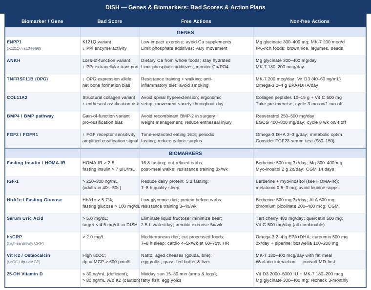
Now that both biomarkers and genes are covered, the table below summarizes the full picture at a glance:
What "Why We Get Sick" Gets Right About DISH
Ben Bikman's Why We Get Sick (BenBella Books, 2020) is not a book about DISH. But it may be one of the most useful books for anyone living with it, because it builds the scientific case for why insulin resistance is not merely a diabetes problem — it is a systems-level metabolic disruption that touches bone biology, inflammation, and ectopic calcification in ways that conventional endocrinology has been slow to integrate into clinical guidance for skeletal conditions.
Bikman, a professor of cell biology and physiology at Brigham Young University, draws on hundreds of peer-reviewed studies to argue that chronic hyperinsulinemia is an upstream driver of most of the major chronic diseases of modernity. Several of his points land with particular force for DISH patients.
1. Insulin Directly Activates Osteoblasts
Bikman explains that osteoblasts — the cells that build new bone — have insulin receptors. When insulin is chronically elevated, these receptors receive a sustained anabolic signal. In the right context (healthy bones under mechanical load), this is beneficial. In the wrong context (entheseal tissue in a metabolically dysfunctional individual), it creates the biologically permissive environment for ectopic ossification. He calls this one of insulin's "side effects at scale."
2. Visceral Fat Is Not Passive Storage
One of the book's central arguments is that visceral adipose tissue is a metabolically active endocrine organ, secreting BMP-4, leptin, IL-6, and other signals that drive bone formation and inflammation. Bikman cites research showing that individuals with the highest visceral fat indices have significantly elevated circulating osteoblast-activating cytokines — making waist circumference not just a cosmetic concern but a direct DISH risk variable.
3. Fructose Raises Uric Acid and Promotes Calcification
Bikman devotes considerable attention to fructose metabolism. Unlike glucose, fructose is processed almost entirely in the liver, producing uric acid as a metabolic byproduct. He cites work by Robert Lustig and others showing that uric acid functions not just as a gout marker but as an active promoter of calcification and oxidative stress. For DISH patients with elevated uric acid, this chapter alone may be worth the price of the book.
4. Standard Labs Miss the Most Important Signal
One of Bikman's most practically important points: fasting glucose and HbA1c can remain in "normal" ranges for years while fasting insulin is significantly elevated — meaning a patient can have meaningful hyperinsulinemia with a clean standard metabolic panel. He argues that fasting insulin should be a routine screening test and gives a clear target (below 6 to 8 μIU/mL fasting). This is directly actionable for DISH patients who have been told their blood sugar is "fine."
5. The Insulin–IGF-1 Amplification Loop
Bikman explains how chronic insulin elevation suppresses insulin-like growth factor binding proteins (IGFBPs), effectively freeing more circulating IGF-1 to act on tissues. This amplification loop means that addressing insulin resistance does double duty: it lowers both insulin and bioavailable IGF-1 simultaneously — two of the seven DISH biomarkers, improved with one intervention.
6. Intermittent Fasting Outperforms Caloric Restriction for Metabolic Reset
The book reviews multiple randomized trials comparing caloric restriction alone to time-restricted eating. Time-restricted eating produces larger improvements in fasting insulin and HOMA-IR for similar caloric reductions, because the fasting window itself — independent of calorie count — allows insulin to fall to baseline, giving insulin receptor sensitivity time to recover. Bikman recommends a minimum 12-hour fast, with 16 hours being optimal for most people seeking metabolic improvement.
7. Berberine as a Low-Risk First Step
Bikman discusses berberine's AMPK-activating mechanism (the same pathway activated by metformin) and its consistent performance in randomized trials for reducing insulin resistance. He is measured in his claims — not overselling supplements — but notes that berberine is one of the few plant compounds with a mechanistic rationale robust enough and a human trial record strong enough to recommend as a metabolic support tool.
8. Magnesium Deficiency Is More Common Than Most Physicians Assume
Bikman cites research showing that up to 50 percent of Americans consume insufficient dietary magnesium, and that magnesium deficiency impairs insulin receptor signaling. He notes that standard serum magnesium tests are unreliable because serum magnesium is tightly regulated even when tissue stores are depleted. He recommends either RBC magnesium testing or simply supplementing with magnesium glycinate as a practical insurance policy, which aligns directly with DISH management.
9. The Role of Sleep in Metabolic Health
Bikman dedicates a chapter to how sleep deprivation — even one to two nights per week of short sleep — substantially raises fasting insulin and reduces insulin sensitivity. The mechanism involves cortisol-driven glucose release and the disruption of adiponectin signaling. For DISH patients juggling pain-disrupted sleep with metabolic goals, this creates a feedback loop worth directly addressing.
10. Inflammation and Insulin Resistance Are Bidirectionally Linked
One of the book's most useful conceptual frames: insulin resistance both causes and is caused by inflammation. TNF-alpha (elevated in chronic inflammation) blocks insulin receptor signaling. Hyperinsulinemia upregulates NFkB, the master inflammatory transcription factor. The implication for DISH is clear: strategies that break this loop — whether dietary, exercise-based, or supplemental — address both biomarkers simultaneously, creating compounding improvements rather than isolated ones.
Complementary Approaches With Relevant Human Evidence
Several non-pharmacological modalities have clinical evidence relevant to spinal stiffness, entheseal inflammation, and pain management in DISH. The four below were selected for meaningful human evidence specific to this clinical context.
Tai Chi
Tai chi is a mind-body practice combining slow, controlled movements with breath coordination and weight shifting. Its relevance for DISH lies primarily in its ability to maintain spinal and joint range of motion through low-load, flowing movement — the opposite of the high-impact loading that can stress ossified entheses. The practice also reduces cortisol and inflammatory markers over time, relevant to the inflammation-insulin loop described above.
A systematic review and meta-analysis published in Journal of Rheumatology examining tai chi across musculoskeletal conditions found consistent improvements in pain, physical function, and stiffness scores. A 12-week randomized controlled trial in adults with chronic spinal conditions showed significant improvements in both spinal flexibility and self-reported pain versus a waitlist control. While DISH-specific RCTs are limited, the condition profile (spinal stiffness, reduced range of motion, pain with movement) maps well to conditions where tai chi evidence is strongest.
Practically: twice-weekly group tai chi classes of 45 to 60 minutes, maintained over 12 weeks, represent the minimum dose used in most positive trials. For home practice, a structured beginner Yang-style short form can be learned from video in 20 to 30 sessions and maintained daily in under 15 minutes. Focus specifically on the cervical rotations and thoracic extension components, which are often the most restricted in DISH patients. Advance movements gradually — never force range of motion at the edge of pain.
Low-Level Laser Therapy / Photobiomodulation
Low-level laser therapy (LLLT), also called photobiomodulation, delivers specific wavelengths of red and near-infrared light to tissue, reducing local inflammation, improving mitochondrial function in stressed cells, and modulating the inflammatory cytokine cascade at treated sites. Its relevance to DISH is primarily for managing pain and stiffness at calcified entheses — particularly in the cervical and thoracic spine — without the systemic risks of NSAIDs.
A meta-analysis of 22 randomized controlled trials, published in BMC Musculoskeletal Disorders (Chow et al., 2009), found significant pain reduction with low-level laser therapy in neck pain from various musculoskeletal causes. Wavelengths of 820 to 1064 nm at 3 to 10 J/cm² per point represent the parameters most consistently associated with benefit in spinal conditions. The evidence is stronger for pain management than for any modification of ossification itself.
Practically: devices cleared for home use in the 630 to 850 nm range are available from several manufacturers (Joovv, Red Light Rising, and others) at $300 to $1200 for panel devices. A protocol of 10 to 15 minutes of near-infrared exposure directly on the affected spinal segments, five days per week, is consistent with published protocols. Alternatively, clinical LLLT sessions through a physiotherapist or sports medicine clinic cost $30 to $80 per session. Start at the lower power density setting and increase gradually; avoid shining directly into eyes.
Yoga
Yoga is relevant to DISH for two reasons: the mobility and stretching components help counteract spinal stiffness, and the documented anti-inflammatory and cortisol-lowering effects of regular practice may modestly reduce the inflammatory component of ossification over time. The critical caveat is that not all yoga styles are appropriate — fast-paced, high-load styles (Ashtanga, hot yoga with extended hyperextension) can aggravate DISH, particularly in the thoracic and lumbar spine.
A randomized controlled trial published in Annals of Internal Medicine (Wieland et al., 2017, referenced in a Cochrane review) found that yoga produced meaningful improvements in chronic low back pain and function compared to usual care. Iyengar yoga — which emphasizes structural precision and uses props (blocks, straps, bolsters) to avoid joint compromise — is the most appropriate style for patients with spinal structural changes.
Practically: two to three weekly classes of Iyengar or Viniyoga (therapeutically adapted yoga) taught by an instructor familiar with spinal conditions is the recommended starting approach. Inform your instructor of your DISH diagnosis. Prioritize thoracic extension over flexion-dominant sequences (which can load anterior spinal osteophytes); avoid deep cervical rotation at end range; use props to reduce joint compression. A 12-week minimum commitment is necessary to observe functional improvements.
Breathing-Based Therapies
DISH that involves the thoracic spine can mechanically restrict chest expansion, reducing respiratory volume and causing the shallow breathing patterns often seen in thoracic hyperostosis. Specific breathing exercises — diaphragmatic retraining, lateral costal breathing, and slow-paced resonance-frequency breathing — address both the mechanical restriction (by actively mobilizing the rib-spine joints within their available range) and the autonomic dysregulation that accompanies chronic pain conditions.
A randomized trial published in Journal of Pain Research demonstrated that slow-paced breathing (at approximately 6 breaths per minute, or resonance-frequency breathing) significantly reduced pain scores and inflammatory markers in adults with chronic musculoskeletal pain over eight weeks. The mechanism involves vagal nerve activation, which reduces the sympathetic-mediated inflammatory response and lowers cortisol. For thoracic DISH specifically, a physiotherapist trained in respiratory rehabilitation can prescribe specific lateral expansion exercises targeting the restricted costovertebral joints.
Practically: daily practice of diaphragmatic breathing for 10 to 15 minutes in a reclined position with hands on lateral ribs is a minimal starting protocol. Breathing apps (Breathwrk, Othership) can guide resonance-frequency breathing at the correct 6-breaths-per-minute target. For significant thoracic restriction, formal respiratory physiotherapy assessment is worthwhile — these clinicians can assess chest expansion measurements and tailor exercises. Combine with the thoracic extension yoga protocol above for compounding benefit.
Conclusion
DISH is not a condition that announces a simple fix. But it is a condition with identifiable drivers — metabolic, genetic, and inflammatory — that are measurable, monitorable, and in many cases responsive to targeted intervention. Tracking your fasting insulin, vitamin K2 status, uric acid, and inflammatory markers gives you a real-time picture of whether the metabolic environment favoring calcification is under control. Understanding your genetic variants in ENPP1, ANKH, OPG, or BMP pathways tells you which mechanisms deserve more attention in your specific case.
The next smart step is straightforward: request a focused lab panel that includes fasting insulin, HOMA-IR, uric acid, hsCRP, 25-OH vitamin D, and HbA1c. From there, work with a physician who is willing to look at the metabolic picture — not just the structural one — and consider one or two of the lifestyle interventions most relevant to your biomarker profile. Progress in DISH is measured in years, but the compounding effect of metabolic optimization is real, well-supported, and within reach.
Musculoskeletal: Bone Conditions Tendon & Ligament Conditions Spine Conditions
Endocrine & Metabolic: Diabetes & Blood Sugar Metabolic Syndrome
Autoimmune: Inflammatory Conditions Connective Tissue Conditions