This article contains AI generated content.
· UpdatedEosinophilic Granulomatosis With Polyangiitis – 5 Genes And 6 Biomarkers To Track
Introduction
Eosinophilic granulomatosis with polyangiitis — EGPA, once called Churg-Strauss syndrome — is one of the more bewildering diagnoses a person can receive. Most people spend years being treated for severe asthma or recurrent sinusitis before the fuller picture emerges: vasculitis affecting small and medium vessels, nerve damage, skin involvement, and in the most serious cases, heart complications. The path from "difficult asthma" to systemic autoimmune vasculitis is disorienting, and the medical response is often broad and aggressive before it is precise.
Generic anti-inflammatory protocols and high-dose corticosteroids are the first tools deployed, and sometimes they work well enough. But EGPA is not a single biological entity. There is an ANCA-positive subtype with a strong vasculitic flavor, and an ANCA-negative subtype dominated by eosinophil tissue infiltration and cardiac risk. Treating these as interchangeable, or following a one-size protocol without looking at the underlying immune drivers, is how patients end up overtreated in one area and under-monitored in another.
What differentiates people who gain genuine control over EGPA — fewer relapses, lower corticosteroid burden, earlier detection of complications — is often a closer relationship with their biological signals. Not a different medication list, but a clearer view of what their labs are actually saying, and what their genetic blueprint suggests about vulnerability. This requires going several layers deeper than standard follow-up panels.
This article is built around two complementary approaches. The primary section covers six biomarkers worth tracking closely in EGPA: markers that reflect disease activity, identify subtype, flag relapse risk, and guide treatment escalation — with specific plans for improving each. The second approach looks at five genetic factors now associated with EGPA susceptibility and severity, with practical guidance for each. Additional sections cover a transformative podcast perspective on immune regulation, and five complementary modalities with genuine clinical evidence for autoimmune and eosinophilic conditions. Better information does not promise a cure, but it consistently leads to better decisions — and for a condition as complex as EGPA, that margin matters enormously.
Summary
This article covers six actionable biomarkers — including absolute eosinophil count, MPO-ANCA, total IgE, IL-5, high-sensitivity CRP, and serum periostin — explaining what each reveals about EGPA subtype and disease activity, how to measure it affordably, and what specific plans (with and without supplements) can move each number in the right direction. A focused genetics section follows, covering five genes (HLA-DRB4, IL5RA, TSLP, IRF4, and PTPN22) with practical, biology-informed strategies for compensating for unfavorable variants. Beyond the lab data, you will find a distillation of key immune-regulation insights from research popularized through the Huberman Lab, five evidence-backed complementary approaches — including the Autoimmune Protocol and breathing-based therapies — and a structured conclusion pointing toward your next smart step. Whether you are newly diagnosed, in remission and trying to stay there, or managing a relapse, this article gives you a more precise map to work from.
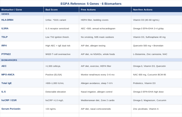
6 Biomarkers to Track in Eosinophilic Granulomatosis With Polyangiitis
Tracking the right biomarkers in EGPA is not just about confirming a diagnosis — it is about understanding which biological processes are most active in your particular case, anticipating flares before they become clinical emergencies, and giving your specialist the data they need to make more targeted decisions. These six markers have the strongest evidence for clinical utility in EGPA and the clearest relationship to patient outcomes.
Biomarker 1: Absolute Eosinophil Count (AEC)
Why it matters and what it reveals
The absolute eosinophil count is the most central laboratory marker in EGPA. An AEC above 1,500 cells per microliter is part of the core diagnostic criteria, and during active disease it commonly rises to 5,000–15,000 cells per microliter or higher. The AEC reflects the degree of eosinophil-driven tissue inflammation — and because eosinophils are cytotoxic when they infiltrate organs, especially the heart (eosinophilic myocarditis), the nerves, and the lungs, sustained elevation correlates directly with organ damage risk. Serial AEC tracking is one of the most reliable indicators of disease activity and treatment response. A falling AEC on therapy is reassuring; a rising AEC during a steroid taper is a clear early warning that the disease is not adequately controlled.
How to measure it
The AEC is derived from a standard complete blood count (CBC) with differential, one of the cheapest tests in medicine. Cost ranges from $15 to $50 in most settings. During active disease or dose changes, testing every 4–8 weeks is reasonable. In stable remission, quarterly monitoring is typical. Some specialized labs offer eosinophil activation markers (eosinophil peroxidase, eosinophil cationic protein), which add granularity at a higher cost of $100–250.
If the AEC is elevated, the plan without supplements
Non-pharmacological strategies focus on reducing upstream drivers of eosinophil production and recruitment. The Autoimmune Protocol (AIP) diet — eliminating grains, legumes, dairy, eggs, nightshades, nuts, and seeds for a minimum of 30–60 days — removes common immune triggers and reduces Th2-skewing dietary antigens. Avoiding NSAIDs is essential: ibuprofen and aspirin can trigger eosinophilic crises in susceptible individuals through the arachidonic acid pathway. Sleep of 7–9 hours per night significantly reduces Th2 cytokine dominance; even one week of shortened sleep measurably elevates IL-5 and IgE. Moderate aerobic exercise (30 minutes, 4 days per week) consistently reduces circulating eosinophil counts over weeks. Reducing mold and dust mite exposure through high-efficiency air filtration (HEPA) is particularly relevant because aeroallergens directly stimulate the epithelial TSLP pathway that initiates eosinophilic responses.
If the AEC is elevated, the plan with supplements or equipment
Omega-3 fatty acids (EPA + DHA): 3–4 grams per day of a high-EPA formula (EPA:DHA ratio of approximately 3:1). EPA competes with arachidonic acid in the cyclooxygenase and lipoxygenase pathways, shifting eicosanoid production away from pro-eosinophilic leukotrienes (LTC4, LTD4) toward less potent LTB5 metabolites. Research published in the Journal of Allergy and Clinical Immunology supports omega-3 effects on eosinophil biology. Frequency: daily, continuous. Reassess every 6 months. Side effects: fishy aftertaste (use enteric-coated), mild blood thinning (note for surgery), occasional GI upset.
Vitamin D3 with K2: 5,000 IU D3 paired with 100–200 mcg MK-7 K2 daily. Vitamin D3 promotes regulatory T cell (Treg) generation, which actively suppresses Th2 activity and eosinophil mobilization. Titrate to serum 25-OH-D3 of 40–60 ng/mL. Test every 6 months. Side effects: rare hypercalcemia at very high doses; K2 directs calcium appropriately, reducing arterial deposition risk.
Quercetin with bromelain: 500–1,000 mg quercetin with 200 mg bromelain (for absorption enhancement), taken away from meals. Quercetin inhibits mast cell degranulation and reduces eosinophil survival signals in vitro. Cycling: 8 weeks on, 2 weeks off. Side effects: occasional headache, possible mild CYP3A4 interaction (discuss with your physician if on ciclosporin or tacrolimus).
HEPA air purifier: A room-level HEPA unit (CADR ≥200) in the bedroom significantly reduces eosinophilic allergen burden during the 7–8 hours of overnight exposure. Single purchase, continuous use. This is not a supplement, but its impact on AEC reduction in allergic eosinophilic disease is underestimated.
Biomarker 2: MPO-ANCA (Myeloperoxidase Antineutrophil Cytoplasmic Antibody)
Why it matters and what it reveals
Approximately 40% of EGPA patients are ANCA-positive, and in most cases this means positivity for MPO-ANCA (producing a perinuclear or p-ANCA immunofluorescence pattern). ANCA positivity is not just a diagnostic detail — it defines a distinct clinical subtype. ANCA-positive patients are at higher risk for glomerulonephritis, mononeuritis multiplex (nerve injury from vasculitis), and higher relapse rates on corticosteroids alone. ANCA-negative patients tend to have more prominent eosinophilic tissue infiltration, particularly in the heart and lungs, without the glomerular injury pattern. Knowing your ANCA status fundamentally changes which organs to monitor most carefully and what treatment escalation may be required. The landmark mepolizumab trial in EGPA (Wechsler et al., NEJM 2017) showed differential responses based on ANCA status, further validating its clinical subtype relevance.
How to measure it
Testing is done via ELISA for MPO-ANCA specifically (not just general ANCA immunofluorescence, which is less precise). Cost ranges from $150 to $400. Testing at diagnosis and at least annually in remission is standard. ANCA titers in EGPA do not correlate as reliably with disease activity as they do in granulomatosis with polyangiitis (GPA) — a negative ANCA does not rule out relapse — but a rising titer in a previously positive patient warrants clinical attention.
If ANCA-positive, the plan without supplements
ANCA-positive status requires expanding the monitoring agenda. Urinalysis with microscopy and serum creatinine should be checked every 3–6 months to catch glomerulonephritis early. Neurological assessments looking for new sensory or motor deficits in distal limbs should be part of every rheumatology visit. Renal biopsy may eventually be needed if proteinuria or hematuria emerges. In practice, ANCA-positive EGPA often requires immunosuppressive therapy beyond corticosteroids alone — rituximab and cyclophosphamide are used in refractory vasculitic cases.
If ANCA-positive, the plan with supplements or equipment
NAC (N-Acetylcysteine): 600 mg twice daily. NAC is a glutathione precursor with antioxidant and anti-inflammatory properties. ANCA-mediated vascular injury involves significant oxidative stress from activated neutrophils, and NAC helps buffer this. Cycling: 8 weeks on, 4 weeks off. Side effects: sulfurous odor, mild GI upset, potential interaction with nitroglycerin.
Curcumin (BCM-95 or Meriva formulation): 500 mg twice daily. Curcumin inhibits NF-κB, the transcription factor central to neutrophil and inflammatory cell activation. Use bioavailable formulations only; standard curcumin has poor absorption. Cycling: continuous with 2-week breaks every 3 months. Important: discuss with your physician before combining with immunosuppressants or anticoagulants, as interactions are possible.
Biomarker 3: Total IgE
Why it matters and what it reveals
Total IgE is almost universally elevated in EGPA, often dramatically so — values of 1,000–5,000 IU/mL are common, and in heavily atopic patients even higher. This reflects the underlying Th2 immune dominance that characterizes EGPA: the same skewing that drives eosinophil overproduction also activates mast cells via IgE-receptor crosslinking, worsening allergic inflammation in the airways and sinuses. The degree of IgE elevation broadly correlates with atopic comorbidity burden (allergic rhinitis, food sensitivities, eczema). Monitoring total IgE over time gives a sense of how successfully Th2 activity is being reduced. Very high IgE (above 5,000 IU/mL) may also indicate candidacy for omalizumab (anti-IgE), though its role in EGPA is less established than mepolizumab.
How to measure it
Total IgE is a simple serum immunoassay available at virtually any lab. Cost: $30–80. Testing at baseline and then every 6–12 months provides useful trend data. Specific IgE panels (food and inhalant) can be added for $150–400 and help identify specific triggers driving the atopic burden.
If total IgE is elevated, the plan without supplements
Identifying and reducing allergen exposures is the most direct non-pharmacological lever. Comprehensive food sensitivity testing followed by a structured elimination and reintroduction protocol can meaningfully reduce overall IgE burden over months. Aggressive environmental allergen control (dust mites, pet dander, mold) through mattress encasements, HEPA filtration, and humidity control below 50% reduces chronic antigenic stimulation. Saline nasal irrigation twice daily removes deposited aeroallergens from the sinonasal mucosa before they can drive further Th2 activation. Sleep duration optimization and circadian alignment (consistent sleep-wake times, minimal blue light after dark) reduce Th2 cytokine bias.
If total IgE is elevated, the plan with supplements or equipment
Targeted probiotics: Specific strains including Lactobacillus rhamnosus GG and Bifidobacterium longum have shown capacity to modulate Th2 responses and reduce atopic sensitization in clinical studies. Dose: 20–50 billion CFU daily. Cycling: 12 weeks continuous, then reassess at 6-month intervals. Side effects: initial bloating (usually resolves in 1–2 weeks), rare systemic infection in severely immunocompromised individuals.
Vitamin D3: As described above, titrating to 40–60 ng/mL 25-OH-D3. VD3 specifically suppresses IgE class switching in B cells by reducing IL-4-driven activation. This is one of the strongest lifestyle-medicine interventions available for Th2 rebalancing.
Quercetin + Bromelain: Same protocol as listed under AEC. The combination also acts as a natural mast cell stabilizer, reducing IgE-triggered histamine and eosinophil-activating factor release.
Biomarker 4: Interleukin-5 (IL-5)
Why it matters and what it reveals
IL-5 is the master cytokine of eosinophil biology. It drives eosinophil differentiation in bone marrow, prolongs eosinophil survival in blood and tissues, and primes eosinophils for activation and degranulation. In EGPA, the entire cascade of eosinophilic inflammation is upstream-regulated by IL-5 — which is precisely why mepolizumab (a monoclonal antibody against IL-5) is approved for EGPA and consistently reduces relapse rates. The mepolizumab EGPA trial demonstrated significant reduction in relapse risk and glucocorticoid dose. When serum IL-5 is measurable and elevated, it confirms that the eosinophilic axis is actively driven — and it provides biological justification for anti-IL-5 therapy.
How to measure it
Serum IL-5 is measured by ELISA but is not yet part of routine clinical panels in most hospitals. Availability is higher in academic medical centers and some specialized immunology labs. Cost: $150–350. Because IL-5 levels are typically very low in healthy individuals and can fluctuate rapidly, the result should be interpreted alongside AEC and clinical status. Some clinical labs offer multiplex cytokine panels ($400–800) that measure IL-5 alongside IL-4, IL-13, and other Th2 markers simultaneously, providing richer context.
If IL-5 is elevated, the plan without supplements
No lifestyle intervention will eliminate IL-5 elevation in active EGPA — this is a medical situation requiring physician management, potentially including biologic escalation. What lifestyle can do is reduce the upstream Th2 signals (epithelial cytokines TSLP, IL-25, IL-33) that drive IL-5 production from Th2 lymphocytes and innate lymphoid cells type 2 (ILC2s). Reducing allergen burden, treating sinonasal disease aggressively (with nasal corticosteroids and saline irrigation), and removing occupational exposures to dust, fumes, and chemical irritants all reduce the epithelial activation that sustains IL-5 output.
If IL-5 is elevated, the plan with supplements or equipment
Omega-3 EPA+DHA: High-dose EPA has shown capacity to reduce IL-5 production in allergic and eosinophilic models by shifting prostaglandin E1/E2 balance and influencing Th2 differentiation. Protocol same as under AEC. This supports but does not replace medical management.
Nasal saline irrigation device (high-volume, low-pressure): Neti pots or Neil Med bottles used twice daily significantly reduce the sinonasal inflammation that drives upstream TSLP, which is one of the primary activators of ILC2 cells (the innate source of IL-5). This is one of the most underutilized mechanical interventions in EGPA sinonasal management. Cost: $15–25 plus saline packets. No significant side effects.
Biomarker 5: High-Sensitivity CRP and ESR
Why it matters and what it reveals
C-reactive protein (measured as high-sensitivity CRP or hsCRP) and erythrocyte sedimentation rate (ESR) are nonspecific but sensitive markers of systemic inflammation. In the context of EGPA, they reflect the intensity of the vasculitic and granulomatous inflammatory process — particularly during the vasculitic phase when small vessel inflammation drives most of the organ damage. hsCRP tends to correlate better with acute-phase inflammation and responds more quickly to treatment changes; ESR integrates over a longer period and reflects fibrinogen elevation. Peter Attia, in his clinical work on longevity and disease prevention, consistently emphasizes hsCRP as one of the most clinically actionable inflammatory biomarkers — ideally below 0.5 mg/L for long-term health, and certainly below 1.0 mg/L. In active EGPA vasculitis, CRP can rise above 50 mg/L.
How to measure it
Both are inexpensive and universally available. hsCRP: $20–50 (slightly more than standard CRP, more sensitive at low ranges). ESR: $10–25. CBC. For EGPA monitoring, testing at every clinical visit during active disease, and every 3–6 months in remission, is reasonable. A rising hsCRP during steroid taper — even before eosinophil count rises — may be the earliest laboratory sign of an impending relapse.
If CRP or ESR is elevated, the plan without supplements
Anti-inflammatory dietary approaches have the most consistent evidence. The Mediterranean diet, or the more aggressive AIP variant for autoimmune conditions, consistently reduces hsCRP over 8–12 weeks. Moderate aerobic exercise (zone 2 cardio: 30–45 minutes at conversational pace, 4 days per week) reduces hsCRP through IL-6 myokine release and adipose tissue reduction. Weight optimization matters here: visceral adipose tissue is a major source of IL-6, which drives CRP synthesis in the liver. Sleep of 7–9 hours with consistent timing reduces overnight IL-6 elevation. Stress management (see MBSR section below) directly reduces cortisol, which at chronic levels paradoxically drives NF-κB-mediated inflammation.
If CRP or ESR is elevated, the plan with supplements or equipment
Omega-3 EPA+DHA: 2–4 g/day as above. Multiple meta-analyses confirm reduction in circulating CRP with omega-3 supplementation, particularly at doses above 2 g EPA per day.
Magnesium glycinate: 300–400 mg before sleep. Magnesium deficiency (highly prevalent) is associated with elevated NF-κB activity and CRP. The glycinate form is best tolerated. Cycling: continuous. Side effects: loose stools if dose is too high; reduce dose if this occurs.
Curcumin (bioavailable form): 500 mg twice daily of BCM-95 or Meriva formulation. NF-κB inhibition reduces IL-6 production and downstream CRP synthesis. Multiple randomized controlled trials support CRP reduction with bioavailable curcumin. Side effects and interactions as noted above.
Resveratrol: 250–500 mg/day with a fat-containing meal. Activates SIRT1 and reduces NF-κB signaling. Cycling: 12 weeks on, 4 weeks off. Side effects: generally mild; avoid with anticoagulants.
Biomarker 6: Serum Periostin
Why it matters and what it reveals
Periostin is an extracellular matrix protein secreted by epithelial and stromal cells in response to IL-4 and IL-13 — the two effector cytokines of Th2 inflammation. Elevated serum periostin indicates that IL-4/IL-13 signaling is active in airway epithelium, which corresponds to the eosinophilic airway component of EGPA. While periostin has been most studied in eosinophilic asthma, where it predicts steroid response and biologic candidacy, its relevance extends to EGPA because the pulmonary component of this disease is fundamentally Th2-driven. High periostin patients may be particularly good candidates for dupilumab (anti-IL-4Rα, blocking both IL-4 and IL-13 signaling), a biologic gaining traction in severe eosinophilic asthma with vasculitic features. Tracking periostin alongside AEC and IgE builds a more complete Th2 immune profile.
How to measure it
Serum periostin ELISA is available in specialized labs and some academic medical centers, though it is not yet a standard clinical panel item in most community settings. Cost: $150–400. Normal is typically below 23 ng/mL (lab-specific cutoffs apply). Because periostin reflects longer-term Th2 activity rather than acute flares, testing every 6–12 months gives meaningful trend data.
If periostin is elevated, the plan without supplements
Periostin elevation signals that IL-4 and IL-13 are driving ongoing epithelial activation. Reducing the upstream sources — allergen exposure, chronic sinonasal inflammation, gut permeability-driven immune activation — is the structural approach. Implementing a comprehensive AIP diet (addressing gut permeability), resolving chronic sinusitis with nasal corticosteroids and irrigation, and aggressive allergen avoidance all target the upstream drivers. Nasal corticosteroid sprays specifically suppress IL-4 and IL-13 production in the nasal mucosa, which is one of the primary sites driving periostin release.
If periostin is elevated, the plan with supplements or equipment
Zinc picolinate: 25–30 mg with food. Zinc is a key co-factor for immune regulatory signaling, promotes Th1/Th2 balance, and supports epithelial barrier integrity. Cycling: 12 weeks on, 4 weeks off (take with 2 mg copper if used long-term to prevent copper depletion). Side effects: nausea if taken on empty stomach.
Vitamin A from food sources: Retinol from liver (beef or chicken liver, 1–2 servings weekly) or vitamin A as retinyl palmitate (2,000–5,000 IU/day, not beta-carotene) supports mucosal immune function, promotes Treg differentiation, and reduces Th2 polarization in mucosal tissues. Side effects: avoid high-dose retinol in pregnancy; headache and skin dryness at very high doses.
Targeted probiotic therapy: As noted above, with specific emphasis on gut microbiome optimization to reduce the gut-to-airway Th2 crosstalk that sustains periostin elevation. Strains with evidence in atopic disease: L. rhamnosus GG, B. infantis.
With these six biomarkers in view, you move from reacting to disease flares to anticipating and interpreting them. The next layer of understanding is genetic — knowing not just what the immune system is doing, but why it is wired to do it in the first place.
What Genetics Research Reveals About EGPA
Genetic research in EGPA is younger and less mature than in more common autoimmune diseases like rheumatoid arthritis or lupus — but it is progressing rapidly. Genome-wide association studies have begun to identify specific loci that reliably confer susceptibility, and understanding these genes helps explain why EGPA often runs in families with strong atopic backgrounds, why certain patients are more vasculitic while others are more eosinophilic, and which immune pathways are structurally biased in affected individuals. Genetic knowledge does not predict whether you will have a flare next month, but it reveals the biological terrain you are working with and informs which compensatory strategies are most relevant.
Gene 1: HLA-DRB4
What it is and why it matters
The HLA-DRB4 allele, particularly the *0101 variant, represents the strongest genetic association with EGPA identified to date. HLA-DR molecules sit on the surface of antigen-presenting cells and control which molecular fragments (peptides) are presented to CD4+ T cells. Specific HLA-DRB4 variants appear to favor presentation of aeroallergen and autoantigenic peptides in ways that bias the immune system toward Th2 differentiation rather than Th1 tolerance — essentially making the immune system more likely to overreact to the environmental antigens that trigger EGPA flares. HLA-DRB4 association is established through GWAS data in European populations, though the functional consequences of the specific variant require continued study.
If HLA-DRB4 is an unfavorable variant, the plan without supplements
You cannot change the HLA variant, but you can manage the antigen load that exploits it. This means more aggressive environmental allergen reduction than the average asthma patient would pursue: HEPA filtration throughout the home, dust mite encasements on all bedding, no carpets in sleeping areas, humidity control below 50%, avoidance of known sensitizing occupational exposures (spray paints, rubber dust, hairdresser chemicals — all associated with EGPA triggers). Nasal saline irrigation twice daily physically removes deposited allergens before they engage antigen-presenting cells. Annual comprehensive allergen testing (inhalant and food) with a specialist is a reasonable monitoring strategy.
If HLA-DRB4 is an unfavorable variant, the plan with supplements or equipment
Vitamin D3 (titrated to 40–60 ng/mL): VD3 modulates the activity of dendritic cells — the primary antigen-presenting cells — and reduces the Th2-skewing antigen presentations that HLA-DRB4 variants facilitate. This is one of the biologically most coherent supplement strategies for HLA-related Th2 bias. Protocol and side effects as above. Reassess serum levels every 6 months.
HEPA air purifier and allergen-impermeable mattress cover (equipment): These mechanical tools reduce the antigen load that HLA-DRB4 would otherwise present inappropriately. No cycling required; these are environmental modifications, not cyclical interventions.
Gene 2: IL5RA (Interleukin-5 Receptor Alpha)
What it is and why it matters
IL5RA encodes the alpha subunit of the IL-5 receptor, expressed on eosinophil precursors and mature eosinophils. Variants in IL5RA affect how sensitively eosinophils respond to circulating IL-5 signals — and higher receptor sensitivity means more robust eosinophil survival and activation even at relatively low IL-5 concentrations. This partially explains why some patients have dramatic eosinophilia with modest IL-5 levels while others tolerate higher IL-5 without the same degree of tissue infiltration. IL5RA variants are directly relevant to treatment: benralizumab (Fasenra) targets the IL-5 receptor alpha directly (rather than IL-5 itself), depleting eosinophils through antibody-dependent cell-mediated cytotoxicity. A favorable IL5RA variant makes you a theoretically stronger candidate for benralizumab.
If IL5RA shows an unfavorable variant, the plan without supplements
The most important non-pharmacological response is to maintain extremely tight control of AEC. Because eosinophils are more receptor-primed in this genetic context, the threshold for tissue damage may be lower — meaning the target AEC in remission should be as close to normal (below 500 cells/μL) as possible. Clinical vigilance around cardiac monitoring (annual echocardiography, troponin levels) is particularly warranted in patients with this variant and a history of eosinophilia.
If IL5RA shows an unfavorable variant, the plan with supplements or equipment
Omega-3 EPA+DHA (high dose, 3–4 g/day): EPA reduces eosinophil IL-5 receptor signaling efficiency in eosinophilic models by altering lipid raft composition in the eosinophil membrane. This is modest but mechanistically coherent and aligns with the broader anti-eosinophilic evidence for omega-3. Protocol as above.
Discuss the IL5RA genetic finding with your rheumatologist or allergist. This is clinically relevant information that may justify earlier transition to IL-5 pathway biologic therapy.
Gene 3: TSLP (Thymic Stromal Lymphopoietin)
What it is and why it matters
TSLP is not itself an eosinophil gene — it is an epithelial alarmin, released from airway and gut epithelial cells in response to mechanical injury, allergens, pollutants, and infectious agents. But TSLP is the master initiator of the Th2 cascade: it activates dendritic cells to program naïve T cells toward Th2 differentiation, and it directly activates ILC2 cells to release IL-5 and IL-13. Variants in the TSLP gene that increase expression or release in response to environmental triggers have been associated with atopic disease and eosinophilic airway conditions. In EGPA, TSLP represents the upstream starting point of the pathological immune cascade — meaning genetic TSLP variants set a lower ignition threshold for the entire disease process.
If TSLP is an unfavorable variant, the plan without supplements
Reducing the environmental triggers that activate TSLP in epithelial cells is the most direct strategy. The most important triggers are: cigarette smoke and second-hand smoke (the single largest activator of airway TSLP), air pollution and diesel particulate exposure, household chemical exposures (cleaning products, off-gassing furniture), and certain occupational dusts. An N95-rated face mask for high-pollution environments is a rational mechanical intervention. Strict no-smoking household policy is non-negotiable. Air purification and mold control address the remainder of the indoor burden.
If TSLP is an unfavorable variant, the plan with supplements or equipment
Vitamin D3 (40–60 ng/mL titration): Vitamin D3 downregulates TSLP expression in bronchial epithelial cells in vitro and in clinical studies of atopic asthma. This makes VD3 particularly relevant for the TSLP-variant profile. Same protocol as above.
Sulforaphane (from broccoli sprouts or standardized supplement): 40–60 mg/day of sulforaphane equivalent. Sulforaphane activates the NRF2 pathway in epithelial cells, reducing oxidative-stress-mediated TSLP release. Clinical studies in allergic airway disease support airway anti-inflammatory effects of sulforaphane. Cycling: 8 weeks on, 2 weeks off. Side effects: sulfurous odor, mild GI upset. Use fresh broccoli sprouts (50–100g daily) as an affordable food-based source if supplements are cost-prohibitive.
Gene 4: IRF4 (Interferon Regulatory Factor 4)
What it is and why it matters
IRF4 is a transcription factor expressed in immune cells including eosinophils, Th2 lymphocytes, and plasma cells. It controls eosinophil differentiation in the bone marrow and is essential for Th2 effector cell programming. IRF4 variants have been identified in GWAS studies of EGPA (as noted in the expanding literature from the European Vasculitis Consortium), and because IRF4 also regulates IgE class switching in B cells, unfavorable variants simultaneously raise eosinophilic and atopic risk. This dual role makes IRF4 variants particularly impactful: they can simultaneously amplify eosinophil output and IgE production, explaining the combination of very high eosinophil counts with very high IgE in some EGPA patients.
If IRF4 shows an unfavorable variant, the plan without supplements
The most rational response to an IRF4 variant is monitoring both AEC and total IgE with higher frequency and lower thresholds for clinical concern. Given IRF4's role in both eosinophil and B cell IgE signaling, this genetic profile argues for particularly aggressive allergen burden reduction (targeting both Th2 T cell activation and B cell IgE class switching). A comprehensive food elimination protocol and environmental allergen testing are worth prioritizing early.
If IRF4 shows an unfavorable variant, the plan with supplements or equipment
Quercetin + Bromelain (as above): Quercetin's mechanism includes interference with STAT6 signaling downstream of IL-4, which partly intersects with IRF4-driven B cell activation. This is not a gene-targeted therapy, but it is a mechanistically coherent support. Protocol as above.
Dietary flavonoids broadly: Foods rich in flavonoids (berries, dark leafy greens, onions, green tea) provide diverse polyphenol support for Th2 modulation at low cost. No specific cycling required; incorporate as permanent dietary staples.
Gene 5: PTPN22 (Protein Tyrosine Phosphatase Non-Receptor Type 22)
What it is and why it matters
PTPN22 encodes a phosphatase that regulates T cell and B cell receptor signaling thresholds. The well-studied W620 variant (rs2476601) reduces the inhibitory signal that normally prevents overly reactive T cell responses. As a result, T cells carrying this variant activate more readily to both self-antigens and environmental antigens. PTPN22 W620 is one of the most broadly shared genetic risk variants across autoimmune diseases — it appears in rheumatoid arthritis, lupus, type 1 diabetes, Hashimoto's thyroiditis, and is also seen in ANCA-associated vasculitides. Its presence in EGPA adds to the understanding of why some individuals develop autoimmune vasculitis rather than atopic disease alone. Importantly, this variant also impairs the ability of the immune system to properly regulate itself after an inflammatory challenge, contributing to disease persistence and difficulty achieving durable remission.
If PTPN22 shows the W620 variant, the plan without supplements
The key lifestyle strategy is gut permeability management — because a permeable gut epithelium continuously supplies microbial and food antigens that an already low-threshold T cell population will respond to inappropriately. This means: removing common gut-disruptive foods (alcohol, gluten, seed oils, ultra-processed foods), eating a whole-foods diet with adequate dietary fiber, avoiding prolonged NSAID use (destroys gut mucosa), and managing physical and psychological stress (cortisol acutely increases gut permeability). In practical terms, this is the AIP elimination protocol, which directly targets gut permeability as a driver of autoimmune T cell activation.
If PTPN22 shows the W620 variant, the plan with supplements or equipment
L-Glutamine: 5–10 g/day in divided doses on an empty stomach. Glutamine is the primary fuel for intestinal enterocytes and is the most evidence-backed supplement for gut barrier restoration. Cycling: 8–12 weeks on, reassess. Side effects: generally excellent tolerability; avoid in hepatic encephalopathy.
Zinc carnosine: 75 mg twice daily. Specifically supports gastric and intestinal mucosal integrity through zinc's role in tight junction protein stabilization. Cycling: 12 weeks. Side effects: mild nausea; take with food.
NAC (N-Acetylcysteine): 600 mg twice daily. Beyond its antioxidant role, NAC supports mucosal glutathione production and reduces the oxidative environment that worsens gut permeability and T cell hyperreactivity. Protocol as above.
Saccharomyces boulardii: 250–500 mg twice daily (5–10 billion CFU). This probiotic yeast specifically supports intestinal tight junction proteins and has been studied in intestinal permeability conditions. Cycling: 8 weeks on, reassess. Side effects: bloating initially; do not use in severely immunocompromised individuals.
Understanding your genetic architecture does not produce immediate treatment changes, but it prioritizes your efforts — knowing which pathways are structurally vulnerable helps you focus the strategies above where they will have the most impact. The following sections offer additional approaches that operate at the intersection of biology and behavior.
What the Huberman Lab Reveals About Managing Immune Dysregulation
The Huberman Lab podcast, hosted by Stanford neuroscientist Andrew Huberman, has produced some of the most widely cited science-communication content on immune regulation, stress biology, and the gut-brain-immune axis. For people with autoimmune and eosinophilic conditions like EGPA, several episodes and research summaries stand out as genuinely framework-shifting. Below are the ten most impactful insights from this body of work for someone managing EGPA.
1. The Gut Is the Immune System's Headquarters
Seventy to eighty percent of immune cells reside in gut-associated lymphoid tissue (GALT). Chronic gut permeability — driven by diet, stress, alcohol, NSAIDs, and dysbiosis — exposes these cells to a relentless stream of microbial antigens, sustaining systemic Th2 and Th17 activation. Healing gut permeability is not a peripheral strategy; it is central to immune recalibration.
2. Chronic Stress Dysregulates the Immune Axis in Non-Obvious Ways
Short-term cortisol is anti-inflammatory. But chronic cortisol — the low-grade, sustained kind from ongoing psychological stress — paradoxically drives NF-κB upregulation, IL-6 elevation, and Th2 skewing. This means stress is not just "bad in general" — it actively worsens the exact immune bias that drives EGPA. Cortisol management is immune management.
3. Sleep Is the Most Powerful Immune Reset Tool Available
Deep non-REM sleep is when the glymphatic system clears cellular waste and when the immune system conducts its most active anti-inflammatory housekeeping. Even a single night of less than six hours raises IL-6, TNF-alpha, and — critically for EGPA — IgE. Consistent 7–9 hour sleep with regular timing is the closest thing to a free immune drug available.
4. Morning Sunlight Exposure Sets the Immune Clock
The immune system operates on circadian time. Natural killer cells, T regulatory cells, and eosinophils themselves follow diurnal patterns. Morning light exposure (10–15 minutes within 30–60 minutes of waking, without sunglasses) sets the circadian rhythm that governs these immune oscillations. Disrupted circadian alignment — common in shift workers and people with irregular sleep — desynchronizes immune cell activity and reduces immune tolerance.
5. Nasal Breathing Produces Nitric Oxide — and That Matters for Airway Inflammation
Nasal breathing generates nitric oxide (NO) in the paranasal sinuses. NO has direct antiviral, antibacterial, and bronchodilatory effects. Chronic mouth breathing — common in EGPA patients with severe sinonasal disease — bypasses this first line of airway defense and may worsen airway eosinophilic inflammation. Training nasal breathing during the day (and using gentle nasal strips at night if needed) is a low-cost, no-risk intervention.
6. Fermented Foods Outperform High-Fiber Foods for Reducing Systemic Inflammatory Markers
A landmark study from the Sonnenburg lab at Stanford demonstrated that a diet high in fermented foods (yogurt, kefir, kimchi, sauerkraut, kombucha) significantly reduced 19 inflammatory markers — including IL-6 and IL-12 — over a 10-week period, outperforming a high-fiber diet alone. For EGPA patients working to reduce systemic Th2 and vasculitic inflammation, adding 1–2 servings of fermented foods daily is a concrete, evidence-based strategy.
7. Zone 2 Cardio Is the Anti-Inflammatory Exercise Dose
Huberman, drawing on work by Iñigo San Millán and Peter Attia, emphasizes that 150–180 minutes per week of zone 2 aerobic exercise (at a pace where conversation is still possible) is the most potent exercise-based anti-inflammatory protocol. At this intensity, muscle-derived IL-6 acts as an anti-inflammatory myokine — not a pro-inflammatory signal — and consistently reduces hsCRP, IL-1β, and TNF-alpha over 8–12 weeks.
8. Cold Exposure Has Measurable Anti-Inflammatory and Eosinophil-Modulating Effects
Deliberate cold exposure (cold showers 2–3 minutes, or cold water immersion 10–15 minutes at 10–15°C) triggers catecholamine release — epinephrine and norepinephrine — which have direct anti-inflammatory actions. Huberman notes that norepinephrine release from cold exposure is dose-dependent and cumulative. For EGPA patients with stable disease, starting with brief cold showers 3–4 times per week is a reasonable protocol; this is not recommended during acute vasculitic flares.
9. Physiological Sighs Rapidly Reduce Cortisol and Slow Inflammatory Signaling
The physiological sigh — a double nasal inhale followed by a long exhale — is the fastest evidence-based technique for real-time stress and cortisol reduction. Huberman and colleagues have published on this technique's capacity to shift the autonomic nervous system from sympathetic to parasympathetic in under 30 seconds. For EGPA patients, incorporating 5–10 physiological sighs before stressful events is a practical immune-supportive tool with no risk or cost.
10. Social Connection Is a Biological Anti-Inflammatory Signal
Loneliness and social isolation reliably elevate NF-κB-mediated gene expression and systemic inflammatory markers. Regular meaningful social connection — not just digital contact — activates oxytocin and serotonin pathways that directly reduce inflammatory signaling. For people managing a chronic disease like EGPA, which often involves fatigue, activity limitations, and psychological burden, prioritizing social engagement is not soft advice — it is a measurable immune intervention.
Complementary and Integrative Approaches With Evidence in Autoimmune and Eosinophilic Disease
The following modalities have meaningful human clinical evidence in autoimmune, eosinophilic, or vasculitic contexts. None replaces specialist medical care, but each represents a rational add-on strategy with a favorable risk-benefit profile.
The Autoimmune Protocol (AIP) — Sarah Ballantyne
The Autoimmune Protocol, detailed in Sarah Ballantyne's book The Paleo Approach, is a structured elimination and reintroduction diet designed specifically to reduce gut permeability, immune activation, and systemic inflammation in autoimmune conditions. It removes grains, legumes, dairy, eggs, nightshades, nuts, seeds, alcohol, and refined sugars — the foods most likely to disrupt gut barrier function and drive T cell activation — while emphasizing nutrient-dense animal proteins, organ meats, bone broth, leafy vegetables, and fermented foods. The rationale is grounded in the gut-immune connection: resolving gut permeability removes the constant antigenic stimulation that sustains autoimmune T cell responses.
A pilot study published in Inflammatory Bowel Diseases (2017) found significant clinical remission in Crohn's and ulcerative colitis patients following the AIP, with histological evidence of improved gut mucosal integrity. While direct EGPA trials do not exist, the immune mechanisms overlap substantially: gut permeability → Th2/Th17 activation → eosinophilic and vasculitic downstream effects.
Practically, implement the AIP for a minimum of 60 days before reintroducing foods systematically, one at a time, every 5–7 days, monitoring for symptom or laboratory changes. Work with a registered dietitian experienced in elimination protocols to ensure nutritional adequacy. The most common mistake is abandoning the protocol at week two when initial detox symptoms (fatigue, irritability) are misinterpreted as reactions rather than transitional effects.
Breathing-Based Therapies
Breathing exercises occupy a uniquely relevant position in EGPA because the condition affects the airways directly. Techniques such as the Buteyko method (focusing on nasal breathing and reduced breathing volume), diaphragmatic breathing training, and slow-paced breathing (5–6 breaths per minute) each address different aspects of respiratory function. Slow breathing activates the vagus nerve and promotes parasympathetic dominance, which reduces mast cell activation and allergic airway reactivity. Nasal breathing during rest and light exercise significantly increases nitric oxide production in the paranasal sinuses, providing local antimicrobial and bronchodilatory effects.
A randomized controlled trial published in Thorax (2006) found that Buteyko breathing technique significantly reduced bronchodilator use and improved asthma-related quality of life in adults with asthma. EGPA's asthma component — often severe and difficult to control — is a directly relevant clinical target. The physiological sigh protocol described in Huberman's work (double inhale, long exhale) provides a rapid vagal activation tool usable during stress episodes.
Practically, start with 10 minutes of diaphragmatic breathing twice daily — lying supine, hand on belly, breathing so the belly rises before the chest. After two weeks, introduce slow-paced breathing at 5–6 cycles per minute (5 seconds in, 5 seconds out). During EGPA asthma exacerbations, do not attempt breath-holding techniques; focus only on slow, relaxed nasal exhalation. Buteyko instruction from a trained practitioner is worth the investment for sustained technique learning.
Mindfulness-Based Stress Reduction (MBSR)
MBSR, the 8-week protocol developed by Jon Kabat-Zinn at the University of Massachusetts, has accumulated substantial evidence for reducing inflammatory markers and improving quality of life in people with chronic immune and inflammatory conditions. Its primary mechanism in autoimmune disease is cortisol normalization and vagal tone enhancement — both of which reduce NF-κB-driven inflammation and Th2 immune skewing. For EGPA patients, the psychological burden of managing a rare, relapsing vasculitis (uncertainty about organ involvement, fear of steroid side effects, disrupted work and social life) is substantial and itself a driver of immune dysregulation.
A meta-analysis by Grossman et al. in the Journal of Psychosomatic Research (2004) found significant improvements in physical and psychological wellbeing across a range of medical conditions. A more recent randomized trial in autoimmune disease contexts demonstrated reductions in IL-6 and CRP following 8-week MBSR protocols. The evidence for direct eosinophil count reduction through MBSR is not established, but the downstream inflammatory pathway modulation is well-documented.
Practically, access the MBSR protocol through an in-person 8-week course (offered at many hospitals and wellness centers) or through validated online platforms. The commitment is approximately 45 minutes of daily practice during the 8-week period. The formal protocol is more effective than informal mindfulness app use, primarily because the structured body scan and mindful movement components specifically target the somatic stress patterns that EGPA patients often carry in posture and breathing patterns.
Saline Nasal Irrigation
Sinonasal involvement — chronic sinusitis, nasal polyposis, rhinitis — is present in 70–90% of EGPA patients and is often the first manifestation of the disease, preceding vasculitic complications by years. Saline nasal irrigation using high-volume, low-pressure devices (Neil Med Sinus Rinse, neti pot) is among the most evidence-supported non-pharmacological interventions in sinonasal disease. It physically removes deposited allergens, inflammatory mediators, eosinophilic debris, and mucus from the nasal cavity before they can perpetuate mucosal immune activation.
A Cochrane-referenced systematic review published in the American Journal of Rhinology (2007) confirmed that daily saline nasal irrigation reduces symptoms, decreases medication use, and improves quality of life in chronic rhinosinusitis — the dominant sinonasal presentation in EGPA. Some protocols add a small amount of sodium bicarbonate to the saline solution to improve mucociliary clearance. In EGPA patients on nasal corticosteroid sprays, irrigating before applying the spray significantly improves medication contact with the nasal mucosa.
Practically, use isotonic or slightly hypertonic saline (the latter is more effective for thick mucus) once in the morning and once before sleep, every day. Use distilled, sterile, or previously boiled water — never tap water directly (risk of rare but serious amoeba exposure). The technique requires positioning the head at an appropriate angle to allow flow through one nostril and out the other. Most patients adapt within 3–5 days and report immediate post-irrigation relief of nasal pressure and congestion.
Microbiome-Directed Therapies
The emerging science of the gut-immune axis is directly relevant to EGPA. In Th2-driven eosinophilic conditions, gut dysbiosis — reduced microbial diversity, depletion of short-chain fatty acid (SCFA) producers like Faecalibacterium prausnitzii and Bifidobacterium species — promotes gut permeability and reduces the regulatory immune signals (IL-10, Treg induction via butyrate) that normally suppress Th2 excess. Conversely, a diverse, fiber-rich microbiome produces butyrate and propionate, which directly promote Treg differentiation and suppress IgE and eosinophil output from bone marrow.
Research published in Cell (2021) demonstrated that a fermented food diet reduced circulating inflammatory markers significantly more than a high-fiber diet alone, while increasing microbiome diversity. This specific finding has direct implications for EGPA management, as both IL-6 reduction and diversity increase are meaningful targets. Targeted interventions — high-dose multi-strain probiotics, prebiotic fiber (inulin, FOS, resistant starch), and fermented foods — work synergistically to shift the gut immune environment.
Practically, the most evidence-supported protocol combines three elements: 1–2 daily servings of fermented foods (kefir, live-culture yogurt, kimchi, or sauerkraut — not pasteurized), a high-fiber plant-diverse diet (aiming for 30+ different plant foods per week), and a targeted probiotic supplement (20–50 billion CFU daily of mixed strains including L. rhamnosus, B. longum, and L. plantarum). Introduce changes gradually — rapid fiber increases in someone with existing gut inflammation can temporarily worsen symptoms. Stool microbiome testing ($100–300 via direct-to-consumer services) can help track microbiome diversity over a 6–12 month protocol.
Conclusion
EGPA is a condition that punishes passivity and rewards precision. Knowing that your AEC is elevated tells you less than knowing why it is elevated, which subtype you belong to, which genetic vulnerabilities are structurally active, and which lifestyle and therapeutic levers are most aligned with your specific immune biology. The six biomarkers and five genetic factors covered in this article are not academic curiosities — they are the signals that differentiate a patient who is managed from a patient who understands their disease.
Start with what is accessible: a CBC with differential, MPO-ANCA, total IgE, and hsCRP are all standard lab tests available at low cost. Bring the trend data to your next rheumatology or immunology appointment. Add lifestyle strategies systematically rather than all at once — one dietary change, one supplement, one breathing practice — and give each at least 8–12 weeks before assessing impact on lab values. If genetic testing interests you, direct-to-consumer panels (with physician interpretation) or clinical GWAS-informed testing can provide relevant insight into HLA and immune-pathway variants. None of this replaces the specialist care that EGPA demands, but all of it can make that care smarter and more targeted. That is the goal worth working toward.
Neurological: Nerve Conditions
Cardiovascular: Heart Conditions Vascular Conditions
Respiratory: Lung Conditions Allergic Respiratory Conditions
Autoimmune: Inflammatory Conditions
Ear, Nose & Throat: Nose & Sinus Conditions
Urological: Kidney Conditions