This article contains AI generated content.
Knee Lymphatic Hygroma: 5 Genes and 7 Biomarkers to Track
Introduction
A lymphatic malformation near the knee doesn't behave like a bruise or a cyst that quietly resolves. It swells with activity, sometimes flares after a minor infection, and rarely follows the tidy timeline that a general pediatric or orthopedic handout describes. If you've been told to "just monitor it" or given a pamphlet written for lymphedema in general, you've probably noticed it doesn't quite fit what you're seeing in front of you.
That mismatch isn't your imagination. Lymphatic hygromas (also called macrocystic or mixed lymphatic malformations) are driven by specific, identifiable biology — usually a localized genetic change in the cells lining the malformed lymphatic channels, plus a set of downstream effects on clotting, inflammation, and lymph flow that can be tracked with real lab values. Generic advice tends to skip both of these layers, because most patient education is written for the average case rather than for the mechanism underneath yours.
This article goes one level deeper. It walks through the biomarkers that actually reflect what's happening inside a lymphatic malformation — clotting markers, inflammatory markers, and the molecular testing that increasingly determines whether targeted medication is an option — and it explains what the genetics research (PIK3CA and related pathway genes) suggests about why these malformations form and behave the way they do. It also looks at where a well-known longevity book intersects, surprisingly directly, with the same biology, and which complementary approaches have actual clinical evidence in lymphatic conditions.
None of this replaces a vascular anomalies specialist, and nothing here promises a cure. But better information changes the questions you're able to ask, the tests you know to request, and the decisions you can make with more confidence. Between the biomarker panel, the genetics, and the practical approaches below, the goal is simple: give you a clearer, more accurate map of the terrain you're actually dealing with.
Summary
Knee lymphatic hygromas are increasingly understood as a molecular problem, not just a structural one — most are driven by a localized (often somatic, meaning confined to the malformed tissue rather than inherited) mutation that overactivates a growth pathway inside the lymphatic vessel walls. That single insight is reshaping both diagnosis and treatment: it's why genetic testing of the lesion itself, not just a blood test, is becoming part of standard workups, and why drugs originally developed for cancer and organ transplant rejection are now used off-label to shrink these lesions.
Below, you'll find the seven lab and imaging markers worth discussing with your care team — from coagulation panels that flag bleeding risk before any procedure, to the molecular test that can determine whether targeted medication is even an option. Then, the genetics section unpacks what PIK3CA and four related genes actually do, why "fixing" them isn't a supplement-and-lifestyle project the way it is for common polygenic traits, and why popular consumer DNA panels won't catch what matters here. A longevity book that keeps coming up in this exact biological context gets its own section, and a final part covers which complementary therapies have real human evidence for lymphatic swelling — and which don't.
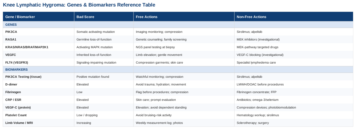
7 Biomarkers Worth Tracking When You're Managing a Knee Lymphatic Hygroma
Lymphatic malformations don't show up cleanly on a standard metabolic panel. The markers that matter here are more specialized, and most primary care visits won't order them unless you ask. What follows are the seven that a vascular anomalies clinic (or a hematologist working alongside one) is most likely to use to understand how active the lesion is, whether it's putting you at risk for bleeding or clotting complications, and whether targeted drug therapy is worth pursuing.
1. Tissue PIK3CA Mutation Status
This is the single most consequential test to come out of the last decade of vascular anomaly research. Roughly 80% of isolated lymphatic malformations carry a somatic (lesion-confined) mutation in the PIK3CA gene, identified in a landmark 2015 study of resected malformation tissue published in The Journal of Pediatrics. This isn't a blood biomarker — it's a molecular biomarker found only in the malformed tissue itself, which is why it requires a sample from the lesion rather than a vein draw.
How to measure it
Testing requires a tissue sample, usually obtained via image-guided core needle biopsy or during a planned sclerotherapy/surgical procedure, followed by next-generation sequencing (NGS) or droplet digital PCR targeted at known PIK3CA hotspots. This is typically only available through a specialized vascular anomalies center, not a general lab. Cost varies widely by country and insurance coverage — often in the $500–$3,000 range when billed separately, though it's frequently bundled into a diagnostic biopsy already being done for other reasons.If the result is positive: the plan without supplements or equipment
No lifestyle intervention, diet, or supplement changes a somatic mutation confined to malformed tissue — it isn't inherited and isn't systemic, so there is nothing to "reverse" through general health measures. The realistic non-drug options are watchful monitoring with imaging, compression garments for symptom control, and sclerotherapy or surgical debulking for localized disease, all directed by the treating specialist.If the result is positive: the plan with medication or equipment
A positive PIK3CA result opens the door to targeted therapy. Sirolimus (an mTOR inhibitor) has shown partial response in the majority of patients with complicated vascular anomalies in a phase II trial published in Pediatrics, typically dosed continuously with periodic blood trough-level checks (target 10–15 ng/mL) rather than cycled on and off. Side effects include mouth sores, elevated cholesterol/triglycerides, delayed wound healing, and increased infection risk, all of which require regular lab monitoring. For more severe or refractory cases, the PI3K-inhibitor alpelisib produced anatomical and functional improvement in patients with PIK3CA-related overgrowth in a 2018 Nature study, though it carries its own monitoring requirements (blood glucose, skin reactions) and is generally reserved for more complex or life-limiting presentations. Both require ongoing specialist supervision — this is not a self-directed regimen.2. D-dimer
Lymphatic and mixed lymphatic-venous malformations are frequently associated with a phenomenon called localized intravascular coagulopathy (LIC) — a slow, contained clotting-and-breakdown process happening within the abnormal channels themselves. Elevated D-dimer is the most sensitive lab sign of this, showing up in about 42% of patients with venous or mixed malformations in a large cohort study, particularly larger and deeper lesions, published in Archives of Dermatology.
How to measure it
A standard blood draw, widely available, typically $20–$80 depending on region and insurance. It's most useful when tracked over time rather than as a single value, since it can fluctuate with lesion activity, minor trauma, or intercurrent illness.If it's elevated: the plan without supplements or equipment
Avoid prolonged limb immobility, stay well hydrated, and avoid trauma or excessive strain to the affected knee, since local injury can trigger a flare in clotting activity. Gentle, regular movement (as opposed to sedentary stretches or high-impact strain) supports normal flow through the limb without provoking the lesion.If it's elevated: the plan with supplements or equipment
Graduated compression garments (equipment, not a supplement) are a mainstay and can meaningfully reduce symptomatic swelling and stabilize LIC markers over weeks of consistent daily wear. Be cautious with fish oil, high-dose vitamin E, and other supplements marketed for circulation — several have mild antiplatelet effects that could theoretically add to bleeding risk in a limb already showing coagulopathy, so don't start them without checking with the hematologist managing your case. In more pronounced LIC, clinicians sometimes use low-molecular-weight heparin or direct oral anticoagulants before procedures — always a prescribed intervention, not a self-directed one.3. Fibrinogen
Fibrinogen is the other half of the LIC picture. Where D-dimer rises, fibrinogen tends to fall in more severe cases — low fibrinogen alongside high D-dimer marks the more clinically significant end of the coagulopathy spectrum and is a key thing surgeons check before any procedure on the lesion.
How to measure it
Included in most coagulation panels ordered alongside D-dimer, roughly $20–$50. It's especially important to check in the weeks before any planned sclerotherapy, biopsy, or surgery involving the malformation.If it's low: the plan without supplements or equipment
There's no dietary or lifestyle fix for consumptive fibrinogen loss driven by an active malformation — this reflects ongoing clotting-and-breakdown activity within the lesion, not a nutritional deficiency. The practical non-medical step is simply flagging it clearly before any procedure, since it changes bleeding risk calculations.If it's low: the plan with supplements or equipment
Severe cases sometimes warrant fibrinogen concentrate or fresh frozen plasma before surgery — strictly a hospital-administered intervention. Compression again plays a supportive role by reducing stagnant pooling within the lesion, which may modestly ease the coagulopathy over time, though it won't normalize significantly abnormal fibrinogen on its own.4. C-Reactive Protein (CRP) and ESR
Lymphatic malformations are prone to cellulitis and intralesional bleeding episodes, both of which cause the lesion to swell suddenly and become painful. CRP and ESR are the standard way to distinguish an inflammatory/infectious flare from baseline lesion swelling, which matters because the two are managed very differently (antibiotics versus watchful waiting or compression).
How to measure it
High-sensitivity CRP runs about $15–$40; ESR is similarly inexpensive, $10–$25. Both are fast, widely available, and useful to check at baseline so you have a personal reference point for comparison during a flare.If it's elevated: the plan without supplements or equipment
Meticulous skin care over the lesion — prompt attention to any cracks, cuts, or skin breakdown — reduces the entry points for the bacteria that trigger cellulitis flares in the first place. Prompt medical evaluation (and antibiotics when appropriate) at the first sign of redness, warmth, or fever is far more useful here than trying to "treat inflammation" generically at home.If it's elevated: the plan with supplements or equipment
Omega-3 fatty acids (roughly 1–2 g combined EPA/DHA daily) have general anti-inflammatory evidence, but given the bleeding-risk caveat noted above with D-dimer/fibrinogen, this is worth clearing with your care team first rather than starting reflexively. Selenium supplementation has more specific supporting data: a series of studies from a German radiation-oncology group found selenium improved outcomes in secondary lymphedema when combined with physical therapy, including a head-and-neck lymphedema trial using 350 micrograms per body surface area for four to six weeks published in this study, with a low reported toxicity profile summarized here. That evidence is in secondary (acquired) lymphedema, not congenital lymphatic malformations specifically, so treat it as a reasonable adjunct to discuss rather than a proven fix.5. VEGF-C (Vascular Endothelial Growth Factor-C)
VEGF-C is the growth factor that drives new lymphatic vessel formation, and it's an active area of research as a marker of how "active" a lymphatic malformation is — animal and cell-culture work has shown that PIK3CA-driven lymphatic malformations depend on VEGF-C/VEGFR-3 signaling, and blocking that signal shrinks malformed vessels in lab models. In humans, elevated VEGF-C has been documented in other lymphatic-adjacent conditions such as lipedema, where it correlates with tissue changes without necessarily normalizing on its own.
How to measure it
This is not a routine commercial lab test — it's typically run as part of a research angiogenesis panel at an academic center, cost variable and rarely out-of-pocket practical ($100–$300 range where available). Treat it as a marker to ask a research-active vascular anomalies center about, rather than something to order yourself.If it's elevated: the plan without supplements or equipment
Gentle limb elevation, regular movement, and avoiding prolonged dependent positioning (standing still for long periods) support lymphatic flow mechanically, independent of the growth-factor biology, and are reasonable low-risk habits regardless of what this specific marker shows.If it's elevated: the plan with supplements or equipment
There is no supplement with solid human evidence for directly lowering VEGF-C in lymphatic malformations. Pneumatic compression devices and photobiomodulation (covered under complementary approaches below) address downstream swelling rather than the growth-factor signal itself, but they're the more realistic equipment-based options currently available outside of a clinical trial.6. Platelet Count (Part of a Complete Blood Count)
Mild thrombocytopenia (low platelets) can accompany more significant localized intravascular coagulopathy, and a low or dropping platelet count alongside worsening D-dimer and fibrinogen is a signal worth escalating to a hematologist quickly, rather than a case of a more severe overlapping condition called Kasabach-Merritt phenomenon, which occurs in a different type of vascular tumor but is worth ruling out if platelets fall sharply.
How to measure it
Included in a standard complete blood count, $10–$30, widely available and often already part of routine bloodwork.If it's low: the plan without supplements or equipment
Avoid activities with high bruising or bleeding risk around the affected knee while platelets are low, and monitor for unusual bruising elsewhere, which would prompt a call to the care team rather than a wait-and-see approach.If it's low: the plan with supplements or equipment
Iron supplementation is only relevant if repeated testing shows concurrent anemia from chronic low-grade blood loss within the lesion — a targeted fix for a specific finding, not a general "platelet booster." Meaningful thrombocytopenia related to the malformation itself is a medical management issue (sometimes involving the same sirolimus therapy discussed above, which can improve LIC parameters as the lesion responds) rather than something addressed with over-the-counter products.7. Limb Circumference and MRI Volumetry
Not a blood test, but arguably the most practical marker for day-to-day tracking: consistent circumference measurements at fixed points on the knee/leg, paired with periodic MRI to assess lesion volume and extent, give you and your clinical team an objective trendline instead of relying on subjective "it looks bigger" impressions. MRI is also how the extent of disease is mapped before any procedural planning.
How to measure it
Circumference tracking at home costs nothing beyond a tape measure and a consistent routine (same time of day, same marked points, logged in a simple chart or app). MRI without contrast typically runs $500–$1,500 in the US depending on facility and insurance, more with contrast; ultrasound is a cheaper interim option ($100–$400) for tracking size between MRIs, though it's less precise for full extent.If volume is increasing: the plan without supplements or equipment
A simple weekly measurement log, taken at the same time relative to activity level (lesions often swell after exertion or heat exposure), turns vague concern into a real trend your specialist can act on. Photographs from a fixed angle add a useful visual reference alongside the numbers.If volume is increasing: the plan with supplements or equipment
Properly fitted compression garments are the most evidence-supported equipment-based option for controlling volume day to day, and pneumatic compression devices are sometimes added for more significant swelling. Escalating volume on serial imaging is generally the trigger for revisiting medical or procedural options (sirolimus, sclerotherapy, alpelisib, or surgery) rather than something to manage indefinitely with compression alone.What the Underlying Genetics Suggest
The biomarkers above tell you what's happening right now. The genetics explain why it's happening at all — and they matter for a very practical reason: unlike common polygenic traits, the gene changes behind a knee lymphatic hygroma are mostly confined to the malformed tissue itself, which changes what "fixing the gene" can even mean.
PIK3CA
As covered above, PIK3CA is the dominant driver in the majority of isolated lymphatic malformations, identified through direct sequencing of resected lesion tissue in this foundational study. The mutation keeps a growth-signaling enzyme (PI3K) switched on inside the lymphatic endothelial cells lining the malformation, driving continued abnormal vessel growth. Human evidence for targeted compensation is strong and growing: sirolimus and alpelisib, discussed above, directly counteract this overactive signal rather than working around it.
RASA1
RASA1 normally acts as a brake on the Ras signaling pathway; loss-of-function mutations remove that brake and are linked to capillary malformation-arteriovenous malformation syndrome, a condition that can include lymphatic-vascular overlap features, identified across 68 families in a 2013 study in Human Mutation. Unlike PIK3CA-driven isolated lymphatic malformations, RASA1 mutations are often germline (inherited, present in every cell), which is why genetic counseling and family screening are typically recommended when it's identified. Human evidence for direct pharmacologic compensation is earlier-stage than for PIK3CA, though MEK inhibitors (which act downstream of Ras) are being explored in related conditions.
RAS-MAPK Pathway Genes (KRAS, NRAS, BRAF, MAP2K1)
A smaller subset of lymphatic and vascular malformations carry activating mutations in this pathway instead of PIK3CA — mechanistically related, since both pathways converge on cell growth and survival signaling, but genetically distinct. This is why molecular testing (not just a clinical diagnosis) increasingly matters: it identifies which pathway is driving a specific lesion, which in turn determines whether a PI3K/mTOR-targeted drug or a MEK-pathway-targeted drug is the more logical option. Evidence here is earlier and more case-based than the PIK3CA literature, and testing for this group is generally done as part of the same broad NGS panel used for PIK3CA rather than as a separate test.
VEGFC
VEGFC encodes the growth factor discussed above under biomarkers. Rare inherited mutations in VEGFC itself (distinct from elevated VEGF-C protein levels seen in active malformations) cause a form of primary lymphedema, established through gene sequencing in affected families. This is a different mechanism from a localized PIK3CA-driven hygroma — it reflects a developmental lymphatic-vessel problem present from birth throughout the lymphatic system, rather than a focal malformation — but the two conditions share enough biology that VEGFC research directly informs how PIK3CA-driven malformations are understood and treated.
FLT4 (VEGFR3)
FLT4 encodes the receptor that VEGF-C binds to, and missense mutations that impair its signaling were identified as a cause of hereditary lymphedema (Milroy disease) in a 2000 study in Nature Genetics — one of the papers that first established the VEGF-C/VEGFR-3 axis as central to lymphatic development. Like VEGFC mutations, FLT4 mutations are typically germline and cause a distinct, more diffuse lymphedema phenotype rather than a focal hygroma, but the receptor they disrupt is the same one that PIK3CA-driven malformations depend on downstream, which is part of why VEGF-C-blocking strategies are being studied as a possible future add-on to mTOR/PI3K inhibitors.
Where Consumer Genetic Testing Fits — and Where It Doesn't
Researchers like Ali Torkamani have done valuable work showing how polygenic risk scores from consumer genome data can flag elevated lifetime risk for common conditions, and figures like Gary Brecka have popularized using broad genetic and biomarker panels to guide general health optimization. Neither approach is built to catch what matters here: a somatic PIK3CA mutation confined to a few square centimeters of malformed tissue won't appear in blood-based consumer DNA testing, because that DNA is normal everywhere else in the body. If you're curious about the genetics of your specific lesion, the useful next step is a tissue-based NGS panel through a vascular anomalies center, not a mail-in saliva kit.
The Longevity Book That Reframes How to Think About This
Peter Attia's Outlive: The Science and Art of Longevity isn't written about vascular malformations, but it spends real time on two things that turn out to be directly relevant here: proactive, marker-driven medicine, and the biology of mTOR — the exact pathway that sirolimus, one of the main treatments discussed above, works on. Reading it with a knee lymphatic hygroma in mind reframes a few things worth pulling out directly.
1. "Medicine 3.0" means acting on markers before a crisis forces your hand
Attia's central argument is that reactive medicine — waiting for a clear diagnosis or a crisis — misses years of useful intervention. Applied here, that means tracking D-dimer, fibrinogen, and limb volume as a baseline habit, not only during a flare, so trends are visible before they become emergencies.2. Rapamycin's discovery story explains why sirolimus works the way it does
The book traces rapamycin from a soil sample on Easter Island to its role as an mTOR inhibitor. That's the same mechanism sirolimus uses against PIK3CA-driven lymphatic malformations — one drug, two very different applications of the same biological lever, which is a useful mental model for understanding why your medical team might reach for it.3. mTOR is a growth-versus-maintenance switch
When nutrients and growth signals are high, mTOR pushes cells toward growth; when suppressed, cells shift toward repair and recycling (autophagy). A PIK3CA-driven lesion is, in effect, a pocket of tissue stuck in permanent "growth" mode — which is precisely what mTOR inhibition is designed to interrupt.4. Rapamycin's metabolic effects are more nuanced than "anti-aging drug"
Attia is careful to note rapamycin isn't a simple longevity supplement — animal studies have shown metabolic tradeoffs, including effects on insulin sensitivity, that depend heavily on dosing pattern, as detailed in this mechanistic study. That nuance is exactly why sirolimus dosing for vascular anomalies is trough-level-guided and physician-managed rather than a fixed low-dose regimen borrowed from longevity forums.5. Inflammation markers deserve routine tracking, not just symptom-triggered checks
The book pushes readers toward checking CRP and related markers as a matter of course. For a lymphatic malformation prone to cellulitis flares, a personal CRP baseline is what lets you and your clinician tell a real infection apart from ordinary lesion swelling.6. Zone 2 aerobic exercise supports circulation without the strain of high-intensity efforts
Attia champions low-intensity, sustained aerobic work for metabolic health. For someone managing swelling and coagulopathy risk in a limb, this style of movement offers a way to stay active without the joint impact or acute strain that higher-intensity training could place on the knee.7. Stability and strength training protect the joints around a compromised limb
Much of the book's "Centenarian Decathlon" framework is about preserving functional strength for daily life. Applied practically, strengthening the muscles around an affected knee — under guidance, and avoiding direct strain on the lesion — helps offset any mechanical disadvantage from chronic swelling.8. Objective data changes the emotional experience of a chronic condition
Attia writes about how uncertainty, more than the medical facts themselves, often drives the hardest parts of managing a long-term condition. Consistent tracking (the biomarker panel above) converts vague worry into specific, answerable questions for your next appointment.9. Sleep quality affects inflammatory tone
Poor sleep is linked to elevated inflammatory markers throughout the book's discussion of metabolic health. For a condition where CRP spikes matter, consistent sleep is a low-cost lever that supports a lower inflammatory baseline, even though it won't touch the malformation directly.10. The right specialist matters more than the right supplement
Attia is emphatic that sophisticated self-tracking is meant to inform conversations with expert clinicians, not replace them. For a rare, molecularly-driven condition like a knee lymphatic hygroma, that point is arguably more true than for almost anything else in the book — the biggest gains come from getting the right vascular anomalies specialist involved, not from optimizing supplements at home.Complementary Approaches Worth Considering
None of the following shrink a lymphatic malformation on their own, and the evidence for most comes from acquired (secondary) lymphedema rather than congenital lymphatic malformations specifically — an important distinction to hold onto. Used as an adjunct alongside medical management, though, a few have genuine human evidence behind them for the swelling and discomfort side of the condition.
Massage Therapy (Manual Lymphatic Drainage)
Manual lymphatic drainage is a specific, gentle massage technique designed to encourage fluid movement through remaining functional lymphatic pathways, which is why it's often suggested for swelling around a lymphatic malformation despite the lesion itself being structurally different from typical lymphedema. The rationale is mechanical: light, rhythmic strokes in the direction of lymph flow can temporarily ease fluid buildup in adjacent, less-affected tissue.
A systematic review and meta-analysis of randomized controlled trials in breast-cancer-related lymphedema found that manual lymphatic drainage did not significantly reduce lymphedema overall, but noted a benefit in patients under 60 and in courses lasting longer than a month or more than 20 sessions, per this analysis. That's a genuinely mixed result, and it comes from breast-cancer populations, not lymphatic malformations.
Realistically, this means treating manual lymphatic drainage as a comfort-oriented adjunct rather than a treatment for the malformation itself — worth trying with a certified lymphedema therapist for a defined trial period (a month, multiple sessions per week), and worth discontinuing without much loss if it isn't providing noticeable relief.
Low-Level Laser Therapy (Photobiomodulation)
Photobiomodulation uses specific low-power light wavelengths applied to the skin over swollen tissue, with a proposed effect on local lymphatic vessel contractility and tissue drainage. It's one of the more evidence-backed physical adjuncts for lymphatic swelling generally, which is why it comes up as an option for knee-area swelling from a lymphatic malformation.
A meta-analysis in breast-cancer-related lymphedema found a meaningful reduction in limb volume across pooled trials, with three high-quality studies showing photobiomodulation outperforming sham treatment, using a protocol of roughly three sessions per week at a laser density of 1.5–2 J/cm² described in this systematic review and meta-analysis. Side effects reported in these trials were minimal — mild, transient warmth at the treatment site.
For a knee lymphatic hygroma, this would realistically be delivered by a physical or lymphedema therapist with the appropriate device, a few times weekly for several weeks, evaluated by circumference tracking (see biomarker 7 above) to see whether it's producing a measurable effect for you specifically, rather than assumed to work simply because it helped in cancer-related lymphedema trials.
Breathing-Based Therapies (Diaphragmatic Breathing)
Deep diaphragmatic breathing changes intrathoracic pressure in a way that mechanically promotes lymph flow through the thoracic duct — the body's main lymphatic drainage channel — a mechanism first described decades ago and still cited in current lymphedema self-management guidance.
A more recent clinical study specifically evaluating diaphragmatic breathing combined with limb coordination training for lower-limb lymphedema following gynecologic cancer surgery found meaningful rehabilitation benefit, reported in this study — notably, a lower-limb population, which is a closer anatomical match to a knee-area lesion than most of the upper-limb lymphedema literature.
This is close to a zero-risk, zero-cost addition: a daily practice of slow, deep diaphragmatic breathing (10–15 minutes, once or twice a day, lying down with knees supported) alongside gentle limb movement. It won't address the malformation's structure, but it's a reasonable low-effort habit with a plausible mechanism and no meaningful downside.
Yoga
Gentle yoga combines controlled breathing, light stretching, and postural work, all of which theoretically support lymphatic flow and joint mobility around a swollen limb — relevant for a knee-area lesion where stiffness and reduced range of motion often accompany swelling.
A randomized controlled pilot trial in breast-cancer-related lymphedema found modest improvements in arm volume and quality-of-life measures after an eight-week program combining a weekly class with daily home practice, detailed in this trial. It was a small pilot study, and again, an upper-limb, cancer-related population — evidence should be read as suggestive, not definitive, for a knee lymphatic hygroma.
A sensible approach is a gentle, restorative style (not hot yoga or deep-stretch styles that strain the joint), ideally with a knee modification discussed in advance with an instructor familiar with lymphatic conditions, practiced a few times a week and adjusted immediately if any pose increases swelling or discomfort.
Conclusion
A knee lymphatic hygroma responds better to specific information than to general reassurance. The biomarker panel above — coagulation markers, inflammatory markers, molecular testing, and simple volume tracking — gives you an objective way to see what's actually changing, and the genetics behind PIK3CA and its related pathways explain why targeted drug therapy has become a real option rather than a distant hope. The complementary approaches with genuine evidence (photobiomodulation and diaphragmatic breathing most notably) are worth adding as low-risk adjuncts, not substitutes, for the medical plan your specialist builds with you.
The next useful step is concrete: ask your care team whether tissue-based genetic testing has been done on your lesion, request a baseline coagulation and inflammatory panel if you don't already have one, and start a simple weekly circumference log today. Bring all three to your next appointment — that combination of data is what turns a vague "keep an eye on it" into an informed, specific conversation about what to do next.
Musculoskeletal: Joint Conditions
Cardiovascular: Blood Vessel Conditions Vascular Conditions