This article contains AI generated content.
· UpdatedKnee Neurofibroma: 4 Genes and 6 Biomarkers to Track
Introduction
Having a neurofibroma in or around the knee puts you in an uncomfortable position. The condition is real, sometimes painful or functionally limiting, and yet the standard clinical response is often minimal. Most patients are told to monitor it, avoid unnecessary surgery, and return if something changes. That guidance is not wrong—but without understanding what actually drives growth, what signals transformation risk, and what you can influence between appointments, monitoring becomes passive and decisions lack foundation.
The frustration is compounded by the fact that most available information about neurofibromas is surface-level. It explains what they are and how they are removed, but says almost nothing about the molecular drivers, what makes one person's tumor stable for decades while another's grows or changes character, or what can be done beyond surveillance and surgery. Generic advice—eat well, reduce stress, stay active—does not address the specific genetic and biochemical architecture of this condition.
The science has moved further than most clinical communication suggests. The genetic cascade that drives neurofibroma formation, particularly through the NF1 gene and its downstream signaling, is now well characterized. Key biomarkers that reflect tumor activity, microenvironment inflammation, and proliferation can be measured and tracked over time. Neither genetics nor biomarkers guarantee a specific outcome, but both offer a considerably more precise picture than symptom observation alone.
This article covers four genes with strong mechanistic evidence in neurofibroma biology—including what to do when your profile is unfavorable—six biomarkers worth monitoring over time, a metabolic framework for tumor biology with direct practical implications, and three complementary approaches with meaningful clinical evidence. None of this replaces specialist care. It is meant to help you engage with that care more intelligently.
Summary
- Four genes shape the biology of this tumor: NF1 (the master regulator), KRAS (the downstream amplifier), CDKN2A (the malignant transformation gatekeeper), and TP53 (the genome's repair guardian). For each, there are specific, practical protocols—with and without supplements—addressing the molecular dysfunction directly, with dosing, cycling schedules, and side effects. - Mast cells are a key and underappreciated driver: NF1 neurofibromas are heavily infiltrated by mast cells that drive both tumor growth and local inflammation. Several of the most practical interventions—quercetin, omega-3s, dietary changes—work partly through mast cell stabilization, a mechanism rarely discussed in standard care conversations. - Six trackable biomarkers give you a longitudinal picture: From affordable standard blood tests (hsCRP, IGF-1) to tissue-based markers (Ki-67) and advanced liquid biopsy panels (ctDNA), these measurements let you track tumor activity, microenvironment inflammation, and transformation risk over time rather than waiting for symptoms to shift. - A metabolic perspective reframes what you can actually control: Jason Fung's The Cancer Code connects insulin, IGF-1, intermittent fasting, and tumor microenvironment biology into a framework with direct relevance to neurofibroma growth dynamics—and puts concrete lifestyle levers in your hands. - Three complementary approaches have meaningful clinical evidence: Photobiomodulation for nerve-related pain, mindfulness-based stress reduction (MBSR) for chronic pain management, and biofeedback for autonomic regulation and symptom control each have realistic application protocols that can be integrated alongside standard care.
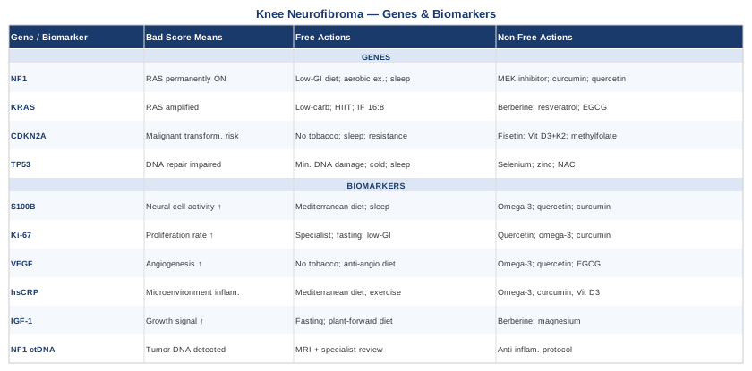
The Genetic Blueprint of Knee Neurofibroma: 4 Key Drivers
Neurofibromas are not random overgrowths. They arise from a chain of molecular events with a well-defined starting point in most cases: dysfunction in the NF1 gene and the signaling cascade it controls. Understanding this cascade—and the cellular checkpoints that either slow or accelerate it—is the most clinically useful genetic lens available right now for anyone managing this condition.
The four genes discussed below are not equally relevant to every case. NF1 is central to the vast majority of neurofibromas, whether or not a formal NF1 diagnosis exists. KRAS-related signaling functions as a downstream amplifier of the same pathway. CDKN2A and TP53 matter most for assessing malignant transformation risk—the progression from a benign neurofibroma to a malignant peripheral nerve sheath tumor (MPNST). Knowing your profile for each is increasingly practical through commercial genomic testing and germline panels; acting on it thoughtfully is the aim.
Researchers like Ali Torkamani have emphasized that genetic variants rarely act in isolation—their impact is shaped by metabolic state, inflammation, sleep quality, and nutrition. Gary Brecka similarly argues that identifying your specific variants is only useful when paired with a concrete compensatory plan. Both perspectives inform the structure of this section: for each gene, a plan exists—with and without supplementation—that directly addresses the molecular dysfunction.
Gene 1: NF1 (Neurofibromin) — The Master Driver
The NF1 gene, located on chromosome 17q11.2, encodes a large cytoplasmic protein called neurofibromin. Its central function is to act as a GTPase-activating protein (GAP) for RAS—it converts active RAS-GTP into inactive RAS-GDP, functioning as a molecular brake on cell proliferation signals.
When NF1 is mutated or deleted (a loss-of-function event), RAS remains locked in its active state permanently. This drives constant activation of two downstream pathways: the RAF-MEK-ERK cascade (the MAPK pathway) and the PI3K-AKT-mTOR pathway. Together, these cascades produce unchecked Schwann cell proliferation—the cellular basis of neurofibroma formation.
In NF1-associated neurofibromatosis, one NF1 copy is lost in every cell at birth. A somatic "second hit" in a Schwann cell, frequently triggered by mast cell-derived stem cell factor (SCF), initiates tumor formation. In sporadic cases, both copies are lost through somatic mutations alone. Biallelic NF1 loss is a prerequisite for neurofibroma development in either scenario.
The classification, diagnostic criteria, and clinical management of NF1 are extensively documented at NIH GeneReviews — Neurofibromatosis 1. A phase 2 randomized trial of selumetinib (a MEK inhibitor targeting the pathway downstream of NF1) published in the New England Journal of Medicine demonstrated significant reduction in plexiform neurofibroma volume, establishing MEK inhibition as the first molecularly targeted therapy for this condition.
One mechanistic detail that rarely makes it into patient conversations: NF1-deficient mast cells produce excess SCF, which acts as a paracrine signal driving the second NF1 hit in neighboring Schwann cells. Reducing mast cell activation is therefore not a general wellness gesture—it is a direct therapeutic mechanism for this specific tumor biology.
If the gene is bad, the plan without supplements
MEK inhibitor therapy (selumetinib/Koselugo): FDA-approved for NF1-associated plexiform neurofibromas in patients aged 2 and older. If you have NF1 and have not discussed this with your specialist, that conversation is overdue regardless of tumor size or current symptoms. Frequency: daily oral dosing per physician prescription. Side effects: acneiform rash, GI upset, elevated creatine kinase; requires regular laboratory monitoring and dose adjustment by a specialist.
Low glycemic diet: Insulin and IGF-1 are upstream activators of the very RAS pathway that NF1 normally suppresses. A diet targeting glycemic index below 50—emphasizing non-starchy vegetables, legumes, whole grains, fatty fish, and limited refined carbohydrates—measurably reduces both markers. Apply daily with no cycling; this is a sustained metabolic baseline, not a short-term intervention.
Aerobic exercise (150-200 minutes per week, moderate intensity): Reduces insulin resistance and circulating IGF-1, creating a less permissive environment for RAS-driven Schwann cell growth. Swimming, cycling, and brisk walking are well-tolerated options that protect the knee joint while delivering systemic metabolic benefit. No cycling needed; consistency is the mechanism.
Regular MRI monitoring: Every 12-24 months for NF1-associated knee neurofibromas, or immediately with any new neurological symptom (weakness, new numbness, rapid growth). No lifestyle measure or supplement substitutes for imaging surveillance—this is the irreplaceable backbone of management.
Avoid growth hormone supplementation: GH and IGF-1 directly amplify the same RAS-PI3K pathway that is already overactive. This applies specifically to anti-aging GH protocols, which are increasingly marketed and prescribed. Their use in anyone with NF1 or an active neurofibroma is contraindicated without careful specialist review.
Sleep optimization (7-9 hours, high continuity): Reduces inflammatory cytokines that activate mast cells—directly addressing one of the key drivers of NF1 neurofibroma growth. Practical targets: consistent sleep timing, room temperature 18-20°C, complete darkness, no alcohol within 3 hours of bedtime. Nightly, no cycling.
If the score is bad, the plan with supplements or equipment
Curcumin with piperine (500-1000mg curcumin, 10mg piperine daily, with a fatty meal): Inhibits NF-κB and the MAPK pathway downstream of RAS. Also reduces mast cell activation and degranulation. Use a phospholipid-complexed or liposomal formulation for meaningful absorption. Cycle: 6 weeks on, 1 week off. Side effects: GI discomfort at higher doses; antiplatelet effect (inform your surgeon); avoid high doses immediately before procedures.
Quercetin (500-1000mg/day in divided doses, with food): Dual-acting as a mast cell stabilizer and mild RAS pathway inhibitor. Given NF1's mast cell biology, this combination of actions makes quercetin particularly relevant and mechanistically specific. Cycle: 8 weeks on, 2 weeks off. Side effects: potential interaction with fluoroquinolone antibiotics and blood thinners; mild GI upset; separate from medications by 2 hours.
Omega-3 fatty acids (EPA+DHA, 2-4g/day with the largest meal): Reduces mast cell degranulation and suppresses the NF-κB-driven inflammatory microenvironment that sustains NF1 neurofibroma growth. Choose a triglyceride-form fish oil for best absorption. No cycling needed. Side effects: antiplatelet effect above 3g/day; always inform surgeon or specialist before any procedure; avoid with pharmaceutical blood thinners without guidance.
Sulforaphane (25-50mg/day from broccoli sprout extract, or daily fresh broccoli sprouts): Activates Nrf2, which partially offsets NF-κB and shows antiproliferative activity in Schwann cell and nerve sheath-related tissue models. No cycling required. Side effects: possible thyroid interaction at very high doses; consult if you have pre-existing thyroid conditions.
Photobiomodulation device (red/near-infrared panel, 660-850nm combined): 10-20 minutes applied to the knee region, 3-4 times per week. Reduces local inflammatory cytokine production and may support peripheral nerve tissue health. Do not apply directly over large, confirmed plexiform neurofibromas without explicit physician guidance. Cost: $150-600 for a quality home panel; professional clinical LLLT is available at physiotherapy centers.
Gene 2: KRAS and the RAS Pathway Amplifiers
While NF1 loss is the initiating event in most neurofibromas, the downstream RAS pathway can be independently activated or compounded by mutations in RAS family genes themselves—most notably KRAS, HRAS, and NRAS. These are the exact oncogenes that neurofibromin normally restrains. When NF1 is lost and a RAS family gene is additionally mutated or when RAS activating mutations drive sporadic cases without NF1 involvement, the MEK-ERK cascade runs at significantly higher throughput.
In sporadic neurofibromas occurring without germline NF1 mutations, activating mutations in KRAS or HRAS are identified in a meaningful proportion of cases. In some NF1-associated tumors, additional somatic RAS mutations create a compounding effect. Either way, the consequence is the same: the proliferation signal is amplified beyond what NF1 loss alone would produce, and the tumor biology becomes more aggressive.
RAS pathway mutations are detectable through tumor genomic profiling panels and increasingly through liquid biopsy. Their identification has direct implications for understanding growth dynamics and for treatment strategy. MEK inhibitors, which target a step downstream of RAS, remain relevant regardless of whether the activating event is NF1 loss or RAS mutation.
If the gene is bad, the plan without supplements
Low-carbohydrate diet (under 100g net carbs/day): RAS-driven tumor cells preferentially use glycolysis. Reducing dietary glucose limits their preferred fuel source while simultaneously lowering insulin and IGF-1—two upstream amplifiers of the RAS-PI3K axis. A whole-food low-carb or Mediterranean low-carb approach is most sustainable. Apply daily without cycling.
Time-restricted eating (16:8 protocol): An 8-hour eating window reduces insulin exposure meaningfully across the day, reducing a key upstream activator of the RAS-mTOR axis. Start with a 12-hour window and progress to 16:8 over 2-4 weeks. Apply 5-7 days per week. No cycling; this is a baseline metabolic strategy.
High-intensity interval training (HIIT): 2-3 sessions per week of 20-30 minutes reduces whole-body insulin resistance more effectively than steady-state cardio, creating a less favorable metabolic environment for RAS-driven proliferation. For knee involvement, prioritize low-impact HIIT options: rowing, cycling intervals, upper-body interval circuits. Side effects: begin gradually; avoid during acute pain flares; ensure adequate recovery between sessions.
Metformin (by prescription): AMPK activation via metformin directly counteracts mTOR activity, a key downstream node in RAS-PI3K signaling. Increasingly referenced in integrative oncology for metabolic tumor suppression support. Daily per physician guidance. Side effects: GI discomfort (take with food), vitamin B12 depletion with long-term use (monitor annually and supplement if needed).
If the score is bad, the plan with supplements or equipment
Berberine (500mg 2-3 times daily with meals): Functions through AMPK activation similarly to metformin, reducing mTOR and partially counteracting RAS-driven cell growth. One of the most accessible and well-studied metabolic modulators. Cycle: 3 months on, 1 month off. Side effects: GI discomfort, constipation, blood glucose lowering (caution with diabetes medications); check for CYP3A4 drug interactions.
Resveratrol (250-500mg/day with a fatty meal for absorption): Activates SIRT1 and AMPK, creating metabolic signaling that counteracts RAS overactivation and reduces mTOR downstream of IGF-1. Cycle: 8-12 weeks on, 2-4 weeks off. Side effects: possible interaction with blood thinners; caution with estrogen-sensitive conditions at higher doses.
EGCG from green tea extract (400-600mg/day with food): RAS pathway inhibitory properties demonstrated across multiple cell models; additionally anti-angiogenic through VEGF suppression. Cycle: 12 weeks on, 4 weeks off. Side effects: rare liver enzyme elevation at high doses; take with food to reduce GI irritation; avoid during chemotherapy without specialist guidance.
R-Alpha Lipoic Acid (300-600mg/day, taken on an empty stomach): Antioxidant support for mitochondrial function that reduces the oxidative stress which amplifies RAS pathway activity in metabolically stressed cells. No cycling required. Side effects: mild blood glucose lowering; possible interaction with thyroid hormone conversion at very high doses; monitor if you have thyroid conditions.
Gene 3: CDKN2A — The Malignant Transformation Gatekeeper
The CDKN2A locus on chromosome 9p21 encodes two distinct tumor suppressors using alternative reading frames: p16 (INK4a) and p14 (ARF). These proteins operate through different mechanisms that converge on the same critical purpose—preventing cells from advancing through cell cycle checkpoints that separate normal growth from uncontrolled proliferation.
p16 inhibits CDK4 and CDK6, kinases that would otherwise phosphorylate the retinoblastoma protein (Rb), releasing E2F transcription factors and opening the door to cell cycle progression. p14 stabilizes p53 by binding and sequestering MDM2, its primary negative regulator. Lose both functions and the brake on cell division is essentially absent.
In neurofibroma biology, CDKN2A matters most for one high-stakes question: malignant transformation risk. The progression from benign neurofibroma to malignant peripheral nerve sheath tumor (MPNST) occurs in approximately 8-13% of NF1 patients over a lifetime and carries a poor prognosis. CDKN2A deletion is identified in approximately 60-70% of MPNSTs—making it one of the strongest molecular markers of the malignant transition. Knowing your CDKN2A status through tumor tissue analysis or germline genomic panel is directly relevant to how closely you monitor and how seriously you take protective lifestyle strategies.
If the gene is bad, the plan without supplements
Eliminate tobacco exposure entirely: Tobacco carcinogens cause direct DNA damage and promote CDKN2A promoter hypermethylation through epigenetic mechanisms—effectively silencing the gene even when it is structurally intact. This is non-negotiable. There is no acceptable dose of tobacco exposure in the context of CDKN2A vulnerability. Daily, permanent.
Maintain healthy body composition: Visceral adiposity drives chronic low-grade inflammation and promotes CDKN2A silencing through DNMT3-mediated epigenetic mechanisms. Target BMI 18.5-24.9 and waist circumference below 90cm for men or 80cm for women. The mechanism is metabolic, so body composition change through diet and exercise—not weight alone—is what matters.
Sleep prioritization (7-9 hours, high continuity): CDKN2A coordinates DNA repair processes that are most active during slow-wave sleep. Disrupted sleep architecture—from alcohol, inconsistent timing, or light exposure—reduces the repair window and allows DNA damage to accumulate unchallenged. Prioritize consistent bedtimes, complete darkness, and a cool room. Nightly.
Resistance training (2-3 times per week, progressive): Evidence suggests resistance training maintains cellular senescence pathways involving p16 and p21. For knee neurofibroma, programming must avoid direct loading through the tumor site—adapt to upper-body, contralateral, or functionally unloaded movements where needed. Consistency over intensity.
If the score is bad, the plan with supplements or equipment
Fisetin (100-200mg/day routine maintenance; or 500-1000mg pulsed across 2-3 consecutive days per month as a senolytic protocol): Fisetin activates AMPK and supports senescence-related pathways that partially compensate for CDKN2A dysfunction. The monthly pulsed high-dose approach mirrors senolytic protocols in longevity research. Cycle: daily routine or monthly pulse; no need to cycle the lower daily dose. Side effects: mild GI discomfort; caution with anticoagulant medications.
EGCG (green tea extract) (400-800mg/day with food): Functions as an epigenetic modifier with evidence for reducing aberrant promoter methylation of tumor suppressor genes including CDKN2A. When CDKN2A is epigenetically silenced rather than genetically deleted, this mechanism is particularly relevant—it directly addresses the silencing process rather than working around it. Cycle: 12 weeks on, 4 weeks off. Side effects: liver enzyme elevation is rare but possible at high doses; always take with food.
Vitamin D3 paired with K2 (3000-5000 IU D3 daily, with 100-200mcg K2 MK-7): Vitamin D receptor activation modulates the CDK4/6-Rb pathway that CDKN2A normally regulates. Monitor serum 25(OH)D and aim for 60-80 ng/mL. K2 prevents soft tissue calcification associated with aggressive D3 supplementation. Side effects: toxicity risk emerges above 150 ng/mL serum; test every 6 months when supplementing above 3000 IU.
Methylfolate and methylcobalamin (B12) (400-800mcg methylfolate, 1000mcg methylcobalamin daily): Essential cofactors for DNA methylation balance. Deficiency—particularly in individuals with MTHFR polymorphisms—can contribute to aberrant epigenetic silencing of CDKN2A. Take daily with no cycling. Always use the pair together: excess folate supplementation alone can mask B12 deficiency symptoms.
Gene 4: TP53 — The Genome's Repair Guardian
TP53 encodes p53, one of the most thoroughly characterized proteins in cancer biology. As a stress-activated transcription factor, p53 responds to DNA damage, hypoxia, RAS hyperactivation, and other cellular insults by triggering one of three protective outcomes: DNA repair, cell cycle arrest, or apoptosis. Its name in the literature—"the guardian of the genome"—reflects a function that is central to preventing the mutation accumulation that drives malignant transformation.
In benign neurofibromas, TP53 mutations are uncommon. Their significance in this context lies in progression risk: TP53 is altered in approximately 20-30% of MPNSTs, particularly in high-grade tumors. Beyond direct mutation, suboptimal p53 pathway function can arise through common polymorphisms (such as the Pro72Arg variant), through overexpression of the negative regulator MDM2, or through chronic oxidative stress that degrades and impairs p53 protein function. These functional impairments matter even when structural testing reports an intact gene—p53 status is as much a question of function as of sequence.
If the gene is bad, the plan without supplements
Minimize DNA damage inputs daily: UV radiation without adequate protection, processed meat carcinogens (nitrosamines), alcohol (acetaldehyde is directly genotoxic), and chronic sleep deprivation all create DNA damage loads that test p53 repair capacity at a scale that an impaired p53 cannot adequately address. Reduce or eliminate each category. This is not optional context—it is the most directly impactful step.
Structured cold exposure: Cold water immersion (10-15°C, 3-10 minutes) or cold showers (3-5 minutes at minimum comfortable temperature) are hormetic stressors that activate heat shock proteins and cellular stress response pathways, including upstream sensors that engage p53 activity. Frequency: 3-5 times per week. Begin gradually over 2-3 weeks; caution with cardiovascular conditions. Side effects: vasovagal response in some individuals; always start with shorter exposures.
Moderate aerobic exercise (150+ minutes per week): Controlled, moderate reactive oxygen species generated during aerobic exercise activate p53 pathway activity through a beneficial hormetic mechanism—the same principle as cold exposure, but through a different physiological route. The critical variable is moderate: excessive training without adequate recovery can create a net negative oxidative burden. Combine daily light activity with 3-4 moderate intensity sessions weekly.
Sleep architecture optimization: DNA repair is most active during slow-wave sleep, which is when p53-driven repair processes are most engaged. Practical targets: consistent sleep timing (within 30 minutes day to day), no alcohol within 3 hours of bedtime, no blue light within 2 hours, room temperature 18-20°C. Nightly.
If the score is bad, the plan with supplements or equipment
Selenium (as selenomethionine, 200mcg daily with food): An essential cofactor for glutathione peroxidase and thioredoxin reductase—antioxidant enzymes that reduce the oxidative stress that damages and functionally degrades p53 protein. Do not exceed 400mcg/day; selenium toxicity risk is real and dose-dependent. No cycling required. Side effects above 400mcg/day include hair loss, nail brittleness, and neurological symptoms; these are dose-related and resolve with reduction.
Zinc (as zinc picolinate or zinc glycinate, 25-30mg daily with food): Structurally required for proper p53 protein folding and its ability to bind DNA. Zinc deficiency—common in individuals with poor diet quality or chronic inflammation—is directly associated with p53 dysfunction. Take with food, separated from iron supplements by at least 2 hours. Cycle: 3 months on, then measure serum zinc and copper. Side effects: long-term high-dose zinc depletes copper; consider 1-2mg copper supplementation if using beyond 3 months.
NAC (N-Acetyl Cysteine, 600-1200mg daily): The most direct glutathione precursor available as a supplement. Reduces oxidative DNA damage that can trigger p53 loss of heterozygosity or progressive functional impairment. This single supplement has the broadest applicability across all four genes discussed in this article. No cycling required. Side effects: occasional GI upset; may affect certain lab values including plasma homocysteine.
Vitamin C (sodium ascorbate or liposomal form, 1-2g daily with meals): Synergistic antioxidant protection working alongside NAC; at consistent doses also supports collagen synthesis in the connective tissue surrounding the tumor site, which can be compressed or distorted by growth. No cycling. Side effects: above 3g/day, osmotic diarrhea may occur—reduce dose rather than cycle; avoid high-dose supplementation with a history of calcium oxalate kidney stones.
The four genes above describe the underlying molecular architecture of knee neurofibroma. The biomarkers below measure how that architecture is currently performing—and whether the biological balance is actively shifting.
6 Biomarkers Worth Tracking
Genetics describes the fixed starting conditions. Biomarkers describe what is actually happening right now—the result of those genetic predispositions interacting with your current metabolism, inflammation levels, diet, sleep, and overall biology. For knee neurofibroma, a targeted panel of six markers covers the most clinically relevant dimensions: neural tissue activity, cellular proliferation rate, angiogenic drive, systemic inflammation, growth factor signaling, and molecular tumor monitoring.
None of these markers is neurofibroma-specific in isolation. Their value comes from tracking trends over serial measurements in the context of a known tumor—not from any single reading. Establish a baseline for each, then measure at consistent intervals.
Biomarker 1: S100B Protein — Neural Tissue Activity
S100B is a calcium-binding protein expressed primarily by Schwann cells and glial cells—the exact cell types that compose neurofibromas. In healthy tissue, S100B expression is tightly controlled. In active or growing neurofibromas, elevated serum S100B reflects increased neural cell turnover and tumor-associated Schwann cell activity. It is used diagnostically via immunohistochemistry on tissue to confirm neural origin, but can also be measured serially in serum as a longitudinal activity marker.
S100B is not specific to neurofibromas—it is also elevated in melanoma, traumatic brain injury, and certain autoimmune conditions. In the context of a known neurofibroma, however, a consistently elevated or rising S100B trend provides useful context alongside imaging and clinical findings.
How to measure it
Serum S100B is available through specialized clinical labs (ARUP Laboratories, Mayo Clinic Laboratories). Cost: $75-150 USD per test. Request through a neurologist, oncologist, or NF1 specialist. Establish a baseline when clinically stable, then repeat every 6-12 months. Normal range: typically below 0.10-0.12 µg/L, though this varies by lab method and assay. Request the same lab and method for serial comparison.
If the score is bad, the plan without supplements
A rising S100B trend across multiple measurements warrants accelerated imaging review—discuss with your specialist whether updated MRI is indicated ahead of the scheduled surveillance interval. Dietary-wise, shift to a Mediterranean pattern, eliminate ultra-processed food and alcohol, and address sleep quality. These changes reduce the inflammatory microenvironment that sustains Schwann cell activation and mast cell activity in the tumor.
If the score is bad, the plan with supplements or equipment
Omega-3 fatty acids (2-4g EPA+DHA/day) reduce neural inflammation and mast cell degranulation. Quercetin (500mg twice daily with food) stabilizes mast cells and reduces NF-κB-driven Schwann cell signaling. Curcumin with piperine (500mg/day) targets the same inflammatory transcription pathway through a complementary route. Photobiomodulation applied to the knee (660-850nm, 10-20 minutes, 3x/week) has shown anti-inflammatory effects on peripheral nerve tissue and may reduce localized tumor microenvironment activation. These are adjunctive; do not delay imaging evaluation based on supplementation alone.
Biomarker 2: Ki-67 Proliferation Index — How Fast Cells Are Dividing
Ki-67 is a nuclear protein present only in actively dividing cells. Measured as a percentage of tumor cells staining positive via immunohistochemistry, the Ki-67 proliferation index directly quantifies the growth rate of the sampled tissue. In benign neurofibromas, Ki-67 is typically well below 5%, often under 1%. In atypical neurofibromas the index rises, and in MPNSTs it can reach 10-40% or higher—making it the single most important proliferative marker on any pathology report for evaluating a peripheral nerve tumor.
This marker requires tissue. It cannot be measured in blood. It is most useful following a biopsy or surgical excision, and provides a snapshot of tumor behavior at the moment of sampling.
How to measure it
Ki-67 immunohistochemistry is performed by a pathologist on biopsied or excised tissue. Cost: typically included within the broader pathology report cost ($300-800 USD depending on the facility and panel). If you have received a pathology report following any knee tumor procedure, confirm specifically that Ki-67 was performed—it is not always automatically included for presumed benign cases. Request it explicitly if uncertain.
If the score is bad, the plan without supplements
A Ki-67 above 5% in a neurofibroma warrants prompt specialist referral and should not be managed with passive watchful waiting. Above 10% is considered indicative of atypical or high-risk status. Accelerate imaging surveillance, seek assessment at a dedicated NF1 or sarcoma center, and discuss MEK inhibitor eligibility or surgical excision. The proliferation rate, not size alone, is the meaningful signal here.
If the score is bad, the plan with supplements or equipment
When Ki-67 is elevated, supplemental strategies are adjunctive—they support medical management, not replace it. MEK inhibitor eligibility is the primary clinical conversation. Alongside that, the full anti-proliferative lifestyle protocol applies: caloric restriction or intermittent fasting, low glycemic diet, regular aerobic exercise, quercetin (500-1000mg/day), omega-3s (2-4g/day), and curcumin—all targeting the growth-promoting signals that translate RAS pathway activation into cellular division events measured by Ki-67.
Biomarker 3: VEGF — Tumor Blood Supply Signaling
Vascular Endothelial Growth Factor (VEGF) drives angiogenesis—the formation of new blood vessels that supply growing tissue. Tumors, including neurofibromas, upregulate VEGF to secure their blood supply as they expand. NF1 loss upregulates VEGF expression directly through RAS-driven transcription, making elevated serum VEGF particularly relevant in NF1-associated tumors. A consistently elevated VEGF in someone with a known neurofibroma reflects enhanced angiogenic drive, which generally corresponds to more active tumor biology.
VEGF is not tumor-specific—it is also elevated in systemic inflammation, wound healing, and following strenuous exercise. Interpretation requires serial measurement in context, not single-point decision-making.
How to measure it
Serum VEGF is available at most clinical labs. Cost: $100-200 USD. Normal range: typically below 115-165 pg/mL (varies by lab method and collection type). Establish a baseline when clinically stable and not immediately following intense exercise or acute illness. Repeat every 6-12 months as a trend marker.
If the score is bad, the plan without supplements
Anti-angiogenic dietary strategy: reduce red and processed meat, saturated fat, refined carbohydrates, and alcohol. Prioritize polyphenol-rich whole foods—berries, dark leafy greens, olive oil, green tea, fatty fish. Eliminate tobacco entirely; nicotine is a potent VEGF upregulator and its presence makes almost any tumor management strategy less effective. Regular moderate-intensity aerobic exercise chronically normalizes VEGF across time even as it may cause transient acute elevation.
If the score is bad, the plan with supplements or equipment
Omega-3 fatty acids (3-4g EPA+DHA/day) suppress VEGF expression through anti-inflammatory eicosanoid pathways. Quercetin (500-1000mg/day) inhibits VEGF signaling at multiple receptor and pathway levels. Resveratrol (250-500mg/day with a fatty meal) has shown VEGF-inhibitory effects in human cell models. EGCG (400-600mg/day with food) is among the best-studied natural VEGF modulators. These approaches are modulatory and best applied as part of a comprehensive strategy—not as standalone responses when VEGF is significantly or rapidly elevated.
Biomarker 4: High-Sensitivity CRP (hsCRP) — Tumor Microenvironment Inflammation
High-sensitivity CRP measures low-grade systemic inflammation. Its specific relevance to knee neurofibroma extends well beyond general health: mast cells are a defining cellular feature of NF1-associated neurofibromas. These immune cells infiltrate the tumor and release histamine, stem cell factor (SCF), TGF-β, and other mediators that drive both tumor growth and local inflammation through direct paracrine signaling to Schwann cells. Systemic inflammation—reflected by elevated hsCRP—recruits and potentiates mast cell activity.
A chronically elevated hsCRP (above 1.0-2.0 mg/L) in someone with a known neurofibroma is not just a general health concern. It is a directly modifiable input into the tumor's microenvironment biology—and one that responds well to non-pharmacological intervention.
How to measure it
Standard blood test, orderable by any physician. Cost: $20-50 USD. Target: below 1.0 mg/L for an optimal risk profile. Important note: hsCRP is acutely elevated by any illness, injury, or intense exercise—always test when clinically stable and after at least 48 hours of recovery. Annual monitoring is a reasonable minimum; twice-yearly if actively implementing lifestyle changes and tracking progress.
If the score is bad, the plan without supplements
The Mediterranean diet has among the strongest population-level evidence for reducing hsCRP—sustained adherence in clinical studies produces 30-50% reductions in inflammatory markers. High olive oil intake, abundant non-starchy vegetables, fatty fish 3+ times weekly, legumes, and minimal ultra-processed food and refined carbohydrates are the core components. Consistent aerobic exercise—5+ hours per week of moderate intensity—independently reduces CRP through its insulin-sensitizing and anti-inflammatory mechanisms. Sleep quality improvement addresses both upstream inflammatory drivers and downstream mast cell activation.
If the score is bad, the plan with supplements or equipment
Omega-3 fatty acids (3g EPA+DHA/day) are among the most evidence-backed CRP-reducing supplements, with multiple meta-analyses showing 15-30% reductions with consistent use. Curcumin with piperine (500-1000mg/day, enhanced bioavailability formulation) produces significant hsCRP reductions in randomized controlled trials—the phospholipid-complexed versions show the most consistent human data. Vitamin D3 (3000-5000 IU/day with blood level monitoring) reduces inflammatory markers, particularly when baseline deficiency exists (below 30 ng/mL); confirm deficiency before aggressive supplementation. Magnesium glycinate (300-400mg before bed) reduces both inflammatory markers and improves sleep quality, addressing two drivers simultaneously. No cycling needed for any of these at standard doses.
Biomarker 5: IGF-1 — The Growth Amplifier
Insulin-Like Growth Factor 1 is produced by the liver in response to growth hormone and is one of the most potent activators of the PI3K-AKT-mTOR pathway—a critical downstream node in the RAS signaling cascade that NF1 normally suppresses. High IGF-1 does not cause neurofibromas. But in the context of already-dysregulated RAS pathway activity from NF1 loss or KRAS mutation, chronically elevated IGF-1 acts as a continuous growth accelerant—turning up the volume on a signal that is already too loud.
Epidemiological data consistently links chronically elevated IGF-1 to increased risk of multiple solid tumors. In NF1, where the RAS pathway is constitutively primed, reducing IGF-1 represents a meaningful and measurable lever.
How to measure it
Standard blood test available at most labs. Cost: $50-120 USD. Test fasting in the morning for consistency across serial measurements. Optimal range is debated; functional medicine clinicians including Peter Attia commonly target 100-180 ng/mL depending on age and sex—younger adults naturally run toward the upper end of this range. Annual measurement is a reasonable baseline; twice-yearly if actively intervening with diet or fasting.
If the score is bad, the plan without supplements
Caloric restriction and intermittent fasting (16:8 daily or a 5:2 weekly approach with significant caloric reduction on two non-consecutive days) are the most potent non-pharmacological reducers of circulating IGF-1. Plant-forward diets lower in animal protein—particularly from dairy and red meat, which strongly drive GH-IGF axis activity—are associated with meaningfully lower IGF-1 in both cross-sectional and intervention studies. Improving sleep quality directly regulates GH pulsatility and downstream IGF-1 output. Apply as a sustained daily lifestyle pattern.
If the score is bad, the plan with supplements or equipment
Berberine (500mg 2-3 times daily with meals) reduces both insulin and IGF-1 through AMPK activation—this is its most directly relevant mechanism for this specific marker. Magnesium glycinate (300-400mg/day) improves insulin sensitivity and reduces the insulin-driven IGF-1 rise. If DHEA is confirmed low by laboratory testing, targeted optimization under physician guidance can improve the IGF-1 to IGFBP-3 ratio, which may be more clinically informative than total IGF-1 alone. Cycle berberine: 3 months on, 1 month off. Side effects: berberine causes GI discomfort in some individuals; hypoglycemia risk with diabetes medications; check for CYP3A4 drug interactions.
Biomarker 6: NF1 Circulating Tumor DNA (ctDNA) — Molecular Tumor Monitoring
Circulating tumor DNA refers to fragmented DNA shed by tumor cells into the bloodstream. Liquid biopsy panels can now detect specific NF1 mutations—or downstream RAS pathway mutations—in a blood sample without requiring surgical access to the tumor. This technology is established practice in several cancers and is increasingly applied at specialist centers for peripheral nerve tumor monitoring.
For knee neurofibroma, the most clinically meaningful application of ctDNA is longitudinal: a detectable NF1 mutation in ctDNA confirms the molecular driver, and a rising ctDNA burden across serial measurements may signal increased tumor activity or early molecular changes preceding imaging-visible transformation. This is an emerging application in benign tumor contexts, but the technology is advancing rapidly and is already available commercially.
How to measure it
Commercial liquid biopsy panels: Foundation Medicine (FoundationOne Liquid CDx), Guardant Health (Guardant360), Tempus xF. Cost: $500-2500 USD depending on panel and insurance coverage. Best ordered through an oncologist or NF1 specialist with a clear clinical indication. Sensitivity is lower for small, slow-growing, or benign tumors—this test is most informative in the context of a growing or symptomatic neurofibroma, or when transformation is clinically suspected. Baseline followed by annual measurement is a reasonable approach for high-risk NF1 cases.
If the score is bad, the plan without supplements
A newly detectable or rising NF1 ctDNA signal warrants prompt specialist review and updated MRI—even if imaging on schedule had been planned for a later date. Rising ctDNA provides a molecular early-warning signal that is worth acting on regardless of whether current imaging shows change. Communicate findings to your specialist and do not wait for the next routine appointment.
If the score is bad, the plan with supplements or equipment
ctDNA-positivity in the context of a growing or changing neurofibroma is primarily a trigger for medical management review—MEK inhibitor eligibility, assessment at a sarcoma center, and discussion of surgical timing. Supplemental support remains relevant as adjunctive care: the full anti-inflammatory and anti-proliferative protocol—omega-3s, quercetin, curcumin, low glycemic diet, intermittent fasting, aerobic exercise—addresses the growth-promoting inputs that translate genetic vulnerability into measurable molecular activity. But supplementation does not replace the specialist conversation that rising ctDNA warrants.
Beyond individual genes and markers, it helps to step back and look at the broader metabolic framework that ties all of these signals together—and that framework has been laid out more accessibly than anywhere else in a single book.
What The Cancer Code Gets Right About Tumor Biology
Dr. Jason Fung is a nephrologist and author known for his work on insulin, obesity, and metabolic disease. In The Cancer Code (2020), he extends this metabolic framework to oncology, proposing that cancer is best understood not as random genetic chaos but as a coherent cellular program activated when the signals that normally coordinate cell behavior break down. Many of his arguments intersect directly with the gene and biomarker content above. Ten of the most relevant points follow.
1. Cancer Is Disrupted Cellular Information, Not Just Random Mutation
Framing cancer purely as random mutation leads to passive management: wait for the mutations to accumulate, then treat the disease that results. Fung proposes a more useful frame: cancer represents cells reverting to an ancient, preserved growth program when the coordination signals that normally govern cell behavior fail. For neurofibromas, the NF1-RAS axis is precisely this coordination system. Understanding NF1 loss as a disrupted information relay—rather than simply "a broken gene"—opens the door to interventions targeting the information environment rather than the mutation itself.
2. Insulin and IGF-1 Are the Most Underappreciated Growth Drivers in Modern Medicine
Fung is direct about this: chronic hyperinsulinemia—driven by refined carbohydrate diets and obesity—elevates IGF-1 and directly activates the PI3K-AKT-mTOR pathway that runs downstream of RAS. For NF1 patients, where the RAS pathway is already constitutively active, insulin and IGF-1 are not background metabolic noise. They are growth amplifiers operating on an already-amplified signal. Reducing them through diet and fasting is one of the most directly relevant interventions available without a prescription.
3. The Tumor Microenvironment Is as Important as the Tumor Cell Itself
Stromal cells, immune cells, and extracellular matrix surrounding a tumor are not passive bystanders—they are active participants in tumor biology. Fung argues that targeting only the cancer cell while ignoring the microenvironment misses half the picture. In NF1 neurofibromas this is mechanistically literal: mast cells, NF1-deficient fibroblasts, and endothelial cells infiltrate the tumor and drive growth through direct paracrine SCF signaling. Reducing the tumor microenvironment's inflammatory activation is a therapeutic target, not just a wellness goal.
4. Intermittent Fasting Is Anti-Tumorigenic Through Multiple Simultaneous Mechanisms
Fasting reduces insulin, IGF-1, mTOR, and inflammatory cytokines simultaneously—four signals that independently promote tumor growth and together create a permissive environment for RAS-driven proliferation. Fasting also activates autophagy, the cellular housekeeping process that clears damaged proteins and dysfunctional organelles. This makes time-restricted eating one of the most mechanistically justified lifestyle practices for anyone managing a tumor-related condition, including neurofibroma.
5. Tumors Preferentially Burn Glucose — and This Can Be Used Against Them
The Warburg effect—tumor cells preferring glycolysis even when oxygen is available—has been documented since the 1920s. Fung contextualizes it practically: reducing dietary glucose deprives tumor cells of their preferred metabolic substrate. A low-carbohydrate or ketogenic dietary pattern is metabolically hostile to glucose-dependent tumor cell growth. It is not a cure, but it represents a meaningful and readily accessible modifier of the environment tumors depend on.
6. Obesity Causes Cancer Through Insulin Resistance, Not Body Weight
Body mass itself is not the driver—it is the insulin resistance and chronically elevated growth factors that accompany excess visceral fat. This distinction matters: thin individuals with poor metabolic health (high fasting insulin, high IGF-1, low insulin sensitivity) carry a comparable risk profile to those with higher body weight and good metabolic markers. Standard BMI tracking misses this completely. Tracking fasting insulin and IGF-1 does not.
7. Chronic Inflammation Sustains Tumors — It Does Not Only Initiate Them
Acute inflammation resolves. Chronic, low-grade inflammation—driven by ultra-processed food, sleep deprivation, insulin resistance, and chronic stress—creates a persistent tumor-permissive microenvironment that continuously supports tumor growth, not just initiates it. For NF1 neurofibromas this is mechanistically specific: chronic inflammation recruits and activates the mast cells whose paracrine signals drive the tumor's Schwann cell proliferation. Reducing hsCRP is not a general wellness goal for these patients—it is a targeted anti-tumor strategy.
8. The PI3K-AKT-mTOR Pathway Is the Convergence Point of All Major Tumor Signals
Fung returns repeatedly to this pathway as the integration node where insulin, IGF-1, RAS activation, and inflammatory signals converge into a single pro-growth output. This is exactly the pathway activated by NF1 loss. Interventions that reduce mTOR activation—fasting, berberine, metformin, low-carbohydrate diet, aerobic exercise—are directly relevant to neurofibroma biology regardless of whether Fung discusses peripheral nerve tumors explicitly.
9. Senescent Cells Create a Pro-Tumorigenic Environment Through the SASP
Senescent cells—those that have stopped dividing but resist death—secrete the senescence-associated secretory phenotype (SASP): a mix of inflammatory cytokines, growth factors, and proteases. Fung details how accumulating SASP from senescent cells creates a fertile local environment for neighboring tumor cells. Senolytic strategies—fasting, fisetin, quercetin—that selectively clear senescent cells are therefore relevant not just to aging but to active tumor microenvironment management.
10. Metabolic Optimization and Surveillance Are Most Powerful Together
Fung's clearest practical argument: surveillance alone—imaging a tumor without actively reducing its growth inputs—is a missed opportunity. The most defensible approach combines regular imaging (to detect growth early when intervention is most effective) with active metabolic optimization (to reduce the signals driving that growth between imaging cycles). This is the coherent strategy that ties together everything discussed in this article.
For anyone managing a knee neurofibroma, The Cancer Code is worth reading not because it addresses neurofibromas specifically, but because it provides a metabolic framework that makes the connections between diet, fasting, inflammation, and tumor biology concrete, logical, and actionable.
Alongside these molecular and metabolic strategies, a smaller set of complementary approaches has enough clinical evidence to be worth considering for the day-to-day functional impact of the condition.
Three Evidence-Backed Approaches for Symptom Management
The following modalities do not treat the neurofibroma itself. They address the functional limitations, pain, and quality-of-life impact that a knee neurofibroma can create—particularly when the tumor causes perineural pressure, neuropathic pain, or restricted mobility. Each has meaningful clinical evidence in relevant contexts. Evidence specific to neurofibroma is limited; extrapolation from peripheral nerve pain and chronic pain conditions is the basis for inclusion here.
Low-Level Laser Therapy (Photobiomodulation)
Low-level laser therapy (LLLT), also called photobiomodulation, uses specific wavelengths of red and near-infrared light (typically 630-850nm) to stimulate mitochondrial activity, reduce oxidative stress, and modulate inflammatory cytokine production in target tissue. In peripheral nerve conditions, LLLT has shown evidence for both neuropathic pain reduction and nerve regeneration support—directly relevant when a knee neurofibroma causes perineural compression or produces neuropathic symptoms such as burning, hypersensitivity, or referred tingling along the distribution of the saphenous or peroneal nerve.
In randomized controlled trials evaluating LLLT for peripheral neuropathic pain, including a systematic review published in the European Journal of Physical and Rehabilitation Medicine, photobiomodulation consistently demonstrated significant reductions in pain intensity compared to sham treatment. Its proposed mechanism—reducing inflammatory cytokine production (particularly TNF-α and IL-6) and supporting mitochondrial ATP production in stressed nerve tissue—is biologically compatible with the neurofibroma context. Evidence specifically in neurofibroma is not yet available, and this limitation should be acknowledged.
Practical application: a red/near-infrared light panel combining 660nm and 850nm wavelengths, at minimum 100mW/cm² output, applied to the knee for 10-20 minutes per session at a distance of 15-30cm, 3-4 times per week. Clinical LLLT through a physiotherapy center with professional-grade devices is the preferred starting point before investing in home equipment. Avoid applying directly over large or plexiform neurofibromas without explicit physician guidance—the interaction of photobiomodulation with viable tumor tissue has not been adequately studied in this specific context.
Mindfulness-Based Stress Reduction (MBSR)
MBSR is a structured 8-week program developed by Jon Kabat-Zinn that combines body scan meditation, seated meditation, mindful movement (gentle yoga), and group discussion to build non-reactive awareness of physical and emotional experience. Its relevance to knee neurofibroma is primarily for chronic pain management—particularly the neuropathic or pressure-related pain that accompanies tumors near peripheral nerves—and for reducing the anxiety, hypervigilance, and catastrophizing that commonly accompany a tumor diagnosis and can independently amplify pain perception.
A landmark randomized trial by Cherkin et al. (PMID 26903537, JAMA Internal Medicine 2016) demonstrated that MBSR produced statistically significant and clinically meaningful improvements in chronic pain, functional limitation, and psychological well-being compared to usual care, with effects maintained at 52-week follow-up. While this trial focused on chronic musculoskeletal pain, the mechanisms—reduced pain catastrophizing, altered central pain processing, reduced cortisol and inflammatory mediators—apply to chronic pain from any source, including neurofibroma-associated neuropathic symptoms.
Practical application: enroll in an 8-week facilitated MBSR course, available in-person through hospital-affiliated mindfulness programs or online through structured platforms such as Palouse Mindfulness (free) or Breathworks. The evidence for structured, facilitated MBSR is substantially stronger than for self-directed app-based meditation; the group format and instructor guidance matter for skill acquisition. Daily practice of 30-45 minutes during the 8-week program, tapering to 20-minute maintenance sessions thereafter. The time investment is real, but so is the evidence.
Biofeedback
Biofeedback uses real-time physiological monitoring—electromyography (EMG) for muscle tension, heart rate variability (HRV) for autonomic regulation, or skin temperature and galvanic skin response—to help individuals learn voluntary control over normally involuntary physiological processes. For knee neurofibroma, its most relevant applications are chronic pain management through progressive muscle relaxation guided by EMG feedback, and autonomic nervous system regulation through HRV biofeedback, which reduces the sympathetic hyperactivation that amplifies pain perception in chronic nerve-related conditions.
Multiple controlled trials and systematic reviews have established biofeedback efficacy for musculoskeletal and neuropathic pain conditions. A meta-analysis of HRV biofeedback specifically found significant reductions in self-reported pain and anxiety across multiple chronic pain populations. EMG biofeedback targeting the musculature of the quadriceps, hamstrings, and calf—which often develop protective guarding patterns around a painful knee tumor—can directly improve functional mobility by interrupting the guarding-pain-immobility cycle that compounds the original tumor-related symptom.
Practical application: begin with a structured course of clinical biofeedback sessions with a certified practitioner (typically 6-12 sessions; available through pain clinics, neurology-linked rehabilitation services, and some physiotherapy centers). This establishes the foundational skill properly before transitioning to consumer-grade HRV biofeedback devices (Polar chest strap paired with the Elite HRV app, or dedicated devices such as HeartMath Inner Balance) for ongoing independent practice. The clinical starting point matters—biofeedback learned only through apps is rarely acquired deeply enough to produce the meaningful physiological control that generates clinical benefit.
Conclusion
Knee neurofibroma is a condition where better information genuinely changes what you can do. The genetic drivers—particularly the NF1-RAS cascade and the cellular checkpoints that determine transformation risk—are now well mapped, and targeted interventions exist for each step of that pathway. The six biomarkers outlined here provide a practical monitoring framework that extends beyond imaging alone, tracking proliferation, angiogenesis, inflammation, and molecular tumor activity across time. The metabolic perspective connects these markers to daily lifestyle inputs in ways that are both scientifically grounded and actionable. And three evidence-backed complementary approaches offer tools for managing the quality-of-life impact of the condition while the larger picture is being addressed.
No single approach here replaces specialist care—particularly from an NF1-specialized center or peripheral nerve tumor service. The next clear step: if you have not yet had a full discussion of your genetic status (NF1 germline testing), MEK inhibitor eligibility, or a baseline biomarker panel, those are the conversations to initiate now. From there, the lifestyle and supplemental strategies described in this article give you concrete, biologically grounded levers to act on—with a clear rationale behind each one.
Musculoskeletal Cancer & Oncology Endocrine & Metabolic
Musculoskeletal: Joint Conditions
Neurological: Nerve Conditions
Autoimmune: Inflammatory Conditions