This article contains AI generated content.
· UpdatedOsteopetrosis Genes and Biomarkers: 7 Genes and 6 Biomarkers to Track
If someone in your family has been told their bones are "too dense" on an X-ray, or a baby has just been diagnosed with osteopetrosis, the explanation you get at a first appointment is usually thin: "it's genetic, we'll run more tests." That sentence is true and almost useless at the same time. It points in a direction without giving you a map.
Generic advice about rare bone disease tends to flatten a genuinely complicated situation. Osteopetrosis is not one disease with one gene and one treatment path — it is a family of at least ten distinct genetic conditions that happen to produce the same basic finding on a bone scan: bone that is too dense because the cells responsible for remodeling it, osteoclasts, are not doing their job properly. Two people with the same diagnosis code can carry different genes, face different prognoses, and qualify for entirely different treatments.
This article goes further than "osteopetrosis is genetic." It walks through the genes most often responsible, explains what each one actually does inside a bone cell, and lays out what a realistic management plan looks like once a specific mutation is identified — including where supplements, medications, or equipment genuinely help, and where they cannot. It also covers the blood tests and biomarkers that let a family and their care team track what is happening between genetics appointments.
None of this replaces a clinical geneticist, a pediatric endocrinologist, or a bone marrow transplant team — for the more severe forms of this disease, those specialists are not optional. But better information changes the questions you ask and how quickly you ask them. What follows covers the genetics first, then the biomarkers worth tracking, then a look at what current research suggests should replace one-size-fits-all thinking about this disease, and finally a set of supportive approaches that can make the medical journey more manageable.
Summary
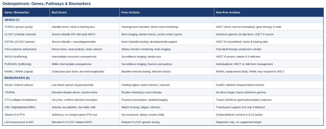
Osteopetrosis has a short list of usual suspects — TCIRG1, CLCN7, OSTM1, CA2, SNX10, PLEKHM1, and the RANKL/RANK pair — and each one breaks a different part of the same machine: the osteoclast's ability to dissolve old bone so new bone can take its place. Below, each gene gets a plain-language explanation of what it disrupts, what a mutation there tends to look like clinically, and a two-track plan — one path that needs no medication or equipment, and one that does, complete with realistic dosing patterns and side effects. After the genetics, six blood-based biomarkers are covered in depth, including a lesser-known enzyme pairing that can flag a mild adult form of the disease years before a fracture forces the question. A closer look at a widely cited research review then explains why specialists increasingly treat osteopetrosis as ten different diseases rather than one — a distinction that decides who actually benefits from a bone marrow transplant and who does not. Supportive therapies used during transplant and around jaw complications round out the picture, giving families something concrete to act on between specialist visits.
The Genes Behind Osteopetrosis and What Each One Means in Practice
Researchers like Ali Torkamani, who has spent years arguing that whole-genome sequencing should be used to catch rare pathogenic variants long before symptoms force the issue, and clinicians like Gary Brecka, who has popularized the idea that a genetic report should drive a specific action plan rather than generic lifestyle advice, both make the same basic point: knowing which gene is broken changes what you do next. Osteopetrosis is close to a textbook case. Mutations in at least ten genes are known to cause it, and together they account for roughly 80% of diagnosed cases, with a handful of genes responsible for the large majority. Below are the seven most clinically important ones, grouped by what they actually do inside the osteoclast, according to the detailed genotype-function review published in Frontiers in Endocrinology and the StatPearls osteopetrosis overview.
TCIRG1 — the acid pump switch
TCIRG1 codes for a subunit of the proton pump that osteoclasts use to acidify the space where they attach to bone. Without acid, old bone mineral cannot dissolve, so the osteoclast sits on the bone surface without resorbing it. Pathogenic variants here account for more than half of all autosomal recessive osteopetrosis cases, according to the GeneReviews entry on TCIRG1-related osteopetrosis, and they typically cause the severe, infantile-onset form that presents with vision loss, low blood counts, and fractures within the first year of life.
If this gene carries a pathogenic variant: the plan without supplements
There is no lifestyle substitute for a functioning proton pump. The realistic non-pharmacological plan is early referral to a bone marrow transplant center, baseline hearing and vision testing (cranial nerve compression is common as bone overgrows the skull's nerve canals), and close monitoring of blood counts, since the bone marrow cavity itself is being crowded out. Genetic counseling for future pregnancies is part of this plan, not an afterthought.
If this gene carries a pathogenic variant: the plan with supplements, medication, or equipment
Hematopoietic stem cell transplant (HSCT) is the only treatment that can meaningfully correct this defect, because osteoclasts are derived from the same blood-forming stem cells that a transplant replaces. Reduced-intensity conditioning protocols have improved survival and engraftment in infants who are otherwise poor candidates for full-intensity chemotherapy, per a study on HSCT with reduced-intensity conditioning for osteopetrosis. Side effects mirror any transplant: infection risk during engraftment, graft-versus-host disease, and a multi-month recovery. Gene therapy approaches that correct TCIRG1 in a patient's own stem cells before reinfusion are in preclinical and early proof-of-concept stages, reviewed in this gene therapy research summary — promising, but not yet a standard clinical option.
CLCN7 — the chloride channel that makes or breaks severity
CLCN7 encodes a chloride channel that works alongside the TCIRG1 proton pump to acidify the resorption space. What makes this gene unusual is that different types of mutations in the same gene cause wildly different diseases: recessive loss-of-function mutations cause a severe infantile form, while a specific dominant-negative mutation causes Albers-Schönberg disease, also called autosomal dominant osteopetrosis type II (ADO2) — a much milder, often adult-diagnosed condition, detailed in the GeneReviews entry on CLCN7-related osteopetrosis.
If this gene carries a pathogenic variant: the plan without supplements
For the milder ADO2 form, the non-pharmacological plan is largely about fracture prevention and surveillance: periodic bone density and skeletal imaging, dental checkups every six months (jaw osteomyelitis is a known complication), avoidance of high-impact contact sports, and awareness that standard osteoporosis drugs like bisphosphonates are inappropriate here, since bone is already over-mineralized rather than under-mineralized.
If this gene carries a pathogenic variant: the plan with supplements, medication, or equipment
Interferon gamma-1b, an FDA-approved therapy originally developed for chronic granulomatous disease, has been studied in non-infantile osteopetrosis and modestly increases bone turnover markers in some patients, based on an open-label pilot study in non-infantile osteopetrosis, though a related trial found it did not meaningfully change resorption markers specifically in ADO2, per this ADO2-specific analysis — a good example of why genotype matters even within one gene. Dosing is typically three subcutaneous injections per week, cycled over months with regular blood count monitoring; expect flu-like symptoms, fever, and injection-site soreness, especially after the first few doses. Vitamin D and calcium should only be supplemented if blood levels show an actual deficiency, since these patients do not benefit from — and may be harmed by — extra calcium loading.
OSTM1 — the chloride channel's essential partner
OSTM1 does not do much on its own; its job is to stabilize the CLCN7 protein and help it reach the right location in the cell. Mutations here cause one of the most severe infantile forms of osteopetrosis, frequently accompanied by primary neurodegeneration that is separate from bone-related nerve compression, an important distinction covered in the same Frontiers osteopetrosis genotype review.
If this gene carries a pathogenic variant: the plan without supplements
Because neurodegeneration in OSTM1-related disease is intrinsic to the mutation rather than caused by bone overgrowth pressing on nerves, the honest non-pharmacological plan centers on early neurological baseline testing, palliative and developmental support planning, and setting expectations with the family that a transplant corrects the blood and bone components of the disease but has limited effect on the neurological trajectory.
If this gene carries a pathogenic variant: the plan with supplements, medication, or equipment
HSCT remains the standard offered intervention for the hematologic and skeletal manifestations, following the same reduced-intensity protocols used for TCIRG1-related disease. Supportive equipment matters here more than pharmacology: low-vision aids, hearing amplification, and early physical and occupational therapy referrals give a child the best functional outcome regardless of the neurological course. Any medication trial (interferon gamma, calcitriol) should be framed to families as supportive, not corrective, for this specific gene.
CA2 — the milder, kidney-and-brain-involving form
CA2 encodes carbonic anhydrase II, an enzyme both osteoclasts and kidney cells use to manage acid-base balance. Its deficiency causes a distinctive triad: osteopetrosis, renal tubular acidosis, and brain calcification, described in detail in this case report on carbonic anhydrase II deficiency. Unlike TCIRG1 or OSTM1 disease, most patients follow an indolent course and are often diagnosed well after infancy.
If this gene carries a pathogenic variant: the plan without supplements
Regular kidney function monitoring (basic metabolic panel, urine pH), a baseline brain MRI or CT to document calcification, growth tracking, and developmental assessment form the core of a non-pharmacological plan. Because the bone phenotype is usually milder, transplant is considered case by case rather than automatically.
If this gene carries a pathogenic variant: the plan with supplements, medication, or equipment
The renal tubular acidosis component responds well to oral alkali therapy — typically potassium citrate or sodium bicarbonate, split into two to three daily doses to keep blood bicarbonate in range. Side effects are usually mild gastrointestinal upset at higher doses; treatment is generally lifelong rather than cycled, with periodic blood gas and electrolyte rechecks to adjust the dose as a child grows.
SNX10 and PLEKHM1 — the trafficking genes
Both of these genes are involved in moving vesicles inside the osteoclast so that acid and enzymes actually reach the bone surface instead of staying trapped inside the cell. SNX10 mutations account for a small but consistent share of intermediate autosomal recessive osteopetrosis, and PLEKHM1 mutations cause a rarer, generally milder intermediate form, both summarized in the genotype-treatment review referenced above.
If this gene carries a pathogenic variant: the plan without supplements
Because these forms tend to sit between the mild adult type and the severe infantile type, the non-pharmacological plan is individualized: some children need transplant-level intervention, others are managed with surveillance imaging, dental care, and fracture precautions similar to the ADO2 plan. This is one area where a second opinion from a center that specifically manages osteopetrosis genotypes is worth the wait.
If this gene carries a pathogenic variant: the plan with supplements, medication, or equipment
When the clinical picture is closer to the severe end, the same HSCT pathway used for TCIRG1 applies, since the defect is still intrinsic to the blood-derived osteoclast. When it is closer to the mild end, management mirrors the ADO2 approach: cautious vitamin D correction only if deficient, dental surveillance, and avoidance of bone-density-altering drugs not designed for this condition.
RANKL and RANK (TNFSF11 / TNFRSF11A) — the signal, not the cell
This pair is genuinely different from the others. RANKL is the signal that bone-forming cells send to tell osteoclast precursors to mature; RANK is the receptor on the osteoclast precursor that receives it. Mutations in either gene cause "osteoclast-poor" osteopetrosis — there simply aren't enough functioning osteoclasts, rather than plenty of osteoclasts that can't do their job. This distinction is documented in studies on RANKL-deficient osteopetrosis and RANK-deficient osteopetrosis with hypogammaglobulinemia.
If this gene carries a pathogenic variant: the plan without supplements
Because RANK mutations can also affect immune signaling, a non-pharmacological plan includes baseline immunoglobulin testing and infection-history review, not just skeletal survey. Families should be told directly that this subtype behaves differently from the others in one crucial way described below.
If this gene carries a pathogenic variant: the plan with supplements, medication, or equipment
RANKL-deficient osteopetrosis generally does not respond to standard bone marrow transplant, because the transplanted cells still lack the external RANKL signal needed to mature into working osteoclasts — the problem isn't in the marrow, it's in the signal reaching it. Research groups are testing recombinant RANKL protein replacement as an experimental alternative, an approach still confined to case reports and early trials rather than routine care. RANK-deficient disease, by contrast, can sometimes still respond to transplant since the defect is intrinsic to the precursor cell itself. This is precisely the kind of nuance a general "osteopetrosis treatment" plan misses, and why exact genetic confirmation changes the conversation with a transplant team rather than just confirming a diagnosis they already suspected.
Understanding which gene is involved changes not just the prognosis conversation but the specific tests worth running in the months between specialist visits — which is where blood-based biomarkers become genuinely useful.
Blood and Bone Markers Worth Tracking Alongside Genetic Testing
Genetic testing tells you which switch is broken; biomarkers tell you how the body is coping with it right now. Peter Attia and Thomas Dayspring have both argued, in the context of more common conditions, that tracking the right handful of numbers regularly beats an annual snapshot — the same logic applies here, scaled to a rare disease where changes in marrow function or bone turnover can happen faster than a yearly visit would catch. Below are six markers worth discussing with a hematologist or endocrinologist familiar with osteopetrosis.
Serum calcium and ionized calcium
Why it matters
Despite having unusually dense bones, people with severe osteopetrosis are paradoxically prone to low blood calcium, because osteoclasts can't release calcium from bone storage when the body needs it. This is most dangerous in infancy, where it can trigger seizures.
How to measure it
A basic metabolic panel with ionized calcium is a standard blood draw available at any hospital lab or pediatrician's office, typically $20–60 out of pocket without insurance, and is usually included at no extra cost during any hospital admission.
If the score is bad, the plan without supplements
Feeding schedule adjustments in infants and monitoring for tremors, irritability, or seizure activity are the immediate non-pharmacological steps, alongside urgent notification of the care team rather than waiting for a scheduled visit.
If the score is bad, the plan with supplements or equipment
Oral or intravenous calcitriol (activated vitamin D) is used to help the gut absorb more dietary calcium since bone can't be relied upon as a reserve; dosing is individualized and requires frequent blood recheck (often weekly at first) because the margin between too little and too much is narrow, and hypercalcemia carries its own risks, including kidney stones.
TRAP5b (tartrate-resistant acid phosphatase 5b)
Why it matters
TRAP5b is released by osteoclasts and normally correlates with how much bone resorption is happening. In osteopetrosis, it does something counterintuitive: it's often elevated because there are plenty of osteoclasts present, they're just not functioning, as shown in patients with Albers-Schönberg disease in a study of serum TRACP5b in ADO2. A high TRAP5b alongside dense bones on imaging is a specific pattern, not a contradiction.
How to measure it
This requires a specialty lab (not every hospital runs it routinely), typically costing $50–150, and is most useful when ordered by a specialist already following the diagnosis rather than as a first screening step.
If the score is bad, the plan without supplements
An elevated TRAP5b in a known osteopetrosis patient doesn't need independent correction — it's tracked over time to see whether a treatment (transplant, interferon gamma) is changing osteoclast activity, so the "plan" is really about consistent retesting intervals rather than intervention.
If the score is bad, the plan with supplements or equipment
If TRAP5b is being used to monitor interferon gamma-1b therapy, no additional supplement targets this marker directly; the therapy itself (three subcutaneous injections weekly) is the intervention, and TRAP5b trends alongside routine blood counts help the treating physician decide whether to continue, adjust, or stop it.
CTX (C-terminal telopeptide of type I collagen)
Why it matters
CTX is a fragment released when collagen in bone is broken down. In most bone diseases a high CTX means too much resorption; in osteopetrosis, CTX is typically suppressed, confirming that resorption is genuinely blocked rather than just slow.
How to measure it
A morning fasting blood draw, run at most major reference labs, generally $40–100. Timing matters — CTX has a daily rhythm, so consistent morning draws make results comparable over time.
If the score is bad, the plan without supplements
A persistently very low CTX confirms the resorption blockade; the practical non-pharmacological response is reinforcing fracture precautions and continuing skeletal surveillance imaging rather than trying to "raise" the number through diet or exercise, which won't move a mechanically blocked pathway.
If the score is bad, the plan with supplements or equipment
CTX is one of the clearer markers used to gauge whether interferon gamma-1b or, ultimately, transplant is restoring some resorption capacity; a rising CTX after transplant is generally a reassuring sign of osteoclast engraftment and is checked periodically (often every few months) during the first post-transplant year.
Complete blood count (hemoglobin, platelets, white cells)
Why it matters
As dense bone crowds the marrow cavity, the space available for making blood cells shrinks. Anemia, low platelets, and low white cell counts are often the first sign of disease progression in infants, and the main measure of engraftment success after transplant.
How to measure it
A standard CBC, among the cheapest and most widely available blood tests, roughly $10–30, and typically repeated frequently (weekly to monthly) in infants with severe disease or post-transplant patients.
If the score is bad, the plan without supplements
Watching for bruising, fatigue, recurrent infection, or pallor between blood draws, and having a low threshold to contact the care team rather than waiting for the next scheduled panel, is the realistic non-pharmacological vigilance plan.
If the score is bad, the plan with supplements or equipment
Severe cytopenias may require transfusion support while awaiting transplant, and iron supplementation is only appropriate if iron studies confirm true deficiency rather than the marrow-crowding pattern, since inappropriate iron supplementation in this context does not fix the underlying marrow space problem.
25-hydroxyvitamin D and parathyroid hormone (PTH)
Why it matters
Vitamin D status and PTH together clarify how the body is trying to compensate for calcium handling problems, and both need to be known before starting any calcitriol dosing decision described above.
How to measure it
A combined panel at most labs, roughly $60–120 total, usually ordered alongside the calcium panel rather than separately.
If the score is bad, the plan without supplements
Safe, moderate sun exposure and dietary review are reasonable first steps only for a mild vitamin D deficiency unrelated to the osteopetrosis itself; they are not a substitute for calcitriol when the deficiency is disease-driven.
If the score is bad, the plan with supplements or equipment
Standard cholecalciferol (vitamin D3) supplementation, typically dosed daily or weekly depending on the deficit, is appropriate for a true nutritional deficiency; recheck levels after 8–12 weeks. This is distinct from the specialist-directed calcitriol dosing used specifically to manage osteopetrosis-related hypocalcemia, and the two should not be combined without physician guidance because of the added hypercalcemia risk.
LDH isoenzymes and AST
Why it matters
This is a newer and less widely known finding: elevated lactate dehydrogenase isoenzymes and aspartate transaminase have been shown to distinguish CLCN7-related Albers-Schönberg disease from other sclerosing bone disorders, according to a study on LDH isoenzymes and AST in Albers-Schönberg disease. For a mild adult-onset form that's easy to mistake for ordinary dense bones on an incidental X-ray, this pairing can help point toward the right genetic test sooner.
How to measure it
A basic liver panel with LDH isoenzyme fractionation, available through most hospital labs, roughly $40–90; isoenzyme fractionation specifically may need to be requested rather than assumed as part of a routine panel.
If the score is bad, the plan without supplements
An unexplained elevation in these markers alongside dense bone on imaging is a reason to request CLCN7 genetic testing, not to pursue liver-focused workup on its own, since the pattern here reflects bone and marrow biology rather than liver disease.
If the score is bad, the plan with supplements or equipment
There's no direct supplement target for this marker pairing; its value is diagnostic rather than a treatment lever, and it's best used to justify prioritizing genetic confirmation and a baseline skeletal survey.
With the genetic and biomarker layers covered, it's worth stepping back to see how current research is reshaping the way specialists think about treating this disease at all — because the answer isn't as uniform as "transplant everyone."
What a Widely Cited Osteopetrosis Research Review Reveals
A detailed genotype-by-genotype review, published as "One Disease, Many Genes: Implications for the Treatment of Osteopetroses", makes a case that challenges the instinct to treat osteopetrosis as a single diagnosis with a single default answer. Here are ten of its most useful, practical takeaways.
1. Osteopetrosis is not one disease — it's at least ten
The review's central argument is that lumping every dense-bone diagnosis together obscures meaningfully different biology, prognoses, and treatment eligibility. A diagnosis code is a starting point for genetic testing, not an endpoint.
2. Osteoclast-rich and osteoclast-poor forms need opposite thinking
In osteoclast-rich forms (TCIRG1, CLCN7, OSTM1, SNX10, PLEKHM1), the cells are present but broken. In osteoclast-poor forms (RANKL, RANK), the cells are functionally absent. Treating both the same way ignores this fundamental split.
3. Frequency doesn't mean exclusivity
TCIRG1 explains the majority of severe cases, but "most common" is not "only cause" — a family with a severe presentation and a negative TCIRG1 test still needs broader panel testing rather than a dead end.
4. Transplant works because of cell lineage, not magic
HSCT succeeds specifically because osteoclasts originate from the same blood stem cells being replaced. This mechanistic detail is the reason transplant helps some genotypes and not others.
5. RANKL deficiency breaks the transplant logic entirely
Because the defect sits in the signal from bone-forming cells rather than in the osteoclast precursor itself, transplanting new marrow doesn't fix a missing RANKL signal — a genuinely counterintuitive finding that should stop a family from assuming transplant is automatically the answer.
6. Neurological involvement isn't always secondary to bone
For OSTM1 and some CA2 cases, neurological symptoms arise from the gene's role in neural tissue directly, not just from bone pinching a nerve — meaning a transplant that fixes the bone doesn't guarantee it fixes the neurology.
7. Earlier genetic diagnosis changes outcomes, not just answers
The review links earlier molecular diagnosis to earlier transplant referral and better transplant outcomes, reinforcing that genetic testing turnaround time is itself a clinical variable worth pushing on.
8. Mild adult forms are frequently missed
ADO2 is often picked up incidentally on imaging done for unrelated reasons and mistaken for a benign finding, meaning a family history of "unusually dense bones" mentioned casually is worth pursuing rather than dismissing.
9. Interferon gamma-1b is a bridge, not a cure
The review is candid that this therapy has modest, genotype-dependent effects and should be framed to families as a way to stabilize disease while awaiting transplant or as a milder-disease adjunct, not as a substitute for definitive treatment.
10. Gene therapy is the next frontier, not yet the standard
Correcting the defect directly in a patient's own stem cells, rather than replacing the entire marrow system with a donor's, is under active preclinical investigation and represents where the field is headed — worth asking about at a research-affiliated transplant center, but not yet available outside of trials.
The genotype-specific view from this research also explains why supportive care during transplant and around dental complications deserves its own attention — those experiences are shared across genotypes even when the underlying genetics differ.
Supportive Therapies That Can Ease the Medical Journey
None of the approaches below correct a genetic defect or replace transplant, medication, or dental surgery. Their value is in reducing distress, supporting recovery, and addressing the dental complications that are common across nearly every osteopetrosis genotype.
Music therapy during transplant
Because HSCT is the primary treatment pathway for most severe osteopetrosis genotypes, the supportive-care evidence built around pediatric and young-adult transplant patients transfers directly to this population, even though it wasn't studied in osteopetrosis specifically. Music therapy uses structured musical engagement, guided by a trained therapist, to reduce distress during the isolating weeks of transplant recovery.
A multi-site randomized trial through the Children's Oncology Group tested a therapeutic music video intervention in adolescents and young adults undergoing HSCT, delivered over six sessions across three weeks with a board-certified music therapist, and found improvements in resilience-related outcomes compared with a low-dose audiobook control, documented in this randomized trial of therapeutic music video during HSCT.
For a family preparing for transplant, asking the treatment center whether a certified music therapist is part of the supportive care team — and requesting sessions during the inpatient isolation period specifically — is a realistic way to apply this; it requires no equipment purchase and carries essentially no physical risk, though it works best as a structured program rather than passive background music.
Massage therapy around transplant
Massage therapy in this context refers to gentle, therapist-administered bodywork adapted for medically fragile patients, used to address the physical discomfort, anxiety, and isolation that accompany weeks of inpatient transplant care — again, a pathway most severe osteopetrosis genotypes pass through.
A multisite randomized trial evaluating complementary therapies, including massage, humor therapy, and relaxation/imagery, in 178 pediatric stem cell transplant patients found measurable reductions in distress associated with the transplant experience, reported in this multisite trial of complementary therapies for pediatric stem cell transplant, with a separate pilot randomized trial specifically testing massage for symptom management in pediatric HSCT reinforcing similar benefit, described in this pilot RCT of massage for pediatric HSCT symptom management.
In practice, this means requesting a pediatric-trained massage therapist through the transplant unit's integrative or child life services, scheduled several times weekly during the inpatient stay, with pressure and technique adjusted for low platelet counts and fragile skin — a detail any qualified pediatric HSCT massage program will already build into its protocol.
Photobiomodulation for jaw complications
Dense, poorly vascularized jaw bone in osteopetrosis carries a meaningfully elevated risk of osteomyelitis and delayed healing after dental procedures — a complication that overlaps closely with medication-related osteonecrosis of the jaw (MRONJ), a condition where low-level laser therapy has an actual evidence base. Photobiomodulation uses low-level laser light to support tissue healing and reduce inflammation at the wound site.
A systematic review of laser photobiomodulation dosimetry and treatment protocols in managing MRONJ found consistent, if still maturing, evidence supporting its use as an adjunct to standard surgical and antibiotic management, summarized in this systematic review of photobiomodulation in MRONJ management. Evidence specific to osteopetrosis-related jaw osteomyelitis is limited to case-level reports rather than dedicated trials, so this should be read as reasonable extrapolation, not condition-specific proof.
For someone with osteopetrosis facing a tooth extraction or jaw infection, the realistic application is asking an oral surgeon experienced in sclerosing bone disorders whether photobiomodulation is available as an adjunct alongside — never instead of — standard surgical and antibiotic care, given how much slower jaw bone in this condition heals compared to typical patients.
Diet and dental care fundamentals
Because jaw osteomyelitis is one of the most common complications across nearly every osteopetrosis genotype, and because dense jaw bone heals unusually slowly after any dental trauma or infection, prevention-focused dental care deserves the same weight as skeletal monitoring — and the principles in Cure Tooth Decay by Ramiel Nagel, which emphasizes nutrient-dense diet as a foundation for dental resilience, are worth mentioning here given how directly teeth are affected in this condition.
That said, Nagel's framework is a nutrition-and-prevention philosophy rather than a clinical trial-backed protocol, and it has not been studied in osteopetrosis specifically; it should be treated as a reasonable complement to — never a substitute for — the twice-yearly professional dental surveillance and conservative, infection-averse dental treatment planning that this condition genuinely requires.
In practice, this means prioritizing minimally invasive dental care, treating small cavities early rather than waiting for extraction to become necessary, and discussing any planned extraction well in advance with both the dentist and the treating hematologist or geneticist so that infection risk and healing time are anticipated rather than discovered after the fact.
Conclusion
Osteopetrosis is genuinely a genetic disease first — the gene involved determines severity, which treatments can work, and which ones structurally cannot, as the RANKL example makes clear. Biomarkers like TRAP5b, CTX, and calcium don't replace that genetic answer, but they give a family and their care team a way to see what's happening in the weeks and months between specialist visits, and the LDH/AST pairing shows that even the biomarker landscape here is still being actively refined. Supportive therapies around transplant and dental care won't change the underlying biology, but they address the real, human weeks spent in hospital rooms and dental chairs that a genetics report never mentions.
The most useful next step is rarely dramatic: confirm the exact gene involved if that hasn't happened yet, ask specifically which of the biomarkers above your care team is already tracking and which ones they aren't, and bring this article's questions — particularly around genotype-specific transplant eligibility — into your next appointment with the geneticist or transplant team. Precise information won't change the diagnosis, but it consistently changes how fast the right decisions get made.