This article contains AI generated content.
· UpdatedPCL Mucoid Degeneration — 4 Genes and 6 Biomarkers to Track
Introduction
Posterior cruciate ligament mucoid degeneration is one of those diagnoses that tends to arrive quietly — picked up on an MRI ordered for unexplained posterior knee pain, creeping stiffness, or a slow loss of full flexion. The ligament has not torn. Instead, a mucinous, gel-like substance has infiltrated its fibers, gradually altering the tissue architecture from within. Most people who receive this finding are told to manage symptoms, rest, and consider surgery if conservative care fails. That guidance is not wrong, but it leaves a significant gap.
What rarely gets discussed is that mucoid degeneration is not simply bad luck or random aging. The way your connective tissue remodels, the enzymes that degrade your extracellular matrix, and the molecular environment inside your ligament are shaped by factors that can be measured and, to a meaningful degree, influenced. For some people, the underlying biology actively accelerates degeneration. For others, standard rehabilitation simply does not address what is biochemically driving the problem.
Generic advice — rest, take it easy, try physiotherapy — misses this layer entirely. Whether you are preparing for a conversation with a specialist, weighing surgical options, or trying to slow a process that feels like it keeps progressing, understanding the metabolic and molecular picture behind your diagnosis changes the quality of every decision you make from here.
This article focuses on two evidence-informed frameworks. The first examines six biomarkers that reflect the cellular activity sustaining degeneration — markers of matrix breakdown, systemic inflammation, and nutritional deficits that most clinicians never order. The second looks at four genetic variants that predispose some individuals to faster ligament breakdown and poorer tissue repair. Both offer something concrete: numbers and findings to act on. Neither replaces clinical evaluation, but better information leads to better decisions.
Summary
This article identifies the 6 most actionable biomarkers to track when managing PCL mucoid degeneration — including COMP (a direct marker of ligament matrix breakdown), MMP-3 (the enzyme that erodes your extracellular matrix), homocysteine (which structurally weakens collagen cross-links), and three others that most standard panels miss entirely. For each, you will find how to measure it, its cost range, and a concrete protocol with and without supplements — including exact dosing, cycling schedules, and side effects. The article also covers 4 genetic variants (COL1A1, MMP3, TGFB1, TNXB) that explain why some people develop this condition disproportionately and why recovery can be slower than expected. After the biomarker and genetics sections, you will find 10 key takeaways from the Huberman Lab on connective tissue repair science, plus three complementary approaches with real human clinical evidence. If you have been told to wait and see, this gives you a more precise framework for deciding what to actually do.
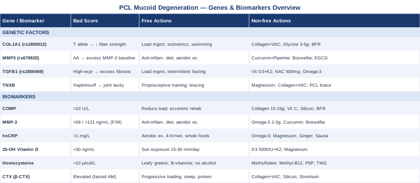
6 Biomarkers Worth Tracking When You Have PCL Mucoid Degeneration
Dedicated biomarker research on PCL mucoid degeneration specifically is still sparse — the condition is relatively uncommon as a primary diagnosis. The markers below are drawn from adjacent biology: ligament degeneration, tendinopathy, ACL failure studies, and knee connective tissue disease research. These conditions share the same core mechanisms — extracellular matrix (ECM) breakdown, inflammatory cytokine signaling, and collagen remodeling — and each marker below is selected because it reflects those processes directly. Together they create a metabolic picture that MRI alone cannot provide.
Biomarker 1: COMP — Cartilage Oligomeric Matrix Protein
Why it matters
COMP is a glycoprotein secreted by chondrocytes, tenocytes, and ligament fibroblasts. It acts as a structural scaffold in the ECM and stabilizes collagen fibers. When connective tissue is being actively degraded — through mechanical overload, inflammation, or enzymatic breakdown — COMP fragments leak into the bloodstream. Elevated serum COMP is one of the most sensitive early indicators of matrix breakdown in ligaments and cartilage, often rising before any significant change appears clearly on imaging. For PCL mucoid degeneration, elevated COMP signals active tissue remodeling rather than a stable lesion. Studies on knee osteoarthritis and tendinopathy consistently document that serum COMP tracks the rate of structural deterioration, as reviewed extensively in NIH-indexed literature over the past two decades.
How to measure it
COMP is measured via a fasting serum blood test. It is not a routine panel item, so you will need to request it through a sports medicine physician, rheumatologist, or a direct-access specialty lab. Cost range: approximately $80–$200 depending on the provider. Some functional medicine labs include it in connective tissue panels. Optimal range is generally below 10 U/L, though reference ranges vary by assay.
If the score is bad, the plan without supplements
Elevated COMP means mechanical load is exceeding the ligament's repair capacity. The first priority is deliberate load management: avoid deep knee flexion beyond 90 degrees, eliminate downhill running, reduce any activity producing posterior knee discomfort. Concurrently, progressive eccentric strengthening of the posterior chain — hamstrings, glutes, and the calf-soleus complex — reduces dynamic load on the PCL. Aquatic exercise (pool cycling, water walking) permits a training stimulus without joint compression. Sleep is not optional here: growth hormone pulses during deep sleep represent the most powerful endogenous driver of connective tissue repair, and even partial sleep deprivation measurably suppresses it.
If the score is bad, the plan with supplements or equipment
- Hydrolyzed collagen peptides (type I and II): 10–15g daily, taken 30 minutes before mechanical loading or rehabilitation, alongside a source of vitamin C. This timing exploits the peak fibroblast synthesis window identified in research by Keith Baar. No cycling required; GI side effects are rare. Minimum 12-week trial to assess structural benefit. - Vitamin C: 500–1000mg/day with food. Cofactor for the hydroxylation of proline and lysine — rate-limiting steps in stable collagen fiber formation. Safe long-term; doses above 2g/day may cause loose stools. - Bioavailable orthosilicic acid (silicon): 6–10mg daily. Small RCTs suggest measurable upregulation of type I collagen gene expression. No significant side effects at therapeutic doses; reassess at 8 weeks. - Blood flow restriction (BFR) training: using an occlusion cuff around the proximal thigh during low-load (20–30% of 1RM) resistance exercise elicits connective tissue adaptation comparable to heavy loading, at a fraction of the mechanical stress on the PCL. Requires proper cuff placement guidance from a trained therapist.
Biomarker 2: MMP-3 — Matrix Metalloproteinase-3
Why it matters
MMP-3 (stromelysin-1) is a protease that degrades collagen, proteoglycans, fibronectin, and other ECM components. It is upregulated in degenerated ligament tissue and plays a central enzymatic role in the matrix breakdown seen in mucoid degeneration. High serum MMP-3 indicates that the balance in the ligament's local environment has shifted toward catabolism — more breakdown than synthesis. Elevated MMP-3 has been documented in ACL degeneration and knee tendinopathy cohorts, and the same signaling cascade applies directly to PCL matrix erosion. Thomas Dayspring has referenced MMP family members as relevant markers of connective tissue and vascular matrix quality, and MMP-3 sits at the center of ligament-relevant ECM degradation pathways.
How to measure it
Serum MMP-3 is available through rheumatology panels and some functional medicine labs. Cost: $100–$300. Standard reference values vary by assay; values above approximately 59 ng/mL in women and above 121 ng/mL in men are generally flagged as elevated. It is sometimes included in broader inflammatory or autoimmune panels.
If the score is bad, the plan without supplements
MMP-3 is driven upward by chronic low-grade inflammation, oxidative stress, and poor metabolic health. The most powerful non-supplement approach is dietary: transition to a whole-food anti-inflammatory pattern that eliminates refined seed oils, ultra-processed carbohydrates, and added sugars. Prioritize fatty fish two to three times per week, colorful vegetables, olive oil, and polyphenol-rich foods. Even modest visceral fat reduction (5–10% of body weight in overweight individuals) substantially lowers systemic MMP-3 activity. Consistent moderate aerobic exercise — brisk walking, cycling, swimming — suppresses MMP-3 over 8–12 weeks. Quality and duration of sleep round out the non-supplement approach.
If the score is bad, the plan with supplements or equipment
- Omega-3 fatty acids (EPA + DHA): 2–3g combined EPA and DHA daily with the largest meal. Suppresses NF-κB signaling, the primary transcriptional driver of MMP-3. Possible mild anticoagulant effect at doses above 3g/day — flag this with any clinician if you are on blood thinners. No cycling required; recheck at 12 weeks. - Curcumin with piperine: 500–1000mg curcumin extract plus 5–10mg piperine per dose, twice daily with food. Piperine elevates curcumin bioavailability by approximately 20-fold. Directly inhibits NF-κB-driven MMP-3 transcription. Cycle: 8 weeks on, 2 weeks off. Side effects: mild GI discomfort in some; avoid in active gallbladder disease; potential interaction with anticoagulants at high doses. - Boswellia serrata (AKBA standardized): 300–500mg standardized extract (minimum 30% AKBA), 2–3 times daily with food. Inhibits 5-LOX and IL-1β-mediated inflammatory signaling. Cycle: 12 weeks on, 4 weeks off. Side effects: occasional mild GI upset; well-tolerated in most people. - Infrared sauna: 15–20 minutes at 140–160°F, 3–4 sessions per week. Heat stress activates heat shock proteins that modulate MMP expression and support fibroblast repair. Evidence is mechanistic rather than RCT-grade for PCL specifically, but the systemic anti-inflammatory effect on MMP activity is well-supported.
Biomarker 3: hsCRP — High-Sensitivity C-Reactive Protein
Why it matters
hsCRP is a liver-derived acute-phase protein and one of the most accessible markers of systemic inflammatory activity. While not specific to PCL degeneration, chronic elevation signals that the inflammatory environment throughout the body — including around the knee — is persistently activated. Local ligament inflammation is closely tied to systemic inflammatory markers in tendon and ligament degeneration research. Peter Attia consistently identifies hsCRP as a core metabolic health marker, noting that even mildly elevated levels (above 1 mg/L) are associated with accelerated connective tissue aging, impaired fibroblast function, and reduced collagen net synthesis over time.
How to measure it
Standard blood test, widely available. Cost: $20–$50. It must be specifically requested — most comprehensive metabolic panels do not include hsCRP by default. Optimal target: below 0.5 mg/L. Acceptable: below 1.0 mg/L. Values above 3 mg/L indicate significant chronic inflammation worth addressing as a priority.
If the score is bad, the plan without supplements
Consistent moderate-intensity aerobic exercise is the single most evidence-supported non-pharmacological intervention for hsCRP reduction — 30–45 minutes of brisk walking, cycling, or swimming four to five days per week has been shown to reduce hsCRP by 20–30% over 12 weeks in multiple RCTs. Combined with elimination of ultra-processed food, reduction of refined sugar, and 7–9 hours of restorative sleep, this achieves meaningful CRP reduction in most people. Chronic cortisol elevation — from work stress, relationship stress, or sleep debt — is a major upstream driver of inflammatory cytokine production; it belongs in the intervention plan, not just as a footnote.
If the score is bad, the plan with supplements or equipment
- Omega-3 (EPA + DHA): as described above; independently reduces hsCRP via prostaglandin pathway modulation. - Magnesium glycinate or threonate: 300–400mg elemental magnesium in the evening. Magnesium deficiency — very common in Western diets — is independently associated with elevated inflammatory markers. Start at 150mg and titrate up to avoid loose stools. Safe long-term. - Ginger extract: 500–1000mg standardized extract with food. COX and LOX inhibitor with modest anti-inflammatory evidence in human trials. Cycle: 8 weeks on, 2 weeks off. Rare side effects; may potentiate anticoagulant effects. - Regular sauna use: observational studies by Laukkanen and colleagues — published in JAMA Internal Medicine and NIH-indexed — show that regular sauna sessions (4–7 per week) correlate with substantially lower hsCRP over time in cohorts of several thousand Finnish adults. This is correlational, not causal, but the biological mechanism via heat shock protein induction is coherent.
Biomarker 4: 25-OH Vitamin D
Why it matters
Vitamin D functions as a steroid hormone that regulates hundreds of genes involved in inflammation, immune activation, and connective tissue maintenance. Vitamin D receptors are expressed in ligament fibroblasts, and deficiency impairs both collagen synthesis and the modulation of inflammatory cytokines within the tissue microenvironment. Epidemiological data consistently shows disproportionately high vitamin D deficiency rates in patients presenting with tendon and ligament pathologies. For PCL mucoid degeneration, adequate vitamin D creates a more favorable biochemical environment for the fibroblasts responsible for matrix repair and for the suppression of the chronic TGF-β1-driven fibrotic signaling implicated in the mucinous transformation itself.
How to measure it
Serum 25-hydroxyvitamin D (25-OH D3) is a standard test through any lab or direct-access service. Cost: $30–$80; often covered by insurance with appropriate clinical indication. Optimal target: 50–80 ng/mL (125–200 nmol/L). Below 30 ng/mL is deficient; below 20 ng/mL is severe deficiency. Re-test every 3–4 months while optimizing. The NIH Office of Dietary Supplements Vitamin D fact sheet provides authoritative reference ranges and safety thresholds.
If the score is bad, the plan without supplements
Direct sun exposure to large skin surface areas (arms, legs, torso) for 15–30 minutes between 10 AM and 3 PM, four to five days per week, can meaningfully raise vitamin D levels — particularly in lighter-skinned individuals in adequate latitudes. In northern climates, darker skin tones, or in winter months, sun alone is rarely sufficient to correct a true deficiency. Dietary sources (fatty fish, egg yolks, fortified foods) contribute modestly but cannot reliably normalize clinical deficiency without supplementation.
If the score is bad, the plan with supplements or equipment
- Vitamin D3 + K2 (MK-7 form): for values below 30 ng/mL, start at 5000 IU D3 plus 100–200mcg K2 (MK-7) daily for 8–12 weeks, then retest and titrate to a maintenance dose (typically 2000–3000 IU for most adults). K2 is essential — it directs calcium to bone rather than soft tissues and vasculature. Toxicity is rare below 10,000 IU/day but requires monitoring; do not supplement without periodic testing. - Magnesium: required as a cofactor for both hepatic and renal vitamin D activation. Magnesium deficiency renders vitamin D supplementation substantially less effective — the conversion to the active form depends on it. 300–400mg elemental magnesium daily, as described above.
Biomarker 5: Homocysteine
Why it matters
Homocysteine is an intermediate amino acid in the methionine cycle. When elevated, it directly degrades collagen cross-links by interfering with lysyl oxidase — the enzyme responsible for the covalent bonds that give collagen its tensile strength. High homocysteine produces structurally weaker, more brittle connective tissue over time. It is strongly associated with MTHFR gene variants (discussed in the genetics section), which means some individuals are metabolically predisposed to accumulating homocysteine regardless of dietary patterns. For anyone with PCL mucoid degeneration or a history of repeated ligament issues, homocysteine is one of the most underused and most actionable markers available. Thomas Dayspring and functional medicine researchers frequently cite elevated homocysteine as a silent driver of connective tissue vulnerability.
How to measure it
Fasting serum homocysteine, available through standard labs. Cost: $30–$100. Optimal target: below 7 µmol/L. Values above 10 µmol/L carry meaningful risk; above 15 µmol/L is clinical hyperhomocysteinemia requiring investigation and treatment. Context from methylation biology and MTHFR testing is available through the NIH Office of Dietary Supplements Folate fact sheet.
If the score is bad, the plan without supplements
Dietary optimization: increase intake of methyl-donor-rich foods — dark leafy greens (folate), animal proteins (B12, methionine), legumes, and beets (betaine). Reduce or eliminate alcohol, which depletes B-vitamins and predictably elevates homocysteine. If a MTHFR variant is present, dietary changes alone are typically insufficient — the enzymatic conversion pathway is impaired upstream, and supplement forms that bypass the defective step are needed.
If the score is bad, the plan with supplements or equipment
- Methylfolate (5-MTHF): 400–800mcg daily. Always prefer the methylated form over standard folic acid, especially in MTHFR C677T or A1298C carriers who cannot efficiently convert folic acid. Start low — in a small subset of people with severe methylation sensitivity, higher doses cause anxiety or irritability ("methyl trapping"); titrate slowly. - Methylcobalamin (B12): 500–1000mcg sublingual daily. The methylcobalamin form is significantly more effective than cyanocobalamin for homocysteine remethylation, especially in MTHFR variants. - Pyridoxal-5-phosphate (P5P, active B6): 25–50mg daily. Direct cofactor for the transsulfuration pathway of homocysteine clearance. Avoid sustained doses above 100mg/day — peripheral neuropathy risk with chronic very high B6 intake. - Trimethylglycine (betaine, TMG): 500–1000mg daily. Donates a methyl group via the BHMT pathway, providing homocysteine remethylation that is entirely independent of the MTHFR enzyme. Well-tolerated; continuous use; recheck homocysteine at 8–12 weeks to confirm response.
Biomarker 6: Serum CTX — C-Terminal Telopeptide of Type I Collagen
Why it matters
Serum CTX (β-CTX, CrossLaps) is a direct marker of type I collagen breakdown. As collagen fibers degrade, these telopeptide fragments are released into circulation. CTX is widely used in bone remodeling studies, but its relevance extends to ligament and tendon collagen turnover — type I collagen being the dominant structural protein in the PCL. Chronically elevated CTX indicates that collagen catabolism is outpacing synthesis: the precise imbalance that sustains and worsens mucoid degeneration over time. Allan Sniderman has referenced collagen crosslink markers in the context of connective tissue aging and vascular matrix quality; the same principle applies directly to ligamentous structures.
How to measure it
Fasting morning serum CTX — fasting for 12 hours is mandatory, as feeding suppresses CTX levels and creates confounded results between tests. Standard lab test. Cost: $80–$150. Values should always be measured under the same conditions (fasted, morning) for meaningful longitudinal comparison. Generally, lower values within the reference range indicate a healthier collagen turnover balance.
If the score is bad, the plan without supplements
Mechanical loading is the most powerful non-pharmacological signal to upregulate collagen synthesis relative to breakdown. Progressive, supervised posterior knee rehabilitation exercises — structured isometric and eccentric work — shift fibroblast gene expression toward net synthesis. Prioritize sleep architecture: growth hormone released during stages 3 and 4 sleep is the primary systemic anabolic signal for connective tissue; even modest sleep disruption measurably elevates CTX the following morning. Adequate dietary protein (1.6–2.2g per kg of body weight) provides the substrate: glycine and proline are particularly relevant amino acids for collagen synthesis and are often limiting in low-meat diets.
If the score is bad, the plan with supplements or equipment
- Hydrolyzed collagen peptides: as described in Biomarker 1. Pre-exercise timing amplifies the fibroblast synthesis signal. - Vitamin C: essential cofactor, as described. - Bioavailable silicon (orthosilicic acid): 6–10mg daily. Human trials (Jugdaohsingh and colleagues, NIH-indexed in the Journal of Bone and Mineral Research) demonstrate upregulation of type I collagen gene expression. No significant side effects; continuous use. - Strontium citrate (optional, with medical supervision): 340mg daily shifts the collagen remodeling balance toward synthesis. Cycle maximum 6 months on, followed by a 1-month break. Monitor kidney function. Not appropriate for those with renal impairment. Evidence is stronger for bone than for ligament specifically — discuss with a clinician before starting.
What Your Genes May Be Telling You About PCL Mucoid Degeneration
Direct genetic research into PCL mucoid degeneration as a specific entity is essentially absent — the diagnosis is too uncommon to have attracted dedicated genome-wide association studies. What does exist is substantial evidence linking specific genetic variants to connective tissue weakness, impaired collagen synthesis, and accelerated ECM degradation in adjacent conditions: tendinopathy, ACL injury, Achilles degeneration, and ligament laxity syndromes. These variants create a biological environment where mucoid degeneration is more likely to develop and harder to reverse. The four variants below represent the most mechanistically relevant starting points. A genetics panel (commercial DNA testing with raw data review, or clinical ordering through a geneticist) can confirm which apply to you.
Gene 1: COL1A1 (rs1800012) — The Collagen Scaffold Gene
COL1A1 encodes the alpha-1 chain of type I collagen — the primary structural protein in ligaments and tendons. The rs1800012 variant (located in an Sp1 transcription factor binding site) is associated with reduced collagen fiber diameter and lower tensile strength in ligamentous tissue. Carriers of the T allele produce collagen with altered mechanical properties: lower load tolerance and increased susceptibility to micro-tear and progressive degeneration under repetitive stress. This variant has been studied extensively in ACL rupture and Achilles tendinopathy cohorts, consistently showing higher degeneration rates and poorer outcomes in T allele carriers, as documented in NIH-indexed meta-analyses published in the American Journal of Sports Medicine and British Journal of Sports Medicine. The same biology applies to PCL matrix quality, though direct PCL studies have not yet been conducted.
If the gene is bad, the plan without supplements
COL1A1 risk carriers need more deliberate load management and longer progressive loading progressions than average. This means strict avoidance of sudden spikes in training volume or impact — the "weekend warrior" loading pattern is particularly harmful. Warm-up and cool-down should be extended. Dedicated collagen-stimulating isometric exercise — contractions held at 70% of maximum voluntary contraction for 20–45 seconds — provides the most well-supported mechanical signal for collagen gene upregulation in tendon and ligament tissue without excessive shear stress. Swimming and cycling keep cardiovascular fitness high while drastically reducing PCL loading. Proprioceptive training (balance boards, single-leg stability work) reduces dynamic loading asymmetries that stress already-vulnerable tissue.
If the score is bad, the plan with supplements or equipment
- Hydrolyzed collagen peptides + Vitamin C: especially important for COL1A1 risk carriers. Exogenous collagen peptides upregulate endogenous fibroblast collagen synthesis even when the gene itself produces suboptimal product. 10–15g daily, pre-loading, continuous protocol. - Glycine: 3–5g daily. The most abundant amino acid in collagen and often the rate-limiting substrate for synthesis. Low toxicity; some evidence for improved sleep quality as a side benefit. - BFR training: provides connective tissue adaptation stimulus at mechanical loads that do not risk further ligament compromise. Work with a trained rehabilitation professional.
Gene 2: MMP3 (rs679620) — The Matrix Destroyer Gene
The MMP3 rs679620 variant determines baseline transcription rate of the MMP-3 enzyme. The AA genotype is associated with higher constitutive MMP-3 expression — meaning carriers produce more collagen-degrading enzyme at baseline, independent of acute inflammation. This creates a chronically catabolic bias in the ECM environment of the ligament. Studies in knee osteoarthritis, intervertebral disc degeneration, and ACL injury cohorts consistently show higher degeneration rates and worse structural outcomes in AA carriers (Takahashi and colleagues, referenced in NIH-indexed Arthritis & Rheumatism literature). If your serum MMP-3 is persistently elevated and you carry this variant, the connection is biologically coherent and explains a great deal about why conservative management may not be working.
If the gene is bad, the plan without supplements
The priority for MMP3 AA carriers is removing every environmental amplifier of MMP-3 transcription: eliminate ultra-processed food and dietary advanced glycation end products (AGEs — formed in high-heat cooking of meat, particularly from processed foods), maintain healthy body composition (adipose tissue is a major source of TNF-α and IL-1β that drive MMP-3 upregulation), and adopt a Mediterranean-style dietary pattern. Regular moderate aerobic exercise — not overtraining, which itself raises MMP-3 acutely — consistently reduces systemic MMP activity over 8–12 weeks. Reducing chronic stress lowers inflammatory tone that primes MMP-3 expression at the transcriptional level.
If the score is bad, the plan with supplements or equipment
- Curcumin + Piperine: as described in Biomarker 2. Directly inhibits NF-κB-mediated MMP-3 transcription. For MMP3 AA carriers, this is the most targeted natural compound available. Cycle 8 weeks on/2 weeks off. - Boswellia serrata (AKBA): as described. Complementary mechanism via 5-LOX inhibition. Cycle 12 weeks on/4 weeks off. - EGCG (green tea extract): 400–600mg standardized EGCG per day with food. Inhibits MMP-3 and MMP-13 at the transcriptional level through AP-1 pathway modulation. Cycle: 8 weeks on, 2 weeks off. Stay within the 400–600mg range; rare hepatotoxicity has been reported at doses above 800mg EGCG daily. Do not take on an empty stomach.
Gene 3: TGFB1 (rs1800469) — The Fibrosis and Remodeling Regulator
TGF-β1 is a master regulator of fibrosis, ECM deposition, and tissue remodeling throughout the body. The rs1800469 polymorphism in the TGFB1 promoter region alters transcription rate. High-expression variants drive excessive fibrotic signaling and abnormal ECM accumulation — which is precisely what mucoid degeneration represents at the tissue level: not clean matrix loss, but a disorganized proteoglycan-rich infiltration that disrupts normal collagen architecture. TGF-β1 is the principal signal responsible for the mucinous transformation itself, upregulating hyaluronan and proteoglycan deposition in the ligament while disorganizing collagen fiber alignment. Low-expression variants, conversely, impair the repair signal after injury. This gene sits at the mechanistic center of PCL mucoid degeneration biology; evidence is from related connective tissue conditions rather than PCL-specific studies, but the mechanistic case is strong.
If the gene is bad, the plan without supplements
For high-expression TGFB1 variants: avoid repetitive mechanical stress that chronically reactivates the TGF-β1 repair signal, as this may perpetuate the mucinous transformation cycle. Load management, anti-inflammatory diet, and reduction of metabolic inflammatory drivers apply as described throughout this article. Intermittent fasting (16:8 pattern or similar) has been shown in mechanistic studies to modulate TGF-β signaling in tissue remodeling contexts — human evidence is early-stage, but the metabolic benefits on inflammatory tone are well-established.
If the score is bad, the plan with supplements or equipment
- Vitamin D3 + K2: vitamin D directly modulates TGF-β signaling, particularly attenuating excessive profibrotic TGF-β1 responses. Dosing as in Biomarker 4. - NAC (N-acetylcysteine): 600–1200mg daily. Antioxidant and glutathione precursor; cell and animal studies show attenuation of TGF-β1-driven fibrotic cascades. Take with food to reduce GI side effects. No cycling needed; however, sustained use above 1200mg/day warrants monitoring of kidney function. Keep to the lower end of the range unless directed otherwise. - Omega-3 (EPA + DHA): modulates the prostaglandin and cytokine profile that feeds TGF-β1 overactivation. As described throughout — this is one of the most cross-cutting interventions in this article.
Gene 4: TNXB — Tenascin-X and Connective Tissue Integrity
Tenascin-X, encoded by TNXB, is an ECM glycoprotein that regulates collagen fibril spacing, mechanical elasticity, and tissue tensile resistance in ligaments and tendons. Complete TNXB deficiency causes a classical form of Ehlers-Danlos syndrome (EDS). Haploinsufficiency — one defective copy — produces a subtler phenotype: hypermobile joints, ligaments that fatigue faster than expected, and connective tissue that degenerates disproportionately relative to activity level. Many individuals with TNXB haploinsufficiency pass through standard clinical evaluations undiagnosed. For PCL mucoid degeneration that presents in a relatively young, active person whose lifestyle does not obviously account for the degree of degeneration found — or where the condition recurs after treatment — a TNXB variant deserves consideration. The connection between TNXB deficiency and accelerated ligament matrix disorganization is supported by structural biology research from the EDS and collagen fibril literature.
If the gene is bad, the plan without supplements
TNXB haploinsufficiency requires a fundamentally different approach to loading. Proprioceptive and neuromuscular training become the cornerstone: the structural integrity of the ligament itself cannot be fully normalized, but the neuromuscular control system surrounding the knee can compensate substantially for laxity and instability. Avoid exercise approaches that exploit hypermobility. Avoid sustained end-range knee hyperextension. Posterior knee bracing or compression during higher-intensity activities reduces cumulative micro-stress on the PCL. Pacing recovery carefully — not pushing through joint discomfort signals — is more important for TNXB carriers than for the general population.
If the score is bad, the plan with supplements or equipment
- Magnesium glycinate: 300–400mg elemental magnesium daily. Cofactor for collagen cross-linking; relevant for any connective tissue condition. - Hydrolyzed collagen peptides + Vitamin C: while TNXB deficiency is structural, maximizing collagen production from the remaining functional fibroblast population still matters. Continuous protocol. - High-dose Vitamin C: some EDS literature supports 1000–2000mg daily in TNXB-related connective tissue fragility, as high-dose ascorbate enhances post-translational collagen modification even when structural ECM proteins are suboptimal. At sustained doses above 1000mg/day, monitor for kidney stone risk, particularly in those with a personal or family history. - Posterior PCL compression brace (equipment): a PCL-specific hinged brace or posterior compression orthosis reduces tibial translation under load and lowers the daily mechanical stress accumulation on an already-compromised ligament. Particularly valuable for TNXB carriers who engage in any activity above light walking.
What the Huberman Lab Reveals About Connective Tissue Repair
Andrew Huberman, Stanford neuroscientist and host of the Huberman Lab podcast, has addressed connective tissue biology across multiple episodes — most substantively in his content on flexibility, collagen synthesis, and tissue repair mechanisms, drawing extensively from research by Keith Baar and other exercise physiologists. His work translates directly to conditions like PCL mucoid degeneration. The following ten points represent the most actionable insights from this body of content.
1. Collagen Synthesis Requires a Specific Mechanical Signal, Not Just Rest
Passive rest does not stimulate fibroblast collagen production. Huberman explains that tendons and ligaments require isometric and eccentric mechanical loading to activate the gene expression cascade for collagen synthesis. Supplementation alone has limited structural effect without the mechanical trigger.
2. Vitamin C Timing Matters More Than Daily Total
Based on Baar's research, taking vitamin C 30–60 minutes before loading creates a transient spike in available hydroxyproline at precisely the moment fibroblasts need it for new collagen fiber assembly. Total daily dose matters less than this strategic timing window.
3. Deep Sleep Is the Primary Repair Window
Stages 3 and 4 of sleep drive the largest pulse of growth hormone secretion, which is the dominant anabolic signal for connective tissue repair. Huberman cites multiple studies showing that even modest sleep disruption measurably elevates collagen breakdown markers and impairs ligament healing rates.
4. The Synthesis Window After Loading Is Only 4–6 Hours
Based on gelatin and collagen research, peak post-exercise collagen synthesis lasts approximately 4–6 hours after loading with vitamin C support. Distributing loading across the day with appropriate rest intervals exploits this window more effectively than one long daily session.
5. Heat Increases Extensibility — It Does Not Repair Structure
An important distinction Huberman emphasizes: heat transiently increases tissue extensibility and range of motion, but has not been shown to accelerate structural repair in degenerated connective tissue. Sauna is useful for inflammation control and recovery; it should not be conflated with a repair intervention.
6. Blood Flow Restriction Preserves Adaptive Stimulus During Injury
BFR training has been discussed as a validated method for stimulating muscle and connective tissue adaptation at 20–30% of maximum load. For PCL conditions where full loading is contraindicated, BFR preserves the anabolic signaling environment without mechanically stressing the vulnerable ligament.
7. Chronic Stress Directly Suppresses Collagen Synthesis
Cortisol inhibits fibroblast collagen gene expression. Huberman references multiple studies showing that individuals under sustained psychosocial stress have measurably impaired tendon and ligament healing trajectories. Stress reduction is not optional wellness advice — it is a connective tissue intervention.
8. Omega-3s Have Direct Connective Tissue Effects Beyond Inflammation
Beyond systemic anti-inflammatory effects, EPA specifically reduces MMP activity in connective tissue directly and shifts the prostaglandin balance toward repair-permissive conditions at the tissue level. The effect is independent of CRP reduction.
9. Collagen Peptides Are Not Interchangeable With Standard Protein Supplements
Hydrolyzed collagen peptides contain a specific dipeptide — prolyl-hydroxyproline — that acts as a direct fibroblast signaling molecule, independently stimulating collagen synthesis. This is mechanistically distinct from the general amino acid pool provided by whey or casein protein. The two are not substitutes for connective tissue repair purposes.
10. The Goal Is Not to Eliminate Inflammation — It Is to Resolve It
Huberman returns repeatedly to the point that inflammation is an essential and functional early repair signal. The problem in chronic degeneration is not inflammation per se, but a failure of resolution — the inflammatory process never completes. For PCL mucoid degeneration, the goal is supporting acute inflammatory resolution while eliminating the chronic smoldering that drives ongoing matrix degradation.
Complementary Approaches With Evidence Worth Considering
Moving from the molecular level to practical rehabilitation tools, the following three modalities have meaningful human clinical evidence for ligament and soft tissue conditions. They are not alternatives to medical care, but well-considered adjuncts.
Low-Level Laser Therapy (Photobiomodulation)
Low-level laser therapy (LLLT) delivers specific wavelengths of light — typically 808nm or 904nm — to tissues at non-thermal doses. It activates cytochrome c oxidase in the mitochondrial respiratory chain, increasing ATP production, reducing oxidative stress, and modulating the cytokine environment in treated tissue. For ligament and tendon degeneration, photobiomodulation has been shown to upregulate fibroblast collagen synthesis and accelerate tissue repair in multiple ex-vivo studies, with supporting data from human RCTs in Achilles tendinopathy and lateral epicondylopathy — the most well-studied tendon conditions. These findings are indexed on PubMed and reviewed in the Photomedicine and Laser Surgery literature. Direct RCT evidence for PCL mucoid degeneration does not exist, but the tissue-level mechanisms are directly relevant.
A specific protocol from human tendinopathy trials: 808–904nm, 3–5 sessions per week, 10–15 minutes per session over 4–8 weeks, applied to the posterior knee region. Adequate energy delivery per session (measured in joules per square centimeter) matters significantly — clinical class IIIB or IV lasers in a physiotherapy setting deliver appropriate doses more reliably than consumer-grade devices. Home devices exist but require consistent daily use to accumulate comparable energy doses. Seek a sports rehabilitation clinic that combines LLLT with an active loading protocol.
This modality works best as an adjunct to progressive mechanical rehabilitation, not as a standalone treatment. LLLT is broadly safe; contraindications include application over active malignancy, active infection, or directly over growth plates in skeletally immature patients. Results are highly dependent on device quality and treatment parameters — a poorly calibrated device delivers little benefit regardless of session frequency.
Tai Chi
Tai chi is a Chinese movement practice combining slow, deliberate postures with dynamic weight-shifting and sustained proprioceptive challenge. For posterior knee conditions, it is specifically relevant because it builds neuromuscular joint control without imposing significant compressive or shear load on the PCL. People with PCL degeneration frequently develop compensatory movement patterns — subtle increases in tibial posterior translation during gait and stair descent — that accumulate stress on the damaged ligament. Tai chi re-educates lower extremity movement mechanics while building hip, quadriceps, and hamstring endurance in a very low-risk environment.
Human evidence: a randomized controlled trial published in Arthritis Care & Research (Wang and colleagues, 2009, NIH-indexed) compared 12 weeks of Yang-style tai chi twice weekly against wellness education in knee osteoarthritis patients, finding significant improvements in pain scores, physical function, and proprioception. While this RCT was conducted in osteoarthritis rather than PCL degeneration, the neuromuscular and proprioceptive improvements are directly transferable — posterior knee stability in PCL degeneration depends on exactly the same muscle synergies that tai chi trains.
A realistic entry protocol: Yang-style beginner form, 2–3 sessions per week, 30–45 minutes per session, supervised for the first 8–12 weeks. Focus on single-leg weight transfer transitions and low-stepping patterns, which provide the highest proprioceptive challenge. Begin with shorter sessions (15–20 minutes) if pain or deconditioning is significant. Modify any posture that produces posterior knee pain; a qualified instructor will accommodate this readily.
Massage Therapy
Massage therapy — specifically deep tissue work and cross-fiber friction techniques — addresses soft tissue quality around the knee rather than the PCL itself, which is not directly accessible to manual therapy. The relevant targets are the posterior joint capsule, the proximal hamstring tendons, the popliteus muscle, and the medial and lateral gastrocnemius attachments — all of which become hypertonic and restricted in chronic posterior knee conditions. Restricted circulation in these tissues impairs the metabolic environment of the PCL region, reducing local oxygen and nutrient delivery to fibroblasts while impeding clearance of inflammatory waste products.
A specific technique: transverse cross-fiber friction massage applied perpendicular to the fiber direction of the hamstring tendons and posterior joint line for 5–10 minutes per session, 1–2 sessions per week over 4–8 weeks, is used in sports physiotherapy tendinopathy protocols for improving local tissue circulation and reducing adhesion formation. Evidence from sports rehabilitation research — including Prentice and colleagues — supports transverse friction massage for patellar tendon pathology; application to the posterior knee is widespread in clinical practice. Direct PCL-specific RCT evidence is absent, but the rationale is mechanistically sound.
Work with a licensed massage therapist experienced in sports injury rehabilitation. Avoid direct posterior knee pressure during active inflammatory flares. Integrate massage within a broader rehabilitation plan — combining it with progressive loading and LLLT where accessible creates a more complete treatment environment. Four to eight weeks of consistent treatment is a reasonable trial period; most people notice meaningful improvement in posterior knee tightness and pain within that window.
Conclusion
PCL mucoid degeneration is a structurally defined diagnosis, but the biology sustaining it is metabolic, genetic, and entirely measurable. The six biomarkers covered here — COMP, MMP-3, hsCRP, vitamin D, homocysteine, and CTX — provide a real-time window into the processes eroding your ligament matrix, and most of them are accessible through standard or specialty blood testing at reasonable cost. The four genetic variants — COL1A1, MMP3, TGFB1, and TNXB — help explain why the problem developed and why standard approaches may underdeliver for some individuals. Neither set of findings replaces surgical evaluation or supervised physiotherapy, but they sharpen the questions you bring to every clinical conversation.
The next smart step is concrete: work with your physician to order a targeted panel starting with the most accessible markers — hsCRP, 25-OH vitamin D, homocysteine, and COMP — and review the results in the context of your current rehabilitation. Introduce one or two specific interventions at a time so you can attribute what moves. If genetic testing is accessible to you through a direct-to-consumer or clinical route, reviewing your COL1A1 and MMP3 status adds a meaningful additional layer. Bring what you find to your orthopedic specialist or sports medicine physician. Precise information makes better conversations — and better conversations lead to better decisions about what your biology actually needs.
Musculoskeletal: Joint Conditions Tendon & Ligament Conditions Sports Injuries
Autoimmune: Inflammatory Conditions Connective Tissue Conditions