This article contains AI generated content.
Peroneal Tendinopathy: 4 Genes and 7 Biomarkers to Track
Persistent pain along the outside of the ankle, behind the fibula, that flares with running, hiking on uneven ground, or simply walking on a cambered street, does not always behave the way "tendinopathy" is described in generic handouts. Rest calms it down for a week, and it returns the moment training resumes. Stretching helps a little. Anti-inflammatories help a little. None of it explains why the tendon stays irritable for months while a training partner with a similar injury recovers in six weeks.
Generic advice about peroneal tendinopathy is built for an average case that may not be the one in front of you. "Reduce load, strengthen the ankle, be patient" is reasonable first-line guidance, but it says nothing about why some tendons remodel efficiently and others stay fragile despite doing everything right. Tendon tissue is not just a mechanical rope; it is a living structure whose repair speed depends on inflammation control, collagen substrate availability, hormonal signaling, and in some cases inherited differences in the collagen and enzyme genes that build and remodel it.
This article looks past the generic advice and toward the underlying biology, using two complementary lenses. The first is a set of blood biomarkers that are measurable today, inexpensively in most cases, and directly tied to how well tendon tissue heals and remodels. The second is a look at the genetic variants researchers have connected to tendon injury risk, with a clear separation between what has solid human evidence and what remains early or preliminary.
None of this replaces a proper clinical diagnosis or a structured loading program. But better information about what is happening inside the tissue, and inside your metabolism, tends to lead to better decisions than another round of generic rest-and-stretch advice. That is the grounded, non-miraculous premise of what follows.
Summary
Peroneal tendinopathy is rarely just a mechanical overuse problem. Underneath the biomechanics sit measurable factors, blood glucose and insulin resistance, lipid particles, thyroid output, uric acid, vitamin D and vitamin C status, and systemic inflammation, that shape how fast a tendon actually repairs itself, and inherited variants in collagen and enzyme genes that influence how resilient the tissue is in the first place. This article walks through the seven biomarkers worth tracking, what each one reveals, how to measure it, and what to do, with or without supplements, if the number is off. It then covers four gene variants with published human evidence in tendon injury, a recent podcast conversation with a leading tendon physiologist that overturns some standard rest-and-ice advice, and a short review of complementary approaches with real, condition-specific research behind them. Read on to see exactly which numbers matter, which genes are worth knowing about, and which of your current habits might be quietly working against tendon repair.
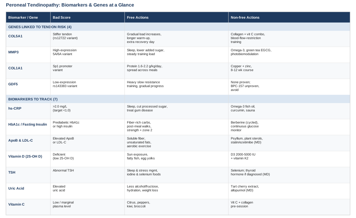
The Biomarkers Worth Tracking for Peroneal Tendinopathy
Tendon is a living, poorly vascularized tissue that depends heavily on the surrounding metabolic environment to repair itself. A tendon exposed to chronic high glucose, low-grade systemic inflammation, or nutrient shortfalls will remodel more slowly than one exposed to a stable, well-supplied environment, even under an identical loading program. The seven markers below are the ones with the clearest tie to tendon biology and the best balance of accessibility and evidence. Peter Attia, Thomas Dayspring, and Allan Sniderman have each pushed clinical practice toward using more precise, particle-based and inflammation-aware markers instead of relying on a single generic cholesterol number, and the same "look deeper than the obvious number" logic applies well here.
1. High-Sensitivity CRP (hs-CRP)
High-sensitivity C-reactive protein is the standard blood marker of low-grade systemic inflammation. Tendinopathy itself involves a local inflammatory and degenerative process, but a person carrying chronically elevated systemic inflammation, from poor sleep, visceral fat, gum disease, or a diet high in refined carbohydrates, gives an already-struggling tendon an even harder environment to repair in.
How to measure it: a standard hs-CRP blood draw, available through a primary care order or direct-to-consumer lab panel, costs roughly 15 to 30 US dollars alone or is often bundled into broader panels for 50 to 100 dollars. Attia and most longevity-focused physicians look for a result under 1.0 mg/L, treating anything persistently above 2.0 mg/L as worth investigating further.
If the score is bad, the plan without supplements: prioritize seven to nine hours of consistent sleep, reduce ultra-processed food and added sugar, address any untreated dental or gum inflammation, lose excess visceral fat if present, and keep training load progressive rather than spiky, since sudden load spikes themselves raise inflammatory markers. Recheck hs-CRP after eight to twelve weeks of consistent change, since it fluctuates with any acute illness or injury and a single reading during a flare-up is not reliable.
If the score is bad, the plan with supplements or equipment: omega-3 fish oil in the range of 2 to 3 grams combined EPA and DHA daily has the best-supported anti-inflammatory effect among common supplements; take it with food to reduce reflux, and expect to reassess at three months rather than expecting a quick shift. Curcumin with piperine (500 to 1000 mg daily) has modest supporting evidence and is generally well tolerated, though it can interact with blood thinners and should be cycled off for a week every couple of months if used long-term. Regular sauna use, three to four sessions weekly at 15 to 20 minutes, has been associated with lower inflammatory markers in observational data, though it should be avoided when acutely dehydrated or unwell.
2. HbA1c and Fasting Insulin
Glucose control matters to tendons more than most people expect. Chronic hyperglycemia drives the formation of advanced glycation end-products that stiffen and disorganize collagen cross-links, and clinical data show a clear association between diabetes, and even prediabetes-range glucose control, and tendon injury risk. One large observational analysis found meaningfully higher odds of lower-limb tendon injury in people with HbA1c already in the prediabetic range, well before a diabetes diagnosis would typically be made (impact of type 2 diabetes on tendinopathy development).
How to measure it: HbA1c is a standard blood test, roughly 10 to 40 dollars depending on whether it is bundled with other panels; fasting insulin is a similarly inexpensive add-on that reveals insulin resistance before HbA1c rises. For a more dynamic view, a continuous glucose monitor worn for one to two weeks (roughly 50 to 100 dollars per sensor) shows how specific meals and training sessions move blood sugar in real time.
If the score is bad, the plan without supplements: shift carbohydrate intake toward fiber-rich, minimally processed sources, walk for ten to fifteen minutes after meals to blunt the glucose spike, and add two to three weekly resistance training sessions plus zone 2 cardio, both of which independently improve insulin sensitivity.
If the score is bad, the plan with supplements or equipment: berberine, 500 mg two to three times daily with meals, has glucose-lowering effects comparable to some first-line medications in trials, but it should be cycled (roughly eight weeks on, one to two weeks off) and avoided in pregnancy or alongside certain medications without medical guidance; gastrointestinal upset is the most common side effect. A continuous glucose monitor is the most useful piece of equipment here, not to fix the number directly but to identify which specific foods and habits are driving the average up, which is often more actionable than the lab value alone.
3. ApoB and LDL-C
This is the marker where the Attia, Dayspring, and Sniderman perspective is most directly useful. Elevated LDL cholesterol does not just matter for arteries; cholesterol-carrying particles accumulate inside tendon tissue, and in cases of marked or prolonged elevation this shows up structurally as tendon thickening or, in more extreme cases, xanthomas, most classically in the Achilles tendon but reflecting a lipid-driven process that affects tendon tissue generally. A study of patients with hypercholesterolemia found a clear positive correlation between LDL-C level and Achilles tendon thickness, while HDL-C and ApoA-I were protective (Achilles tendon thickness and LDL-cholesterol in hypercholesterolemia). Sniderman's body of work argues that ApoB, which counts the actual number of atherogenic particles rather than estimating cholesterol mass, is the more accurate and more clinically actionable number, particularly when LDL-C and ApoB disagree (apoB, non-HDL-C, and LDL-C as cardiovascular risk markers).
How to measure it: a standard lipid panel is typically free or low-cost through insurance, or 20 to 40 dollars direct-to-consumer; adding a direct ApoB assay costs another 20 to 50 dollars and is worth requesting specifically, since it is not part of a routine panel by default. Advanced options include an NMR lipoprotein particle panel (roughly 100 to 150 dollars) and, for people with a strong family history, a one-time Lp(a) test and a coronary calcium score for broader cardiovascular context.
If the score is bad, the plan without supplements: increase soluble fiber intake (oats, legumes, psyllium-rich foods), replace saturated fat sources with unsaturated fats, maintain regular aerobic exercise, and lose excess weight if present, all of which meaningfully shift ApoB and LDL-C over eight to twelve weeks.
If the score is bad, the plan with supplements or equipment: psyllium husk (5 to 10 grams daily with water) and plant sterols (2 grams daily with meals) both have reasonable trial support for lowering LDL-C modestly and carry minimal side effects beyond occasional bloating. For persistently high ApoB despite lifestyle change, this moves from a supplement decision to a medical one; statins or ezetimibe are genuinely effective options but require a physician's judgment, not self-directed use, and should be discussed in the context of overall cardiovascular risk, not tendon health alone.
4. Vitamin D (25-OH D)
Vitamin D receptors are present in tendon tissue, and deficiency has been linked to impaired collagen formation, increased oxidative stress, and worse healing outcomes after tendon surgery. A large retrospective analysis found a clear association between vitamin D deficiency and distal biceps tendon injury (vitamin D deficiency and distal biceps tendon injury), and a broader scoping review found deficiency consistently linked to delayed healing and worse functional recovery after tendon repair procedures, even if the exact mechanism in non-surgical tendinopathy is still being worked out.
How to measure it: a 25-OH vitamin D blood test runs roughly 40 to 50 dollars, or is often included in an annual physical at no extra cost.
If the score is bad, the plan without supplements: 15 to 20 minutes of midday sun exposure on bare skin several times weekly can meaningfully raise levels in most latitudes for part of the year, balanced against reasonable sun-exposure caution, and fatty fish and egg yolks provide modest dietary contribution.
If the score is bad, the plan with supplements or equipment: vitamin D3, 2000 to 5000 IU daily depending on baseline deficiency severity, taken with a fat-containing meal for absorption, paired with a small dose of vitamin K2 (around 100 mcg) since the two work together in calcium handling. Retest after 10 to 12 weeks rather than daily dosing without follow-up, since vitamin D is fat-soluble and can accumulate; sustained levels above roughly 100 ng/mL warrant dose reduction and medical review.
5. TSH (with Free T4 and T3 if abnormal)
Thyroid hormone acts directly on tenocytes, the cells responsible for building and maintaining tendon matrix. Case reports and mechanistic studies show thyroid receptors on tendon cells, with thyroid hormone supporting tenocyte proliferation and limiting apoptosis; tendinitis has even been documented as a presenting symptom of otherwise undiagnosed hypothyroidism (hypothyroidism presenting as tendinitis, influence of thyroid hormones on tendon homeostasis).
How to measure it: TSH alone is a low-cost blood test, roughly 20 to 50 dollars; if TSH is abnormal, free T4 and free T3 add useful detail and are worth requesting in the same draw rather than as a separate visit.
If the score is bad, the plan without supplements: prioritize consistent sleep and stress management, both of which influence thyroid regulation, and ensure adequate dietary iodine and selenium from food sources like seafood, dairy, eggs, and Brazil nuts (in modest quantity).
If the score is bad, the plan with supplements or equipment: selenium, around 100 to 200 mcg daily, has some evidence for supporting thyroid function, particularly in autoimmune thyroid patterns, but should not exceed roughly 400 mcg daily due to toxicity risk, and is best taken as a defined course with periodic reassessment rather than indefinitely. True hypothyroidism identified on labs is a medical diagnosis requiring physician-directed thyroid hormone replacement; this is not an area for self-supplementation once a clinical diagnosis is confirmed.
6. Uric Acid
This is a specifically relevant marker for peroneal tendinopathy, more so than many general tendon discussions acknowledge. Elevated uric acid, even below the threshold of a clinical gout diagnosis, has been linked to disrupted tendon stem and progenitor cell function and to monosodium urate crystal deposition directly within tendon tissue. Foot and ankle tendons, including the peroneal tendons, are among the sites most frequently affected by urate deposition in imaging studies of gout patients (tendon involvement in the feet of patients with gout, dual-energy CT study), and asymptomatic hyperuricemia has been associated with impaired tendon stem cell function and higher tendon rupture risk (asymptomatic hyperuricemia and Achilles tendon rupture).
How to measure it: a standard blood uric acid test costs roughly 10 to 20 dollars and is often already included in a metabolic panel.
If the score is bad, the plan without supplements: reduce alcohol, especially beer, reduce high-fructose beverages, moderate intake of organ meats and shellfish, maintain good hydration, and lose excess weight if present, all of which lower uric acid meaningfully over weeks to months.
If the score is bad, the plan with supplements or equipment: tart cherry extract (480 to 960 mg daily, or roughly one cup of tart cherry juice) has trial support for modestly lowering uric acid and reducing gout flare frequency, with minimal side effects beyond occasional GI upset; it is reasonable to use for a defined 8 to 12 week block and reassess. If uric acid is high enough to reflect diagnosed gout, urate-lowering medication such as allopurinol is a physician-directed decision, not a supplement substitute.
7. Vitamin C (Plasma Ascorbate)
Vitamin C is not a marginal nutrient here, it is a required cofactor for the enzymes that stabilize collagen's triple-helix structure. Without adequate vitamin C, collagen synthesized in response to loading is structurally weaker, regardless of how well the training program is designed. A controlled trial found that consuming vitamin C-enriched gelatin roughly one hour before intermittent exercise nearly doubled a marker of collagen synthesis compared with placebo (vitamin C-enriched gelatin supplementation augments collagen synthesis), a finding with direct practical relevance to a structured peroneal tendon rehab program, discussed further below.
How to measure it: plasma vitamin C is not part of routine panels and needs to be specifically requested, typically 30 to 70 dollars through a specialty or direct-to-consumer lab; many people reasonably skip formal testing and instead assume marginal status if diet is low in fruits and vegetables.
If the score is bad, the plan without supplements: citrus fruit, bell peppers, kiwi, and broccoli provide substantial vitamin C; two to three servings daily is generally enough to reach adequate status without any supplement.
If the score is bad, the plan with supplements or equipment: vitamin C, 500 mg to 1 gram, taken alongside 15 grams of hydrolyzed collagen or gelatin, 30 to 60 minutes before a tendon-loading rehab session, is the specific protocol with trial support. This is worth cycling around actual training days rather than taking daily indefinitely; doses above 2 grams can cause gastrointestinal upset, and very high sustained intake carries a small kidney stone risk in people already predisposed to them.
Taken together, these seven markers describe a metabolic and inflammatory backdrop that either supports or undermines tendon repair, independent of the loading program itself. Genetics adds a second, complementary layer, since it explains part of why two people with an identical biomarker profile can still repair tendon tissue at different rates.
What Genetics Research Suggests About Tendon Injury Risk
Genetic research on tendon injury is younger and smaller in scale than the metabolic literature above, largely built on candidate-gene case-control studies out of research groups in South Africa and the UK rather than large population biobanks. Ali Torkamani's work on polygenic risk and Gary Brecka's popularization of actionable gene panels have both pushed the idea that a known gene variant is useful mainly when paired with a concrete compensating action, not as a fixed label. That framing applies directly here: none of the four variants below are destiny, and all of them are, at most, modifiers of risk that interact heavily with training load and the biomarkers already discussed.
COL5A1
COL5A1 codes for collagen V, a minor but structurally critical collagen that regulates the diameter and assembly of the much more abundant collagen I fibrils that make up most of tendon tissue. Certain COL5A1 variants (commonly studied around the rs12722 marker) are associated with a stiffer, less compliant tendon structure, and case-control studies have linked specific genotypes to increased risk of Achilles tendon pathology and ACL rupture (COL5A1 polymorphisms and Achilles tendon pathology risk, gene polymorphisms and mechanical properties of tendon structures). This is one of the better-replicated findings in tendon genetics, though it remains based on candidate-gene studies rather than large-scale genome-wide data, so it should be read as a real signal rather than a settled fact.
If the gene is bad, the plan without supplements: since a stiffer tendon tolerates sudden load changes poorly, the practical compensation is extending warm-up time before activity, progressing training load in smaller increments than a "normal" tendon might need, and building in an extra recovery day after any session involving unfamiliar terrain or a jump in intensity.
If the score is bad, the plan with supplements or equipment: the vitamin C plus collagen loading protocol described above (15 g hydrolyzed collagen with 500 mg to 1 g vitamin C, 30 to 60 minutes pre-session) is a reasonable, low-risk way to support matrix remodeling regardless of genotype. Blood flow restriction training equipment (a calibrated cuff system, used two to three times weekly) allows tendon loading stimulus at lower absolute joint stress, which may suit a genetically stiffer tendon profile; it should be introduced gradually and avoided in anyone with a history of blood clots without medical clearance.
MMP3
MMP3 codes for stromelysin-1, an enzyme that breaks down and remodels extracellular matrix components, including the proteins collagen depends on for organized assembly. A common promoter variant (the 5A/6A polymorphism, rs679620) affects how much MMP3 enzyme gets expressed, and research has found that this variant interacts with COL5A1 genotype to modify Achilles tendinopathy risk, with the two genes appearing to compound each other's effect rather than acting independently (MMP3 variants and Achilles tendinopathy, interaction with COL5A1, MMP3 gene variants and patellar tendon properties).
If the gene is bad, the plan without supplements: since MMP3 expression is itself pushed higher by systemic inflammation, the most direct non-supplement lever is the same inflammation-control work described under hs-CRP above, consistent sleep, controlled training load, reduced processed sugar intake, since a person carrying a higher-expression MMP3 variant has less margin for additional inflammatory load before matrix breakdown outpaces repair.
If the score is bad, the plan with supplements or equipment: omega-3 fatty acids (2 to 3 g daily) and green tea catechins (2 to 3 cups daily, or a standardized EGCG extract) have shown matrix-metalloproteinase-modulating effects in early laboratory research; the human tendon-specific evidence is preliminary, so this should be viewed as a low-risk, plausible adjunct rather than a proven fix. Photobiomodulation devices (red or near-infrared light therapy, discussed further below) have more direct human tendinopathy evidence and are a reasonable equipment-based option, typically two to three sessions weekly for four to six weeks.
COL1A1
COL1A1 codes for one of the two chains that make up collagen type I, the dominant structural protein in tendon. A regulatory variant near an Sp1 transcription factor binding site in the gene's promoter region affects the ratio of collagen I chains produced, which has been studied more thoroughly in bone mineral density research but has also been examined in ligament and tendon injury cohorts as part of the same broader research effort connecting collagen gene variants to connective tissue injury risk (tendon and ligament genetics narrative review). The tendon-specific evidence here is less extensive than for COL5A1, so it is worth treating as a contributing factor rather than a primary driver.
If the gene is bad, the plan without supplements: since collagen I synthesis is heavily protein-substrate dependent, ensuring total daily protein intake in the 1.6 to 2.2 g per kilogram of bodyweight range, distributed across meals, gives the gene's transcriptional output more raw material to work with regardless of the specific ratio it produces.
If the score is bad, the plan with supplements or equipment: alongside the same collagen and vitamin C protocol, copper (1 to 2 mg daily) and zinc (15 to 30 mg daily) are relevant here because they are cofactors for lysyl oxidase, the enzyme responsible for cross-linking collagen fibers into a strong structure. Zinc should not be taken long-term above roughly 40 mg daily without added copper, since chronic high-dose zinc can induce a copper deficiency; a reasonable approach is an 8 to 12 week course rather than indefinite daily use.
GDF5
GDF5 (growth differentiation factor 5) plays a role in joint and tendon-to-bone interface development, including the enthesis, the specific transition zone where the peroneal tendons attach to bone. A promoter variant (rs143383) has been linked in research to reduced GDF5 expression and has shown associations with osteoarthritis and, more tentatively, with stress fracture risk; the direct link to tendinopathy specifically is still early and should be read as a hypothesis worth watching rather than an established finding (tendon and ligament genetics narrative review).
If the gene is bad, the plan without supplements: because GDF5 is most relevant at the bone-tendon junction, heavy slow resistance training, progressed gradually over months rather than weeks, is the most defensible non-supplement approach, since this loading pattern is specifically associated with favorable enthesis adaptation in the broader tendon rehabilitation literature.
If the score is bad, the plan with supplements or equipment: there is no supplement with solid human evidence for directly raising GDF5 expression. Peptides like BPC-157 are frequently discussed online in this context, but human clinical evidence for tendon healing is essentially absent, it is not an approved or regulated therapeutic in most countries, and it should be treated as unproven rather than incorporated into a plan. The more grounded option is simply prioritizing the isometric and eccentric loading protocols described in the next section, which act on the tendon-bone junction directly through mechanical signaling rather than a supplement pathway.
Genetics and biomarkers describe the internal environment a tendon is trying to heal within. The next piece is what to actually do with a tendon once it is already irritated, and that is where a recent conversation with a leading tendon physiologist reframes some long-standing rehab habits.
The Tendon Science Conversation Worth Knowing About
Dr. Keith Baar, a physiologist at UC Davis whose research focuses specifically on tendon and ligament adaptation, laid out a detailed, evidence-based framework for tendon rehab in a lengthy conversation on The Tim Ferriss Show. Much of it directly challenges the standard rest-ice-and-wait approach still handed out for tendinopathy, including peroneal tendinopathy, and is worth understanding in some depth because several of the specifics are directly actionable.
1. RICE Can Slow Healing, Not Just Delay It
The classic Rest, Ice, Compression, Elevation approach creates what researchers call stress-shielding: by removing load entirely, it prevents the weaker, damaged regions of tendon from receiving the mechanical signal they need to remodel. Laboratory data cited in the conversation found that just three days of immobilization caused measurable collagen loss and a significant drop in tendon mechanical strength.
2. Early, Controlled Loading Beats Prolonged Rest
Rather than waiting out an injury, beginning pain-controlled loading one to two days after an acute flare, kept within a 0 to 2 out of 10 pain range, was associated with meaningfully faster return to activity compared with a multi-day delay. The tissue needs a mechanical signal to know what to rebuild, and rest alone does not provide one.
3. Isometrics May Outperform Eccentrics for the Reason Everyone Assumed Was Different
Eccentric loading protocols have been the default tendinopathy prescription for two decades, but the actual benefit may come from the reduced velocity of the contraction, not the eccentric action itself. A zero-velocity isometric hold distributes load evenly across the tendon, forcing weaker, previously "stress-shielded" regions to share the work instead of letting the strongest part of the tendon absorb everything.
4. A Small, Specific Loading Dose Is Enough
The protocol described is notably modest: about 10 minutes of total activity per session, four sets of an isometric hold, 10 seconds for relatively healthy tissue or up to 30 seconds for a more chronic injury, with two minutes of rest between sets. This can be repeated again after a 6 to 8 hour refractory window, meaning two sessions in a day is plausible for someone motivated to progress faster.
5. How the Hold Is Performed Changes the Outcome
An "overcoming" isometric, pushing against an immovable object such as a wall or door frame, developing force gradually over about three seconds, holding for the target duration, then releasing slowly, produces a lower wear-and-tear cost for a similar remodeling signal, making it a better starting point for a genuinely irritated tendon than a loaded, "yielding" isometric against a weight.
6. Pain Quality Is the Real Guide, Not Pain Absence
A warm, diffuse, muscle-soreness-like sensation during loading is acceptable and expected; sharp, localized, ice-pick-like pain is the signal to stop immediately. This distinction matters more than chasing a pain-free session, which is often an unrealistic bar early in rehab.
7. Inflammation Is Not the Enemy, Uncontrolled Inflammation Is
Inflammation is part of how tendon tissue adapts and is not something to suppress indiscriminately with anti-inflammatory medication throughout a rehab process. The goal described is a favorable signal-to-wear ratio, using velocity, load, and duration to maximize the adaptive stimulus while minimizing unnecessary additional tissue damage.
8. Collagen and Vitamin C Timing Is Specific, Not Just a General Habit
Taking hydrolyzed collagen (around 15 grams) with vitamin C 30 to 60 minutes before a targeted loading session appears to direct amino acids toward the tissue being actively loaded, rather than acting as a generic daily supplement with diffuse effect. Collagen source matters too; bovine or fish-derived hydrolyzed collagen is preferable to homemade bone broth, which can carry variable heavy metal content.
9. Some Common Medications Raise Tendon Rupture Risk Substantially
Fluoroquinolone antibiotics (the "cipro" class) were associated with a roughly 3.5-times increase in tendon rupture risk, and angiotensin receptor blockers, a very widely prescribed blood pressure medication class, with a roughly 7.6-times increase. Anyone with existing peroneal tendinopathy who is prescribed either class of medication has a reasonable basis to discuss alternatives with their prescriber, not to stop a prescribed medication unilaterally.
10. Post-Injury Loading Position Matters as Much as Whether You Load
After a significant tendon injury or repair, loading in a shortened muscle position (for example, keeping the foot in a relaxed, plantar-flexed position for an Achilles-region repair, and by direct extension a comparably protected position for a peroneal tendon issue) allows a gentle, gradually built isometric signal without stressing the tissue being protected, even while still wearing a protective boot or brace.
None of this is a substitute for a clinician who has actually examined the tendon, particularly to rule out a partial tear or subluxation before beginning any loading protocol, but the overall message, that early, precise, dosed loading generally beats prolonged rest, lines up with where current tendinopathy rehabilitation research is heading.
Complementary Approaches With Real Supporting Evidence
Beyond biomarkers, genetics, and loading protocols, a small number of complementary approaches have condition-relevant human evidence worth knowing about. These are additions to a structured rehab plan, not replacements for one, and the evidence base for each varies in strength.
Photobiomodulation (Low-Level Laser Therapy)
Photobiomodulation uses specific wavelengths of red and near-infrared light to stimulate cellular metabolism in the mitochondria of tendon and surrounding tissue, with the aim of reducing pain and supporting a more favorable local inflammatory environment during tendon healing. For peroneal tendinopathy, where the tendon sits relatively close to the skin surface behind the ankle, this is one of the more mechanically plausible light-based therapies, since penetration depth is less of a limiting issue than it is for deeper tissue.
A systematic review and meta-analysis of randomized controlled trials found that low-level red and near-infrared photobiomodulation produced superior pain relief compared with minimal intervention in chronic tendinopathy, with more treatment sessions associated with greater benefit, though the review also noted that high-quality, large-scale evidence is still limited (photobiomodulation for pain and function in tendinopathy, systematic review and meta-analysis).
A realistic approach is a course of two to three sessions weekly for four to six weeks, using a device with parameters matched to published dosing ranges, ideally under guidance from a physical therapist or sports medicine clinician who has access to a clinical-grade unit rather than a low-powered consumer device, since underdosing is a common reason for disappointing results.
Massage Therapy and Soft Tissue Mobilization
Deep friction massage and instrument-assisted soft tissue mobilization aim to increase local mechanical load and blood flow to tendinopathic tissue and may help reduce molecular cross-linking abnormalities that accumulate in degenerative tendon tissue, offering a plausible complement to active loading exercise for the peroneal tendons.
A systematic review of deep friction massage for tendinopathy found it to be a long-used, biologically plausible treatment, though the review also emphasized that evidence quality varies considerably across studies (deep friction massage for tendinopathy, systematic review), and a broader review of instrument-assisted soft tissue mobilization found it may help reduce pain in tendinopathy populations, again while noting the need for more rigorous trials before strong recommendations can be made (instrument-assisted soft tissue mobilization, systematic review and effect-size analysis).
In practice, this works best as a 5 to 10 minute addition before or after an active loading session, performed by a clinician familiar with the peroneal tendon's anatomy along the fibula, rather than as a standalone treatment, and should never be aggressive enough to provoke sharp pain during or after the session.
Biofeedback for Peroneal Muscle Activation
Electromyographic biofeedback provides real-time visual or auditory feedback on muscle activation, which is directly relevant here because the peroneus longus and brevis are central to dynamic lateral ankle stability, and many cases of chronic ankle instability, closely linked to recurrent peroneal tendon irritation, involve delayed or reduced peroneal muscle activation rather than a purely structural tendon problem.
Research on EMG biofeedback for gait and ankle muscle retraining has shown it can produce significant increases in relevant muscle activation compared with standard training alone, using a straightforward setup where the muscle's electrical activity is displayed on a screen during a task (EMG-based real-time feedback and ankle joint training). Direct trials in chronic ankle instability populations specifically are fewer than in stroke rehabilitation, so this application is reasonable but somewhat extrapolated rather than fully condition-specific.
A practical version is a portable surface EMG unit used during standard peroneal strengthening exercises (resisted eversion, single-leg balance work) for two to three sessions weekly, giving direct visual confirmation that the peroneal muscles, not compensating structures, are doing the work.
Tai Chi for Balance and Neuromuscular Control
Tai chi's slow, weight-shifting movements demand continuous proprioceptive input and ankle stabilization, making it a plausible tool for the neuromuscular control deficits that often accompany chronic ankle instability and recurrent peroneal tendon irritation, rather than for the tendon tissue itself directly.
A randomized controlled trial of a 12-week tai chi intervention in patients with functional ankle instability found significant improvements in postural stability measures, including reach distances on the Star Excursion Balance Test, compared with controls (12 weeks of tai chi in patients with chronic ankle instability, randomized controlled trial), a meaningful finding given how closely ankle instability and peroneal tendon problems tend to travel together clinically.
A realistic protocol is two to three 30 to 45 minute sessions weekly for at least eight to twelve weeks, ideally in a beginner-friendly class or guided video series, since the balance demands can be surprisingly challenging for someone with an already-irritable ankle and should be introduced gradually rather than at full intensity from the first session.
Bringing It Together
Peroneal tendinopathy that lingers despite reasonable rehab effort is rarely explained by loading mechanics alone. The seven biomarkers covered here, inflammation, glucose and insulin handling, lipid particles, vitamin D, thyroid function, uric acid, and vitamin C status, describe the metabolic terrain a tendon has to heal within, and each one comes with a clear, low-risk first step before reaching for anything more aggressive. The four gene variants add a layer of "why me," not as a fixed sentence but as a nudge toward specific compensations, more gradual loading, closer inflammation control, or particular nutrient cofactors, that make sense regardless of whether a genetic test ever confirms them. Layered on top of that, a shift toward early, dosed, precisely-performed loading instead of prolonged rest, and a small set of complementary tools with real, if still-developing, evidence, gives a more complete picture than the generic "rest, stretch, and wait" advice most people start with.
None of this requires acting on everything at once. A sensible next step is picking two or three of the biomarkers most likely to be relevant given your history, ordering the bloodwork, and pairing that information with an honest look at how your current loading pattern has actually progressed over the last few months. From there, a conversation with a sports medicine physician or physical therapist who can examine the tendon directly, review the numbers with you, and rule out anything structural, is the most productive next move.
Endocrine & Metabolic: Diabetes & Blood Sugar Thyroid Conditions Metabolic Syndrome
Autoimmune: Inflammatory Conditions