This article contains AI generated content.
· UpdatedPopliteal Pterygium Syndrome — 3 Genes And 5 Biomarkers To Track
Introduction
Popliteal pterygium syndrome is one of those diagnoses that tends to stop at the surface: the webbing, the cleft, the surgical timeline, the specialist referral. Families and individuals navigating it often receive a clear anatomical picture of what is happening but far less clarity on why — and even less on what, beyond the operational roadmap, might genuinely influence outcomes. That gap between structural diagnosis and biological understanding is exactly where this article begins.
Part of what makes PPS so difficult to navigate is that the same diagnostic label can cover a surprisingly wide spectrum. Two people with confirmed PPS may have vastly different severity of webbing, different degrees of palate involvement, and different surgical healing trajectories. That variability is not random — it is, at least in part, written in the genome and in the downstream biological environment that genome is operating in. Understanding which gene is involved, what it regulates, and what the body is doing with that information today changes the picture in meaningful ways.
Generic advice for rare congenital conditions tends to be either overly cautious or frustratingly vague. The real value lies in precision: which mutation, which pathway, which measurable markers reflect how the body is managing tissue and repair right now. This article is not about curing a genetic condition or overpromising on what any supplement can accomplish. What it offers instead is a more complete and honest map of the molecular landscape of PPS — and where evidence-supported action may shift outcomes in a practical direction.
Two angles form the backbone of what follows. The first is genetic and epigenetic: the three genes most central to PPS, what they do in the body, and how lifestyle and targeted nutrition may support their downstream networks. The second is biomarker-based: five measurable values that give real-time information about tissue health, inflammation, methylation status, and healing capacity — all of which matter enormously for anyone managing a condition where surgery is part of the picture. Together, these two approaches offer something that a single appointment rarely provides: a more complete map of what is happening, and what can reasonably be done about it.
Summary
This article covers three genes central to popliteal pterygium syndrome — IRF6, the primary driver; RIPK4, the gene behind the recessive form; and TP63, the upstream regulator — along with specific, actionable plans for each, both with and without supplementation. It then shifts to five trackable biomarkers — TGF-β1, 25-OH Vitamin D, homocysteine, hsCRP, and collagen remodeling markers — with measurement costs, optimal targets, and step-by-step protocols to address poor scores. Beyond that, you'll find ten epigenetics insights from cutting-edge research that apply directly to life with a rare genetic condition, plus four complementary approaches with real human clinical evidence for managing the tissue, pain, and psychological load that PPS brings. If you've been told there's nothing more to do beyond surgery and monitoring, this article will change that picture.
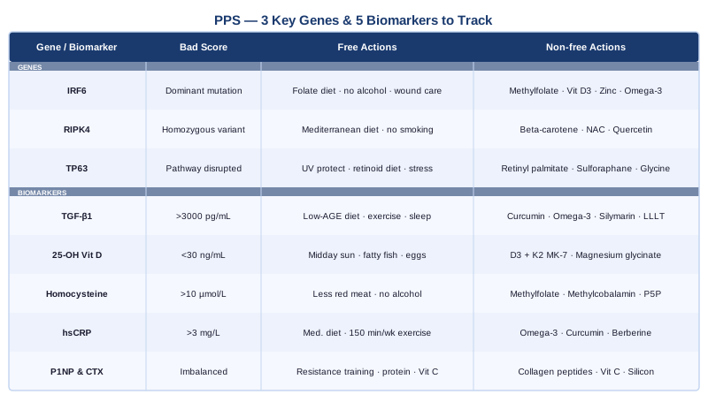
The Genetics Behind Popliteal Pterygium Syndrome
Understanding the genetic architecture of PPS is not an academic detour — it is the foundation of any serious effort to manage the condition more precisely. For parents of children with PPS making decisions about future pregnancies, for adults with the condition optimizing their surgical outcomes, and for anyone wanting to understand why this condition behaves as it does, the molecular picture matters in ways that a clinical description alone cannot capture. The three genes below represent the current state of the science: one primary driver, one autosomal recessive contributor, and one upstream regulator that ties the whole pathway together.
IRF6 — The Primary Driver
Interferon regulatory factor 6 (IRF6) is the gene most definitively linked to popliteal pterygium syndrome. Located on chromosome 1q32.2, it encodes a transcription factor that directs how epithelial cells proliferate, differentiate, and fuse during fetal development. When IRF6 functions normally, it coordinates the timing and spatial organization of tissue fusion events — the palate, the lips, the perineum, the popliteal fossa. When it carries a disabling mutation, those fusion events can fail or occur abnormally, producing the range of features that characterize PPS.
The relationship between IRF6 and PPS was confirmed by Kondo and colleagues in a landmark 2002 study published in Nature Genetics, which identified IRF6 mutations in both Van der Woude syndrome and popliteal pterygium syndrome families. PPS and VWS are now understood as allelic conditions — caused by mutations in the same gene, but with the location and nature of the mutation influencing which syndrome results. Mutations disrupting the DNA-binding domain of IRF6 tend to produce more severe phenotypes consistent with PPS, while those affecting the protein-interaction domain more often produce the milder VWS presentation. Understanding which type of mutation is present is directly relevant to prognosis and recurrence counseling.
What makes IRF6 especially important to understand is that its function extends beyond embryonic development. It regulates keratinocyte differentiation — the process by which skin cells develop specialized roles — throughout life. It also interacts extensively with the TGF-β signaling pathway, which governs wound healing, fibrosis, and tissue remodeling after every surgical intervention. This means that IRF6 is not a gene whose consequences are limited to the womb; it continues to influence how connective tissue responds to every injury, suture, and scar.
If IRF6 Is Mutated — The Plan Without Supplements
No supplement reverses a constitutional IRF6 mutation. That needs to be stated clearly. What can be addressed, however, is the downstream biological environment that the mutation operates in. Practical, non-supplement steps that genuinely matter include:
Adequate dietary folate: IRF6 operates within a developmental network that is highly sensitive to folate availability. A diet consistently rich in leafy greens, legumes, and organ meats provides the methylation substrate this network needs. This is particularly critical during pregnancy in families with known IRF6 variants.
Avoiding recognized IRF6 pathway disruptors: Alcohol and therapeutic retinoids (at high doses) are documented disruptors of IRF6-dependent epithelial signaling in developing tissue. Eliminating alcohol and being cautious with retinoid-based dermatological products is prudent for individuals with IRF6 mutations.
Moist wound healing protocols: Because IRF6 influences keratinocyte behavior throughout life, individuals with IRF6 mutations may have subtle alterations in scar formation patterns. Using silicone gel sheets, maintaining moisture at healing sites, and working with surgical teams on extended post-operative scar protocols addresses this practically.
Genetic counseling: PPS follows an autosomal dominant inheritance pattern — each child of an affected parent has a 50% probability of inheriting the mutation. Preconception genetic counseling, and discussion of preimplantation genetic testing (PGT-M) where available, is the highest-impact single action for affected adults planning families.
Frequency: Dietary and wound care practices are daily and lifelong. Genetic counseling is typically a structured process of two to four consultations.
If IRF6 Is Mutated — The Plan With Supplements and Targeted Support
For those who want to specifically address the downstream biology of IRF6 dysfunction, the following have mechanistic plausibility and reasonable safety profiles — though direct evidence in PPS populations is limited and extrapolated from adjacent science.
L-methylfolate (5-MTHF), 400–800 mcg/day: The bioactive form of folate bypasses MTHFR conversion inefficiency and directly supports IRF6-adjacent developmental and methylation networks. Frequency: daily, continuous. Side effects: minimal at standard doses; pair with methylcobalamin to avoid masking B12 deficiency.
Vitamin D3, 2000–5000 IU/day (adjusted to serum levels): Vitamin D receptor signaling modulates keratinocyte differentiation and post-surgical immune-mediated tissue repair — both downstream of IRF6. Target serum 25-OH Vitamin D: 40–60 ng/mL. Frequency: daily. Cycling: retest every 3–6 months and adjust dose. Side effects: minimal within this range; pair with Vitamin K2 MK-7 (100–200 mcg/day) to direct calcium appropriately.
Zinc bisglycinate, 15–25 mg/day: A cofactor for multiple transcription factors including those in the IRF6-TP63 network, and essential for wound healing and keratinocyte integrity. Frequency: daily, 5 days on, 2 days off. Side effects: monitor copper status with long-term use; the zinc-to-copper ratio should remain balanced.
Omega-3 fatty acids (EPA + DHA), 2–4 g/day: Anti-inflammatory; modulate TGF-β signaling downstream of IRF6 and support the resolution phase of wound healing. Frequency: daily, continuous. Side effects: mild antiplatelet effect, occasional GI discomfort; choose third-party tested sources.
RIPK4 — The Autosomal Recessive Form
Receptor-interacting serine-threonine kinase 4 (RIPK4) is the gene responsible for the autosomal recessive variant of PPS, sometimes designated Bartsocas-Papas syndrome. Kalay and colleagues identified RIPK4 mutations in affected families in a study published in the American Journal of Human Genetics in 2012, establishing RIPK4 as a second distinct genetic entry point to a PPS-like phenotype. Unlike IRF6-driven PPS, which requires only one defective copy to produce the condition, RIPK4-related disease requires inheriting two non-functional copies — one from each carrier parent.
RIPK4 functions within the NF-κB signaling pathway and is critical for keratinocyte differentiation and ectodermal development. It operates in the same general developmental territory as IRF6 — regulating epithelial maturation and tissue fusion — which explains why mutations in either gene produce overlapping clinical features. However, RIPK4-related PPS tends to be significantly more severe: homozygous loss-of-function mutations produce a phenotype that is often lethal or severely disabling, affecting multiple ectodermal systems simultaneously.
The clinical implication for families is this: distinguishing between IRF6-driven and RIPK4-driven PPS changes the genetic risk calculation dramatically. Two carrier parents (autosomal recessive pattern) have a 25% chance of an affected child with each pregnancy, compared to the 50% risk in dominant IRF6 families. Getting a confirmed molecular diagnosis is therefore not just academically interesting — it directly informs family planning decisions.
If RIPK4 Variants Are Present — The Plan Without Supplements
For heterozygous carriers and individuals with milder presentations, the focus shifts to managing the NF-κB-mediated inflammatory biology that RIPK4 normally helps regulate.
Minimizing NF-κB activators through diet: Refined carbohydrates, trans fats, and heavily processed foods chronically activate NF-κB — the same signaling hub RIPK4 participates in. A Mediterranean dietary pattern, rich in antioxidants, polyphenols, and fiber, is the most evidence-based dietary approach for suppressing excessive NF-κB activity.
Eliminating smoking: Cigarette smoke is one of the most potent environmental activators of NF-κB, with direct, documented effects on keratinocyte behavior and skin epithelial integrity. For RIPK4 variant carriers, this is non-negotiable.
Preconception genetic counseling for carrier couples: Preimplantation genetic testing for monogenic disorders (PGT-M) is available for RIPK4 variants and can identify unaffected embryos before transfer in IVF cycles. For couples where both partners are confirmed RIPK4 carriers, this is the highest-impact option available.
Frequency: dietary changes are daily and ongoing. Genetic counseling is a structured preconception process.
If RIPK4 Variants Are Present — The Plan With Supplements and Targeted Support
Vitamin A as beta-carotene, 5000–10000 IU/day: Vitamin A is a key regulator of keratinocyte differentiation and supports the epithelial maturation pathways adjacent to RIPK4 function. Beta-carotene is significantly safer than retinol for long-term supplementation, as it does not accumulate to hepatotoxic levels. Frequency: daily. Side effects: benign yellowing of skin at high doses (carotenemia); safe during pregnancy at these levels, unlike preformed retinol.
NAC (N-acetylcysteine), 600–1200 mg/day: A precursor to glutathione that modulates NF-κB activity and supports cellular antioxidant defense. Growing evidence supports NAC in conditions involving dysregulated NF-κB signaling and keratinocyte vulnerability. Frequency: daily. Cycling: 5 days on, 2 days off. Side effects: possible GI discomfort; thins mucus — relevant if there are concurrent respiratory considerations.
Quercetin with bromelain, 500 mg/day: A flavonoid with documented NF-κB inhibitory activity. Bromelain improves absorption meaningfully. Frequency: daily. Cycling: 8 weeks on, 4 weeks off. Side effects: mild; may interact with certain antibiotics and anticoagulants.
TP63 — The Upstream Regulator
Tumor protein p63 (TP63) sits upstream of IRF6 in the developmental gene cascade and directly activates IRF6 expression in epithelial tissue. Research published in Human Molecular Genetics established that p63 protein binds to an enhancer region within the IRF6 gene locus and drives its transcription during the critical window of epithelial tissue development. This means TP63 and IRF6 are not parallel actors — they are hierarchically linked, with TP63 functioning as a master regulator of the very gene most responsible for PPS.
Mutations in TP63 itself cause a family of ectodermal dysplasia syndromes — EEC syndrome, AEC syndrome, ADULT syndrome — some of which share features with PPS including palate defects and ectodermal abnormalities. Even without a TP63 mutation, understanding its role matters for PPS management because TP63 activity is sensitive to environmental inputs, particularly retinoic acid signaling (connected to vitamin A), which directly modulates how strongly TP63 drives IRF6 expression.
TP63 also plays a critical role in DNA damage response and maintenance of epithelial stem cell populations throughout life. This makes it relevant long after embryonic development: individuals with PPS who undergo repeated surgical interventions have a biological interest in supporting robust TP63-dependent epithelial regeneration at every healing site.
If the TP63 Pathway Is Affected — The Plan Without Supplements
Limit UV exposure at surgical sites: UV radiation induces DNA damage that aberrantly activates TP63 in ways that can dysregulate downstream epithelial signaling. Consistent sun protection over healing skin is particularly important in the first 12 months after surgery, when scar tissue is still being remodeled.
Prioritize dietary retinoic acid precursors: Liver, eggs, grass-fed dairy, and brightly colored vegetables all provide pre-formed vitamin A and carotenoids that feed directly into the retinoic acid signaling axis connected to TP63. A varied whole-food diet consistently outperforms supplementation for this purpose.
Chronic stress management: Prolonged elevation of cortisol suppresses TP63-regulated epithelial repair mechanisms through glucocorticoid receptor signaling. This is not a peripheral concern — the stress burden carried by families managing a rare pediatric condition is real and its biological effects on tissue repair are measurable. This is addressed further in the epigenetics and complementary sections below.
If the TP63 Pathway Is Affected — The Plan With Supplements and Targeted Support
Retinyl palmitate, 2500–5000 IU/day: A stabilized form of preformed vitamin A that feeds directly into retinoic acid signaling supporting TP63 activity. Safer for long-term use than high-dose retinol. Frequency: daily. Cycling: 8–12 weeks on, 4 weeks off. Side effects: teratogenic at high doses — critical to avoid during pregnancy; liver function should be monitored with long-term use above 5000 IU.
Sulforaphane (from broccoli sprout extract), 20–50 mg/day: Activates the NRF2 pathway, which interacts with TP63 in coordinating epithelial stress responses. Human evidence for NRF2 activation via sulforaphane is solid; specific TP63 interaction evidence is primarily preclinical but mechanistically coherent. Frequency: daily. Cycling: continuous, well tolerated. Side effects: minimal; occasional GI adjustment in the first week.
Glycine, 3–5 g/day: Provides the amino acid substrate for collagen synthesis downstream of TP63-regulated tissue remodeling. Easily supplemented directly or obtained from bone broth. Frequency: daily, continuous. Side effects: none at these doses.
Epigenetics: What Modifies Gene Expression When the Blueprint Is Altered
Genetics sets the blueprint; epigenetics determines how that blueprint is read on any given day. For PPS — where a single gene mutation produces a wide phenotypic spectrum — epigenetic regulation is increasingly recognized as a key modifier of severity and recovery.
Folate and methylation capacity are the most directly relevant epigenetic factors for PPS families. Folate provides methyl groups essential for DNA methylation — the mechanism that regulates gene expression genome-wide. The MTHFR gene variants C677T and A1298C, which Ali Torkamani at Scripps Research and practitioners like Gary Brecka have worked to bring into mainstream health awareness, reduce how efficiently the body converts dietary folate into its active form, 5-methyltetrahydrofolate. For families with IRF6 mutations, carrying MTHFR variants may compound the epigenetic maintenance burden on the IRF6-adjacent gene network. MTHFR testing prior to future pregnancies is worth discussing with a geneticist or integrative physician. The intervention is simple: replace standard folic acid with L-methylfolate (400–1000 mcg/day), bypassing the impaired conversion step entirely.
TGF-β signaling is a second epigenetic modifier of particular relevance to PPS. TGF-β controls fibrosis — the deposition and remodeling of scar tissue — and its activity level is regulated both by genetics and by epigenetic inputs. In individuals with IRF6 mutations, TGF-β signaling can become chronically dysregulated, contributing both to the fibrous pterygia that define the condition and to the scarring dynamics that follow surgery. Lifestyle interventions that modulate TGF-β epigenetically — omega-3 fatty acids, vitamin D, curcumin — are covered in detail in the biomarker section below.
Vitamin D receptor (VDR) signaling is the third epigenetic axis worth understanding. Vitamin D binding to its nuclear receptor produces methylation and acetylation changes at hundreds of gene loci — including several in the IRF6 and TP63 developmental network. Optimizing serum vitamin D to 40–60 ng/mL is one of the most practical and broad-reaching epigenetic interventions available to anyone managing a genetic condition, and it is underused in rare disease populations.
With the genetic picture now in place, the practical question becomes: what is actually measurable in real time that reflects how these pathways are performing? That is exactly what the next section answers.
Five Biomarkers That Give You a Real-Time Window Into PPS Biology
Genetics tells you the blueprint. Biomarkers tell you how the building is performing today. For a condition involving connective tissue integrity, epithelial repair, and surgical recovery, five specific blood markers give the most useful and actionable ongoing picture of what is happening at the biological level. Each one is measurable through standard or specialty labs, interpretable by an engaged clinician, and addressable through practical protocols.
1. TGF-β1 — The Fibrosis Signal
Why it matters: Transforming Growth Factor Beta 1 is the primary molecular driver of fibrosis — the process by which the body deposits and contracts scar tissue. In PPS, where fibrous pterygia are a defining feature and where post-surgical scarring is a consistent clinical concern, TGF-β1 is a central player. Chronically elevated TGF-β1 is associated with hypertrophic scarring, excessive keloid formation, and worse functional outcomes following connective tissue surgery. Peter Attia and Thomas Dayspring, who have both written extensively about applying biomarker precision to tissue health, apply the same principle consistently: measure the drivers of dysfunction, not just the consequences.
How to measure it: TGF-β1 is measured via serum ELISA through specialty laboratories; LabCorp and Quest Diagnostics both offer this panel on physician request. Cost: approximately $80–250 USD. Optimal target: below 2300 pg/mL in most reference ranges. Values above 3000 pg/mL with clinical signs of fibrosis are worth discussing with a specialist.
If TGF-β1 Is Elevated — The Plan Without Supplements
Reduce dietary advanced glycation end products (AGEs): Grilled, fried, and highly processed foods spike TGF-β1 through a well-characterized AGE-receptor-mediated mechanism. Shifting to steaming, poaching, and slow cooking meaningfully reduces the daily AGE load and downstream TGF-β1 activity.
Moderate aerobic exercise, five days per week: 30–45 minutes of moderate-intensity cardio (walking briskly, cycling, swimming) consistently lowers systemic TGF-β1 in human intervention studies. High-intensity exercise transiently raises it — moderation, not maximum intensity, is the relevant variable here.
Sleep quality optimization: Poor sleep directly and acutely elevates TGF-β1. Seven to nine hours per night, with consistent sleep timing and dark, cool sleeping conditions, reduces fibrotic signaling in measurable ways.
If TGF-β1 Is Elevated — The Plan With Supplements and Equipment
Curcumin as phytosome or with piperine, 500–1000 mg/day: Among the most studied natural TGF-β1 inhibitors, with human trial meta-analyses supporting its anti-fibrotic effect in multiple tissue types. Frequency: daily. Cycling: 8 weeks on, 3 weeks off. Side effects: mild anticoagulant effect at high doses; take with food to minimize GI irritation.
Omega-3 fatty acids (EPA + DHA), 2–4 g/day: Directly modulates TGF-β1 expression through prostaglandin E3 signaling. The effect is dose-dependent and supported across multiple human trials in inflammatory and fibrotic conditions. Cycling: continuous. Side effects: mild antiplatelet effect; select molecularly distilled, third-party tested products.
Silymarin (milk thistle extract standardized to 70–80% silymarin), 140 mg three times daily: Human evidence for TGF-β1 suppression in hepatic and systemic fibrotic conditions is growing. Particularly relevant for post-surgical recovery periods. Cycling: can be taken continuously; well tolerated long-term. Side effects: occasional mild GI adjustment.
Photobiomodulation device (630–660 nm, red light), 3–5 sessions per week: Growing clinical evidence supports LLLT's ability to modulate TGF-β1 activity in skin and connective tissue at the site of application. Home devices range from $150–800 USD; see the complementary approaches section for detailed protocols. Best applied to healing scar sites, not acutely open wounds.
2. 25-OH Vitamin D — The Epigenetic Regulator
Why it matters: Vitamin D functions more as a steroid hormone than a conventional vitamin — its receptor, once bound, regulates the expression of over 2000 genes, including several directly involved in keratinocyte differentiation, IRF6 pathway support, and post-surgical immune-mediated tissue repair. Peter Attia consistently lists 25-OH Vitamin D as one of the essential markers that virtually every patient should track, precisely because its effects are broad, its deficiency is common, and its optimization is straightforward. For PPS specifically, low vitamin D correlates with impaired wound healing, higher peri-surgical infection risk, and reduced connective tissue quality.
How to measure it: Standard blood test, available through any clinical lab. Cost: $30–80 USD. Optimal level per current evidence: 40–60 ng/mL (100–150 nmol/L). Most adults without supplementation fall between 15 and 30 ng/mL — technically within "normal" reference ranges but significantly below optimal.
If Vitamin D Is Low — The Plan Without Supplements
Midday sun exposure: 15–30 minutes of direct midday sun exposure with arms and legs uncovered produces 10,000–20,000 IU of vitamin D in fair-skinned individuals. Darker skin tones require longer exposure. This is the most natural and effective route — though not always practical given geography, surgical recovery, or season.
Dietary vitamin D sources: Wild-caught fatty fish (salmon, sardines, mackerel), egg yolks from pasture-raised hens, and grass-fed dairy all contribute meaningfully. Diet alone is unlikely to fully optimize serum levels, but it reduces the supplementation burden.
If Vitamin D Is Low — The Plan With Supplements and Equipment
Vitamin D3, 3000–5000 IU/day: Adjust dose based on serum testing and retest every 3–6 months. Always pair with Vitamin K2 as MK-7, 100–200 mcg/day to direct calcium to bone rather than soft tissue. Cycling: continuous, with dose adjustment per testing. Side effects: hypercalcemia possible at sustained high doses above 10,000 IU/day without monitoring; this risk is low within the 3000–5000 IU range.
Magnesium glycinate or malate, 200–400 mg/day at night: Magnesium is an essential cofactor for converting vitamin D into its biologically active form. Without adequate magnesium, vitamin D3 supplementation has limited downstream impact. Cycling: continuous. Side effects: loose stools at high doses; glycinate form is generally the best tolerated.
3. Homocysteine — The Methylation Marker
Why it matters: Homocysteine is an amino acid that accumulates in the blood when the body's methylation capacity is impaired — typically due to MTHFR gene variants, inadequate B vitamin intake, or both. Gary Brecka, who has made this marker central to his public-health work on genetic methylation, argues compellingly that elevated homocysteine is one of the most underappreciated markers in medicine, reflecting impaired epigenetic maintenance across the entire genome. For PPS specifically, high homocysteine signals that the methylation machinery supporting IRF6-adjacent developmental gene networks is operating below capacity — and it is also an independent predictor of poor tissue healing and vascular health.
How to measure it: Standard blood test; widely available. Cost: $20–60 USD. Target: below 8 µmol/L. Many practitioners following the work of Brecka and others now consider below 7 µmol/L optimal. Values above 10 µmol/L are clinically significant and warrant active intervention.
If Homocysteine Is Elevated — The Plan Without Supplements
Reduce excessive methionine loading: Very high red meat intake raises methionine, the direct precursor to homocysteine. Balancing protein sources — incorporating eggs, fish, legumes, and vegetables — naturally moderates homocysteine accumulation.
Eliminate alcohol completely: Alcohol rapidly depletes B vitamins — particularly B6, B9, and B12 — driving homocysteine sharply upward. For individuals with IRF6 mutations, alcohol is doubly problematic given its documented interference with IRF6-dependent epithelial signaling.
If Homocysteine Is Elevated — The Plan With Supplements
L-methylfolate (5-MTHF), 400–1000 mcg/day: The most direct and impactful single intervention for elevated homocysteine, bypassing MTHFR conversion inefficiency entirely. Frequency: daily, continuous. Side effects: very low; can feel mildly activating in sensitive individuals.
Methylcobalamin (active B12), 500–1000 mcg/day: Works synergistically with methylfolate in the homocysteine recycling pathway. Essential to include whenever methylfolate is supplemented to avoid relative B12 depletion. Cycling: continuous. Side effects: essentially none.
Pyridoxal-5-phosphate (P5P, active B6), 25–50 mg/day: The third component of the homocysteine recycling triad. Works through a distinct biochemical route (transsulfuration) that complements folate and B12 recycling. Cycling: continuous; avoid sustained doses above 200 mg/day due to peripheral neuropathy risk at high doses.
4. High-Sensitivity C-Reactive Protein (hsCRP) — The Inflammation Gauge
Why it matters: hsCRP is the most widely validated marker of systemic low-grade inflammation. For anyone with PPS preparing for surgery, recovering from it, or managing connective tissue health over the long term, chronically elevated hsCRP is a direct risk factor for impaired wound healing, higher post-surgical complication rates, and increased fibrotic scarring. Peter Attia includes hsCRP as a standard component of his preventive medicine panel precisely because it is cheap, reliable, and broadly predictive of tissue and vascular health outcomes.
How to measure it: Standard blood test available at any clinical lab. Cost: $15–50 USD. Target: below 1.0 mg/L. Values of 1–3 mg/L indicate moderate risk; above 3 mg/L is high risk. Testing should be performed at least two weeks away from any acute infection or illness, both of which transiently spike CRP to levels unrelated to chronic baseline.
If hsCRP Is Elevated — The Plan Without Supplements
Mediterranean dietary pattern: High in vegetables, olive oil, fatty fish, nuts, and legumes — this dietary pattern is the most extensively studied dietary intervention for reducing hsCRP. Meta-analyses of randomized trials document reductions of 20–30% in CRP with consistent adherence. This single change, sustained over months, can move hsCRP from the moderate to the low-risk zone without any supplementation.
150 minutes of moderate aerobic exercise per week: The WHO physical activity recommendation is not arbitrary — it reflects the level at which human studies consistently demonstrate meaningful hsCRP reduction. Consistency matters more than intensity.
Sleep duration and quality: Sleeping six hours or less per night is independently associated with hsCRP values nearly twice those seen in individuals sleeping seven to nine hours. This is a metabolic and immunological effect, not simply correlation.
If hsCRP Is Elevated — The Plan With Supplements and Equipment
Omega-3 fatty acids (EPA + DHA), 2–4 g/day: One of the most consistently effective anti-inflammatory supplements in human trials, with documented hsCRP-reducing effects across multiple populations. Frequency: daily, continuous. Side effects: mild.
Curcumin as phytosome, 500 mg twice daily: Human meta-analyses support its ability to reduce hsCRP in inflammatory conditions across multiple tissue types. Cycling: 8 weeks on, 3 weeks off. Side effects: mild; take with food.
Berberine, 500 mg twice daily with food: Modulates NF-κB and NLRP3 inflammasome pathways that drive chronic CRP elevation. Human trial evidence is growing. Cycling: 8–12 weeks on, 4 weeks off. Side effects: GI discomfort in some individuals; not for use during pregnancy.
5. Collagen Remodeling Markers — P1NP and CTX
Why it matters: Procollagen Type I N-terminal Propeptide (P1NP) and Cross-Linked C-Telopeptide (CTX) are markers of collagen synthesis and degradation, respectively. Together, they give a picture of how actively the body is building and breaking down collagen — the primary structural protein in the connective tissues most directly affected by PPS. An imbalance between synthesis (P1NP) and degradation (CTX), or globally depressed collagen turnover, is relevant to understanding the body's repair capacity — and particularly useful for monitoring around surgical interventions. Allan Sniderman and Thomas Dayspring, known for their rigorous approach to biomarker interpretation in tissue health contexts, emphasize that structural protein turnover markers offer mechanistic information that standard inflammatory panels miss.
How to measure it: Both P1NP and CTX are ordered as serum bone remodeling markers through standard clinical labs. Cost: $50–150 USD each. Reference ranges: P1NP typically 15–74 mcg/L in adults (varies by sex and lab); CTX typically below 0.48 ng/mL in premenopausal women and younger adult males. Testing fasting in the morning for CTX improves consistency.
If Collagen Markers Are Imbalanced — The Plan Without Supplements
Resistance training, 2–3 sessions per week: Weight-bearing and load-bearing exercise is the most potent non-pharmacological stimulus for collagen synthesis, measurably raising P1NP within 8–12 weeks in human studies. Even moderate resistance work — bodyweight exercises, resistance bands — produces meaningful effects.
Protein adequacy: Collagen synthesis requires adequate total protein intake (approximately 1.2–1.6 g/kg body weight per day for adults managing connective tissue conditions) and, specifically, glycine-rich protein sources — slow-cooked meats, skin-on poultry, bone broth — that preferentially supply the amino acids used in collagen triple-helix formation.
Dietary vitamin C: Vitamin C is the essential cofactor for prolyl and lysyl hydroxylase enzymes that cross-link collagen chains and give them their structural integrity. Daily citrus, bell peppers, and berries reliably meet this need.
If Collagen Markers Are Imbalanced — The Plan With Supplements
Hydrolyzed collagen peptides, 10–15 g/day: Human trials — including research by Shaw and colleagues published in the American Journal of Clinical Nutrition — demonstrate that collagen peptides taken with vitamin C before exercise increase collagen synthesis in tendons and connective tissue. Best taken 30–60 minutes before resistance training. Cycling: continuous. Side effects: minimal.
Vitamin C, 500–1000 mg/day: The cofactor for the enzymatic reactions that produce functional collagen. Cycling: continuous. Side effects: GI discomfort at very high doses; 500–1000 mg is well tolerated by most.
Orthosilicic acid (bioavailable silicon), 10–20 mg/day: Emerging evidence in human trials supports silicon's role in stimulating collagen Type I synthesis and improving connective tissue matrix quality. Cycling: continuous. Side effects: very low.
Moving from what is measurable to what shapes expression at a deeper level, the next section draws on recent epigenetics research that reframes what it means to live with a genetic condition — and where the levers for change actually are.
What Epigenetics Research Is Teaching Us About Living With a Genetic Condition
The Huberman Lab podcast episodes on gene expression, epigenetics, and health optimization — including conversations with Dr. Rhonda Patrick and several solo deep-dives on methylation and lifestyle biology — collectively offer one of the most practically useful public frameworks for understanding how genetics and environment interact. The central claim, now backed by a growing body of human research, is this: having a gene variant is not the same as being locked into its consequences. The downstream biology is more plastic than most people are told.
Here are the ten most relevant insights from this body of work, translated into terms that matter specifically for PPS.
Gene Expression Is More Flexible Than Most People Are Told
Even with a fixed germline mutation in IRF6, the expression of every gene in the surrounding network — including those involved in wound healing, inflammation, and tissue remodeling — responds to lifestyle inputs in real time. The mutation is fixed; the environment it operates in is not.
Methylation Is the On/Off Switch of the Genome
DNA methylation determines which genes are read actively and which are silenced. Low dietary folate, elevated homocysteine, and chronic B vitamin insufficiency all compromise methylation capacity in ways that are measurable through the biomarkers above — and fully reversible with targeted intervention. For PPS families, the IRF6-adjacent gene network depends on adequate methylation to function correctly.
MTHFR Variants Are Common and Routinely Underaddressed
The C677T and A1298C variants of MTHFR reduce folate conversion efficiency by 30–70% in affected individuals. Gary Brecka has argued publicly and compellingly that many people with unexplained health trajectories — including those managing rare genetic conditions — are running impaired methylation that a simple supplement switch could improve dramatically. Replace standard folic acid with L-methylfolate, pair with methylcobalamin, and monitor homocysteine as the feedback marker.
Vitamin D Functions as an Epigenetic Regulator
When vitamin D binds its nuclear receptor, it changes the methylation and acetylation status of hundreds of gene loci — including several in the IRF6 and keratinocyte differentiation pathways relevant to PPS. Optimizing to 40–60 ng/mL is one of the broadest epigenetic levers available without a prescription.
Chronic Inflammation Drives Epigenetic Dysregulation — Not Just the Reverse
Elevated hsCRP and IL-6 do not only reflect gene expression dysregulation — they actively modify gene expression through NF-κB-mediated epigenetic changes. Reducing systemic inflammation is both a symptom-management strategy and a gene-regulation strategy. These are not separate goals.
Exercise Is an Epigenetic Medicine
Regular moderate aerobic exercise triggers epigenetic changes — documented through methylation array studies in human subjects — that suppress pro-inflammatory gene expression and upregulate tissue repair and antioxidant pathways. This is not metaphor. It is measurable biology, accessible without a prescription, and relevant to every person with PPS.
Sleep Is When Epigenetic Maintenance Happens
DNA repair enzymes and epigenetic maintenance mechanisms are concentrated in slow-wave (deep) sleep. Chronic sleep deprivation measurably increases error rates in epigenetic maintenance. For anyone managing a genetic condition, this is not optional biology — it is foundational.
The Gut Microbiome Produces Epigenetic Signals
Short-chain fatty acids produced by gut bacteria — butyrate in particular — are natural histone deacetylase inhibitors, meaning they broadly support epigenetic stability and gene expression fidelity. A high-fiber diet is the most effective way to maintain the microbiome diversity that produces them. Diversity of plant foods is the single most consistently supported dietary recommendation for microbiome health across current human research.
Earlier Intervention Changes Outcomes More Than Later Intervention
Developmental epigenetics research consistently shows that nutritional and lifestyle optimization applied earlier in life has larger and more durable effects than the same interventions applied later. For children with PPS, early attention to folate, vitamin D, omega-3s, and sleep — while avoiding the disruptors discussed in the genetics section — is supported by this principle. The earlier the metabolic environment is optimized, the more influence it exerts on downstream gene expression.
Chronic Stress Methylates the Wrong Genes
Sustained psychological stress induces epigenetic changes in stress-response gene promoters that accumulate and compound over years. For families managing a rare pediatric condition — navigating surgical systems, genetic uncertainty, developmental monitoring, and everything that comes with it — the biological consequences of that chronic stress are real and measurable in the genome. Stress management is addressed directly in the complementary section below, and it belongs in the same biological conversation as folate and vitamin D.
Complementary Approaches With Evidence for Tissue Health and Wellbeing in PPS
The following approaches have meaningful human clinical evidence for the tissue, pain, and psychological dimensions of managing a connective tissue condition involving surgery and chronic care. None replaces surgical or genetic specialist management — but each addresses real needs that conventional appointments rarely have time to cover.
Low-Level Laser Therapy (Photobiomodulation) for Scar and Wound Healing
Low-level laser therapy delivers specific wavelengths of red (630–660 nm) and near-infrared (810–850 nm) light to tissue, stimulating mitochondrial cytochrome c oxidase, increasing cellular ATP production, and reducing reactive oxygen species. For PPS, its most relevant applications are post-surgical wound healing acceleration and scar tissue modulation — two areas where the existing evidence base is strongest. The biological relevance is direct: PPS involves fibrotic connective tissue formation, and LLLT has documented effects on TGF-β1 activity at the tissue level, the same fibrotic driver measured in the biomarker section.
Multiple systematic reviews in dermatology and surgery journals confirm LLLT's efficacy for wound healing in epithelial tissues, with the strongest evidence at wavelengths of 630–660 nm for superficial skin and 810–850 nm for deeper connective tissue. A controlled trial published in Photomedicine and Laser Surgery demonstrated significantly reduced hypertrophic scar formation in post-surgical patients receiving LLLT versus sham treatment. The effect size is modest to moderate, which is consistent with what a mechanistically sound physical intervention without systemic effects can realistically achieve.
For individuals with PPS who have undergone popliteal web release, palate repair, or other reconstructive surgery, LLLT can be applied to healing sites beginning approximately day 3–7 post-operatively (once the acute inflammatory phase has passed), at a dose of 3–6 J/cm², three to five times per week for 4–6 weeks. Always coordinate with the surgical team before initiating LLLT over a healing surgical site. Home devices designed for wound and scar applications range from $150–800 USD. The key variable is wavelength — verify the device specification before purchase.
Scar Massage for Connective Tissue Remodeling
Scar massage — also called scar mobilization or myofascial scar therapy — is a manual technique that applies targeted pressure and movement to healing scar tissue to mechanically modify its structure and reduce adhesion formation. For PPS, where fibrous pterygia and surgical scar formation are central clinical concerns, scar massage directly addresses the mechanical and biochemical properties of developing scar tissue. The mechanism involves breaking down excessive collagen cross-links, improving tissue extensibility, stimulating normal collagen fiber alignment, and modulating local TGF-β1 activity at the scar site — all of which are directly relevant to the biology discussed in this article.
A systematic review published in Physical Therapy found significant supporting evidence for scar massage in improving scar pliability, reducing scar height, and reducing associated pain and paresthesia in post-surgical patients. The most studied protocol involves applying firm, circular pressure with the pads of two fingers directly to the closed, healed scar for 10–15 minutes, twice daily. Silicone gel sheet application before or after massage sessions further improves outcomes in human trials, with the combination outperforming either intervention alone.
For PPS patients, scar massage should begin only after full wound closure — typically 3–6 weeks post-surgery — and with explicit clearance from the operating surgeon. A physiotherapist experienced in pediatric or post-surgical scar management is the ideal practitioner for initial technique instruction. At-home maintenance at twice daily, 10–15 minutes per session, is both practical and sustainable over the 12+ months that scar remodeling continues.
Mindfulness-Based Stress Reduction (MBSR) for Stress and Pain
Mindfulness-Based Stress Reduction is a structured 8-week program combining formal mindfulness meditation, body scan practice, and mindful movement, developed at the University of Massachusetts Medical Center and now available through medical centers, community programs, and free online resources globally. For individuals and families managing PPS, MBSR addresses the chronic stress burden that standard clinical care rarely has resources to address — and, as detailed in the epigenetics section, that stress has measurable consequences on gene expression, cortisol regulation, and tissue repair capacity.
A study by Kaliman and colleagues published in Psychoneuroendocrinology demonstrated that a single day of intensive mindfulness practice produced measurable changes in the expression of stress- and inflammation-related genes, including reduced histone deacetylase activity and normalized cortisol gene regulatory response — some of the same epigenetic mechanisms discussed throughout this article. Multiple randomized trials of the full 8-week MBSR course document significant reductions in perceived pain, anxiety, and self-reported health impact across chronic disease populations.
The practical application is straightforward: one 8-week MBSR course, with 45 minutes of daily home practice and weekly group sessions. The Palouse Mindfulness program (developed by a University of Massachusetts MBSR teacher) provides a free, complete 8-week course online. Effects on anxiety and pain perception are typically detectable by week four. MBSR is appropriate for older children (from approximately age 10 onward) with developmental adaptation; adult caregivers benefit from participating independently of, or alongside, the patient.
Breathing-Based Therapies for Perioperative Anxiety and Recovery
Slow, controlled breathing — specifically coherence breathing at five seconds per inhale and five seconds per exhale — activates the parasympathetic nervous system via baroreceptor and vagal afferent pathways, producing measurable reductions in cortisol, improvements in heart rate variability (HRV), and suppression of inflammatory cytokine release. For individuals with PPS who undergo planned surgical procedures, the perioperative anxiety period is a significant biological stress event with real downstream effects on surgical outcomes and recovery trajectory. Breathing-based interventions are among the most accessible, evidence-supported, and risk-free tools available for managing this.
A randomized controlled trial published in BMC Anesthesiology found that structured preoperative breathing training significantly reduced preoperative anxiety scores, postoperative analgesic requirements, and reported recovery quality in elective surgical patients compared to standard care. HRV improvement — one of the most reliable markers of parasympathetic nervous system activity — has been documented in human trials of coherence breathing protocols as short as four weeks of daily practice. For children undergoing PPS-related surgery, developmentally adapted coherence breathing (with simple visual guides or app-based audio pacing) is practical from age six onward.
The application protocol requires no equipment and no prescription: begin 10–20 minutes of daily coherence breathing (5-second inhale, 5-second exhale through the nose) at least two weeks before any planned surgery, and continue through the post-operative recovery period. Apps such as Breathwrk or Othership provide accessible guided protocols. Adults who extend to 20 minutes of daily practice maximize the HRV benefit, which compounds meaningfully over weeks.
Conclusion
Popliteal pterygium syndrome is, at its molecular core, a condition of disrupted epithelial gene regulation. IRF6 is the primary driver, RIPK4 underlies the recessive form, and TP63 connects the whole pathway upstream. None of these mutations can be changed with current clinical tools — but the biological environment they operate in is measurable, modifiable, and worth paying close attention to.
The five biomarkers covered here — TGF-β1, vitamin D, homocysteine, hsCRP, and collagen remodeling markers — offer an ongoing, real-time picture of how tissue health, inflammation, and repair capacity are actually performing. They are affordable, interpretable, and directly actionable. Getting a baseline measurement of each, particularly before and after surgical procedures, gives information that generic follow-up appointments cannot.
The next smart step is not necessarily a new supplement stack or a new appointment. For many readers, it starts here: order a baseline panel of these five markers, bring the results to the clinical team managing PPS care, and start a conversation grounded in data. Better information leads to better decisions — and that, more than any single intervention, is what changes outcomes over time.