This article contains AI generated content.
· UpdatedPopliteal Vein Thrombosis: 5 Genes And 6 Biomarkers To Track
Introduction
A blood clot forming in the popliteal vein — the vessel running behind your knee — is not a random event. For most people who experience popliteal vein thrombosis, there is an underlying biological story that made it possible: a combination of inherited tendencies, acquired shifts in how the blood behaves, and environmental exposures that tipped the balance toward clotting. Understanding that story is one of the most useful things you can do, whether you are recovering from a first clot, trying to prevent a recurrence, or simply concerned about your risk.
The standard advice given after a DVT diagnosis — wear compression stockings, avoid long flights, stay hydrated, take your anticoagulant — is not wrong. But it is also incomplete. It treats the clot as the problem and misses the question that matters most: why did your blood clot in that location, at that time, when most people in a similar situation did not? The answer is almost always rooted in specific, measurable imbalances and, sometimes, in your genetic code.
This article takes a different approach. Rather than describing popliteal vein thrombosis in general terms, it focuses on the six most clinically useful biomarkers you can test right now to understand your clotting profile, and the five genetic variants most strongly associated with venous thromboembolism. For each biomarker, you will find details on how it is measured, what an abnormal result might mean, and concrete plans for improvement — with and without supplements. The genetics section does the same for inherited risk.
The goal here is not to replace your hematologist or vascular medicine specialist. It is to give you the kind of precise biological context that makes those conversations far more productive. Better information leads to better decisions — and understanding your own thrombotic risk at this level is genuinely actionable.
Summary
This article explores two parallel tracks for understanding and reducing your risk of popliteal vein thrombosis. The first is biomarker tracking: six specific blood tests — D-dimer, fibrinogen, homocysteine, Factor VIII activity, Protein C and S, and Antithrombin III — that together reveal how your coagulation system is functioning and whether it is tilted toward excessive clotting. For each, you will find optimal target ranges, practical lifestyle-based improvement plans, and supplement protocols with dosing, cycling, and side effect notes. The second track covers genetics: five gene variants — Factor V Leiden, Prothrombin G20210A, MTHFR C677T, PAI-1 4G/5G, and PROC/PROS1 — that shape your baseline thrombotic risk and directly inform what interventions make the most sense for you personally. Beyond these two tracks, the article also summarizes key insights from Dr. Malcolm Kendrick's landmark book The Clot Thickens, which challenges mainstream thinking on clotting and cardiovascular disease in important ways, and outlines four complementary approaches — yoga, breathing therapy, mindfulness, and massage — that have meaningful human clinical evidence. Together, these tools offer a coherent, personalized framework for working with your medical team to reduce long-term thrombosis risk.
6 Biomarkers That Reveal Your Clotting Risk
Most standard blood panels after a DVT focus on confirming the diagnosis and monitoring anticoagulant therapy. They rarely examine the upstream question: which biological imbalances made the clot possible in the first place? The six biomarkers below do exactly that. Some are inexpensive and widely available. Others require specialized testing. All are worth knowing.
1. D-Dimer
D-dimer is a small protein fragment released into the bloodstream when a blood clot is broken down by the body's fibrinolytic system. It is the most widely used biomarker in acute DVT diagnosis — a normal D-dimer essentially rules out an active clot in low-to-intermediate risk patients — but its usefulness extends well beyond the emergency room. In people who have already been treated for popliteal vein thrombosis, persistently elevated D-dimer after completing anticoagulation is one of the strongest predictors of recurrence. Research shows that patients with elevated D-dimer at the time of stopping anticoagulants face a two- to threefold higher risk of recurrent VTE compared to those with normal levels. References covering this evidence are indexed in PubMed.
It is important to understand that D-dimer is highly sensitive but not specific — it rises in many conditions beyond thrombosis, including infection, pregnancy, recent surgery, cancer, and inflammation. This means a high D-dimer alone does not prove active clotting, but in the context of known DVT history, it is a signal worth taking seriously.
How to Measure It
D-dimer is measured through a standard venous blood draw and is widely available in hospital laboratories and most private labs. Cost ranges from $20 to $80 depending on the setting. Quantitative (numerical) results are more useful than qualitative (positive/negative) results for ongoing monitoring. The generally accepted normal threshold is below 500 ng/mL (or 0.5 mg/L FEU), though age-adjusted thresholds are increasingly used in clinical practice.
If the Score Is Bad: The Plan Without Supplements
Elevated D-dimer after completing anticoagulation treatment warrants a direct conversation with your physician about extended anticoagulation or closer surveillance imaging. From a lifestyle standpoint, the most impactful non-supplement interventions are: regular low-impact aerobic activity (30–45 minutes of walking or cycling most days of the week improves fibrinolytic activity); weight management (obesity independently elevates D-dimer); treating active infections or inflammatory conditions; and avoiding prolonged immobility. Quitting smoking has one of the most immediate effects on D-dimer reduction. Stress management also matters — cortisol elevates fibrinogen and, secondarily, D-dimer.
If the Score Is Bad: The Plan With Supplements or Equipment
Graduated compression stockings (class II, 20–30 mmHg) worn daily reduce venous stasis and have been shown to lower D-dimer levels over time in post-DVT patients. Omega-3 fatty acids (fish oil, 2–4 g EPA+DHA daily) have modest fibrinolytic and anti-inflammatory effects; cycle on for 3 months, then retest D-dimer before continuing. Nattokinase (2,000 FU daily, taken away from meals) is a fibrinolytic enzyme from fermented soybeans that directly degrades fibrin; use only under medical supervision when taking anticoagulants, as the combination can significantly increase bleeding risk; cycling recommendation is 8 weeks on, 4 weeks off; side effects include bruising and gastrointestinal discomfort.
2. Fibrinogen
Fibrinogen is the soluble plasma protein that is converted into fibrin — the mesh-like scaffold of blood clots. Elevated fibrinogen levels mean more raw material for clot formation, and they independently predict venous thromboembolism, arterial events, and all-cause cardiovascular mortality. Fibrinogen also functions as an acute-phase reactant, rising in response to inflammation, infection, and metabolic dysfunction. This dual role makes it both a direct clotting risk factor and a proxy for systemic inflammatory burden — something that practitioners like Thomas Dayspring consistently highlight when building comprehensive cardiovascular risk profiles.
Optimal fibrinogen levels are generally considered to be in the range of 200–350 mg/dL. Many laboratories flag values above 400 mg/dL, but from a preventive medicine perspective, levels above 350 mg/dL in someone with DVT history deserve attention. Levels above 500 mg/dL are associated with substantially elevated risk.
How to Measure It
Fibrinogen is measured by a standard laboratory blood test (the Clauss method is the clinical standard). It is often included in extended coagulation panels or can be ordered separately. Cost: $20–$60. Critically, it should be tested when not acutely ill, as any inflammatory state will artificially inflate the result. Retest at baseline (when well) for the most clinically useful reading.
If the Score Is Bad: The Plan Without Supplements
Aerobic exercise consistently lowers fibrinogen — meta-analyses confirm that regular physical activity reduces fibrinogen by approximately 14–25 mg/dL on average. The effect is dose-dependent: at least 150 minutes per week of moderate-intensity exercise is needed for meaningful benefit. Smoking cessation is essential; smoking raises fibrinogen significantly and independently. Dietary pattern matters: anti-inflammatory diets rich in vegetables, fatty fish, olive oil, and low in processed foods and refined carbohydrates reliably reduce fibrinogen over time. Weight loss in overweight individuals also produces consistent fibrinogen reductions.
If the Score Is Bad: The Plan With Supplements or Equipment
Omega-3 fatty acids (2–4 g/day EPA+DHA) reduce fibrinogen levels over 6–12 weeks — this finding is consistent across multiple trials; retest at 12 weeks to assess individual response. Niacin (vitamin B3) in extended-release form (500–1,500 mg, titrated slowly over weeks) lowers fibrinogen in addition to improving lipid profiles; this requires physician supervision due to hepatotoxicity risk at higher doses; cycle with quarterly liver enzyme monitoring. Curcumin (500–1,000 mg with piperine for absorption, twice daily) has anti-inflammatory properties that secondarily lower fibrinogen; 8-week cycles are appropriate; mild GI side effects are possible and usually self-limiting. Compression stockings remain a foundational mechanical adjunct.
3. Homocysteine
Homocysteine is an amino acid produced during the metabolism of methionine. Under normal conditions, it is efficiently recycled back to methionine or converted to cysteine — processes that require adequate folate (B9), vitamin B12, and B6. When any of these pathways falter due to dietary deficiency, genetic variation (see MTHFR in the genetics section), or both, homocysteine accumulates in the blood.
Elevated homocysteine (hyperhomocysteinemia) damages the vascular endothelium, promotes platelet aggregation, enhances thrombin generation, and impairs Protein C activation — all mechanisms that directly increase DVT risk. Multiple large epidemiological studies have found that homocysteine levels above 15 µmol/L significantly increase venous thromboembolism risk. Optimal levels are considered to be below 10 µmol/L by most precision medicine practitioners.
How to Measure It
Fasting homocysteine is measured by a standard blood test available at most labs. Cost: $30–$90. Some standard panels include it; otherwise it must be ordered specifically. Collect the sample fasting for the most accurate result. Retest 8–12 weeks after starting any B-vitamin protocol to assess individual response.
If the Score Is Bad: The Plan Without Supplements
Dietary changes are the first lever. Increasing intake of leafy greens (rich in folate), animal proteins providing B12, and foods rich in B6 (poultry, fish, potatoes, bananas) can meaningfully lower homocysteine in mild to moderate cases. Reducing alcohol consumption is important — alcohol depletes B vitamins and elevates homocysteine. In cases of very high levels, additional investigation for underlying kidney disease or thyroid dysfunction is warranted, as both can elevate homocysteine independently.
If the Score Is Bad: The Plan With Supplements or Equipment
This is among the most evidence-supported supplement interventions for thrombosis risk reduction. Methylated folate (5-MTHF) at 400–1,000 mcg daily is the bioavailable form of folate that bypasses MTHFR enzyme dysfunction; it is strongly preferred over folic acid, especially for those with MTHFR variants. Methylcobalamin (B12) at 500–1,000 mcg daily (sublingual for best absorption) is the active form preferable to cyanocobalamin. Pyridoxal-5-phosphate (P5P, active B6) at 25–50 mg daily completes both the remethylation and transsulfuration pathways. Betaine (TMG) at 500–1,500 mg daily provides an alternative methylation route (the BHMT pathway) that lowers homocysteine independently of B vitamins and is particularly useful when B vitamins alone are insufficient. The combination of these four is well-tolerated and supported by multiple randomized trials. Take continuously with quarterly homocysteine monitoring. Important: excessive B6 above 100 mg/day long-term can cause peripheral neuropathy — stay within the recommended range.
4. Factor VIII Activity
Factor VIII is a procoagulant protein that plays a central role in the intrinsic coagulation cascade, amplifying the generation of thrombin. Persistently elevated Factor VIII activity — defined as above 150 IU/dL — is a clinically significant, independent, and chronically underrecognized risk factor for venous thromboembolism. People with Factor VIII above this threshold have approximately 4–5 times the DVT risk compared to those with normal levels, and the risk continues to rise with higher levels.
Factor VIII can be elevated due to inherited factors, ABO blood group (people with non-O blood types have naturally higher Factor VIII), active inflammation, liver disease, pregnancy, or obesity. Despite this, it is not always included in standard DVT workups — and this omission means many high-risk individuals go unidentified. Several hematologists and coagulation specialists whose research is catalogued through PubMed consider it one of the most important underutilized thrombophilia markers in clinical practice.
How to Measure It
Factor VIII activity is measured by a coagulation laboratory test (one-stage clotting assay or chromogenic assay). It requires a specialized coagulation panel rather than a standard CBC. Cost: $50–$150. Ideally tested when not acutely ill and at least 3–6 months after any clot or surgery, as acute-phase elevation can obscure the true baseline. Normal range: 50–150 IU/dL.
If the Score Is Bad: The Plan Without Supplements
Sustained aerobic exercise training over several months reliably reduces Factor VIII levels through improved endothelial function and reduced systemic inflammation. Losing excess weight reduces both Factor VIII and fibrinogen. Treating underlying inflammatory conditions that drive acute-phase elevation is essential — these include autoimmune diseases, chronic infections, and metabolic syndrome. Managing insulin resistance and blood glucose dysregulation also contributes to lower Factor VIII over time. Consistent hydration reduces blood viscosity, which does not lower Factor VIII directly but meaningfully reduces thrombotic risk when levels are elevated.
If the Score Is Bad: The Plan With Supplements or Equipment
Omega-3 fatty acids (2–4 g/day EPA+DHA) reduce systemic inflammation with secondary effects on Factor VIII at persistently high levels; take daily, retest at 12 weeks. Berberine (500 mg twice daily with meals) improves insulin sensitivity and has emerging evidence for endothelial protection; cycle 12 weeks on, 4 weeks off; avoid during pregnancy; may interact with certain medications — check with a pharmacist. Graduated compression stockings and, for high-risk periods such as post-surgery or prolonged immobility, intermittent pneumatic compression devices reduce venous stasis mechanically — particularly important when Factor VIII is elevated, since the combination of high Factor VIII and slow venous flow creates sharply elevated thrombotic probability.
5. Protein C and Protein S
Protein C and Protein S are natural anticoagulants produced by the liver. They work as a team: thrombin binds to thrombomodulin on the endothelial surface to activate Protein C, which then uses Protein S as a cofactor to inactivate Factors Va and VIIIa — two of the most powerful procoagulant drivers in the coagulation cascade. This system is one of the body's primary brakes on excessive clot formation. When either protein is deficient, that brake weakens, and the coagulation cascade runs with less restraint.
Heterozygous Protein C or Protein S deficiency carries a 5–10 times increased lifetime risk of venous thromboembolism. These deficiencies can be inherited (autosomal dominant) or acquired — both Protein C and S levels drop with liver disease, vitamin K antagonist therapy (warfarin), sepsis, disseminated intravascular coagulation (DIC), and some malignancies. This makes timing of testing critically important: levels should never be assessed during acute illness, active anticoagulant therapy, or immediately following a clot event.
How to Measure It
Both Protein C and Protein S are measured by functional (activity) and/or antigenic (immunological) assays; functional assays are preferred for clinical decision-making. They are typically ordered as part of a thrombophilia panel. Cost: $100–$300 for the combined panel. Optimal timing: at least 3 months after completing anticoagulation and when not acutely ill. Normal Protein C activity: 70–140%. Normal free Protein S activity: 60–130%.
If the Score Is Bad: The Plan Without Supplements
If deficiency is confirmed by repeat testing at the appropriate time (off anticoagulants, not acutely ill), it is most likely inherited. The primary management is risk stratification and extended or lifelong anticoagulation — a decision made collaboratively with a hematologist. Lifestyle priorities focus on minimizing acquired suppression of these levels: avoiding hepatotoxic substances (excessive alcohol), supporting liver health, avoiding prolonged immobility, quitting smoking, and eliminating estrogen-containing contraceptives, which further suppress Protein S and are typically contraindicated in confirmed deficiency.
If the Score Is Bad: The Plan With Supplements or Equipment
Vitamin K2 (MK-7 form) at 90–200 mcg daily supports the carboxylation and activation of Protein C and S, both of which are vitamin K-dependent proteins. Critical note: if taking vitamin K antagonist anticoagulants like warfarin, do not supplement with vitamin K without physician guidance — it will directly antagonize the anticoagulant effect. For those on direct oral anticoagulants (DOACs) or not on anticoagulants, Vitamin K2 is safe and mechanistically plausible. Milk thistle (silymarin) at 140–420 mg twice daily supports hepatic synthesis of these proteins if liver function is suboptimal; cycle 12 weeks on, 4 weeks off. Magnesium glycinate at 200–400 mg nightly supports endothelial function as a foundational adjunct. Compression stockings remain important for any confirmed thrombophilia.
6. Antithrombin III (AT-III)
Antithrombin III (AT-III) is the most important physiological inhibitor of thrombin and Factor Xa — the two central enzymes driving clot formation. AT-III works continuously and quietly to neutralize procoagulant activity, keeping the coagulation system in balance. Importantly, heparin — including low-molecular-weight heparin — exerts its anticoagulant effect by binding to AT-III and dramatically amplifying its inhibitory capacity. This means AT-III levels are relevant not just for baseline risk assessment but also for understanding why standard anticoagulation may be less effective in some patients.
Hereditary antithrombin deficiency is rare (1 in 500–5,000 people) but represents the most thrombogenic of all the inherited thrombophilias, with lifetime DVT risk exceeding 50% in untreated individuals. Acquired AT-III deficiency occurs in liver disease, nephrotic syndrome (AT-III is lost in the urine), DIC, and paradoxically during heparin therapy itself. Testing should be done when the patient is clinically well, off heparin, and not in an acute-phase state.
How to Measure It
Antithrombin activity is measured by a chromogenic or clot-based functional assay. It is typically included in comprehensive thrombophilia panels. Cost: $50–$150 if ordered separately. Normal activity: 80–120%. Values persistently below 60% strongly suggest hereditary deficiency.
If the Score Is Bad: The Plan Without Supplements
Confirmed hereditary AT-III deficiency is a serious condition requiring formal hematology consultation and typically long-term anticoagulation. Acquired deficiency should prompt investigation and treatment of the underlying cause — particularly liver disease or nephrotic syndrome. Dietary protein adequacy matters here in a meaningful way: AT-III is itself a protein, and adequate amino acid intake is required for its synthesis. Reducing excessive sodium, which worsens proteinuria in nephrotic syndrome, helps preserve AT-III levels. Avoiding dehydration and any period of prolonged immobility is especially important in AT-III deficiency, where the margin for thrombotic risk is already thin.
If the Score Is Bad: The Plan With Supplements or Equipment
AT-III concentrate is a pharmaceutical product used in clinical settings for severe deficiency, particularly perioperatively — this is a medical therapy, not a supplement. From a supportive standpoint, quality protein intake at 1.2–1.6 g per kg of body weight per day from high-quality sources (eggs, fish, poultry, legumes) supports hepatic AT-III synthesis. Resveratrol (250–500 mg daily with a fatty meal) has laboratory-level evidence for upregulating AT-III gene expression; human evidence is limited and exploratory; 12-week cycles are a reasonable approach. Compression stockings and intermittent pneumatic compression devices during travel, surgery, and immobility represent the most evidence-based mechanical interventions for managing risk in confirmed deficiency.
With a clear picture of your biomarker profile, the logical next step is to understand whether your genetics are loading the dice in the same direction — or in some cases, creating the entire predisposition.
5 Key Genes That Shape Your Clotting Tendency
Genetic testing for thrombophilia has become more accessible and more actionable over the past decade. Understanding which variants you carry does not change your DNA, but it does change how aggressively you should address other modifiable risk factors, how your physicians should approach anticoagulation decisions, and which lifestyle and supplement strategies are most appropriate. The five variants below account for the majority of identifiable inherited thrombophilia in people of European descent, though prevalence varies across ethnic groups.
Gene 1: Factor V Leiden (F5 R506Q)
Factor V Leiden is the most common inherited thrombophilia, present in approximately 5% of Europeans and 1–2% globally. It is a point mutation (R506Q) in the F5 gene that makes Factor Va resistant to inactivation by activated Protein C (APC resistance). The result: the procoagulant signal runs longer than it should. Heterozygous carriers face 3–5 times the normal DVT risk; homozygous individuals face 10–80 times the risk. Factor V Leiden is particularly relevant in popliteal vein thrombosis because associated clots tend to be proximal — above the calf — and carry higher pulmonary embolism risk.
Gary Brecka has highlighted Factor V Leiden as one of the most clinically underdiagnosed genetic variants, noting that standard workups often miss it until after a serious thrombotic event. Testing is straightforward: PCR-based genetic testing from saliva or blood, available through both medical and consumer genomics platforms.
If the Gene Is Bad: The Plan Without Supplements
Heterozygous carriers with no history of DVT are managed primarily through lifestyle risk reduction: maintaining a healthy weight, regular physical activity (improving venous flow velocity), avoiding prolonged immobility during flights and desk work, using compression stockings during high-risk periods, and avoiding estrogen-containing contraceptives and hormone replacement therapy. Physicians typically recommend prophylactic anticoagulation during surgery, prolonged hospitalization, or other identifiable high-risk situations. Women who are pregnant and carry Factor V Leiden require hematology consultation for pregnancy-specific management.
If the Gene Is Bad: The Plan With Supplements or Equipment
Nattokinase (2,000 FU daily, taken between meals) improves fibrinolysis and may partially compensate for impaired clot dissolution — use only when not taking anticoagulants, and discuss with a physician first; avoid perioperatively; cycle 8 weeks on, 4 weeks off; monitor for easy bruising. Ginkgo biloba (120 mg standardized extract daily) has antiplatelet and blood viscosity-reducing effects; 12-week cycles; avoid concurrent use with anticoagulants. Graduated compression stockings (20–30 mmHg class II) should be worn daily, especially during travel or prolonged sitting — this is a consistent recommendation for all FVL carriers with DVT history. Omega-3 fatty acids (2–3 g/day EPA+DHA) reduce platelet aggregation and inflammation as ongoing maintenance; take daily with no mandatory cycling.
Gene 2: Prothrombin G20210A (F2)
The prothrombin G20210A mutation involves a G-to-A change at position 20210 in the 3' untranslated region of the F2 gene. This single change leads to increased production and elevated plasma levels of prothrombin (Factor II) — the precursor to thrombin, the enzyme driving fibrin clot formation. Heterozygous carriers have 2–4 times the normal VTE risk. It is the second most common inherited thrombophilia after Factor V Leiden, found in about 2–3% of Europeans. The combination of both Factor V Leiden and the prothrombin mutation in the same individual (compound heterozygosity) multiplies risk dramatically and almost always warrants long-term anticoagulation.
If the Gene Is Bad: The Plan Without Supplements
The lifestyle approach closely mirrors Factor V Leiden management: regular aerobic activity, weight maintenance, compression stockings during risk periods, and strict avoidance of estrogen-based hormones. The duration of anticoagulation after a first DVT in a prothrombin mutation carrier is typically longer than in someone with no identifiable thrombophilia — this should be discussed explicitly and revisited with your hematologist, particularly given the HERDOO2 and Dash prediction scores for recurrence risk.
If the Gene Is Bad: The Plan With Supplements or Equipment
Lumbrokinase (20 mg, 2–3 times daily, taken at least 30 minutes before meals) is a fibrinolytic enzyme with strong evidence in some studies for reducing fibrin and improving fibrinolysis; use only under physician guidance and never concurrently with anticoagulants; 8–12 week cycles with monitoring. Vitamin E (mixed tocopherols) at 200–400 IU/day has mild antiplatelet effects; use cautiously with anticoagulants as it can potentiate bleeding risk; take daily with food. High-dose omega-3s (3–4 g/day) in combination with consistent daily movement represent the most accessible non-anticoagulant maintenance strategy for carriers.
Gene 3: MTHFR C677T
MTHFR C677T is a single nucleotide polymorphism in the MTHFR gene that reduces the activity of the methylenetetrahydrofolate reductase enzyme — a key player in folate metabolism. The homozygous TT genotype (two copies of the T allele) reduces enzyme activity by approximately 70%. As a result, the conversion of folate to the form needed to recycle homocysteine back to methionine is impaired. This leads to elevated plasma homocysteine — itself a direct risk factor for DVT through endothelial damage, enhanced thrombin generation, and impaired Protein C activation.
Both Gary Brecka and Ali Torkamani at the Scripps Research Translational Institute have highlighted MTHFR as significantly underappreciated in conventional thrombosis workups. It is estimated that 40–60% of the global population carries at least one C677T variant. The important clinical insight here is that thrombotic risk from MTHFR is primarily mediated through elevated homocysteine rather than the gene variant itself — meaning that keeping homocysteine in check through targeted supplementation effectively addresses most of the downstream risk.
If the Gene Is Bad: The Plan Without Supplements
Dietary optimization is essential and genuinely impactful in MTHFR carriers. Regular consumption of leafy greens (spinach, asparagus, broccoli) for natural food-form folate, animal proteins for B12, and B6-rich foods (fish, poultry, bananas) directly supports the pathways that MTHFR impairs. Avoiding alcohol (which depletes folate and B12) and smoking (which depletes multiple B vitamins) is critical. Exercise has favorable effects on homocysteine via multiple metabolic pathways. Reducing overconsumption of methionine-heavy protein sources while maintaining adequate total protein helps reduce homocysteine production burden.
If the Gene Is Bad: The Plan With Supplements or Equipment
This is arguably the most supplement-responsive genetic variant covered in this article. 5-MTHF (methylfolate) at 400–800 mcg daily bypasses the impaired MTHFR enzyme entirely — this is the critical distinction from folic acid, which requires functional MTHFR activity to become usable. Taking folic acid in MTHFR homozygotes can allow unmetabolized folic acid to accumulate, which may interfere with folate receptor function. Methylcobalamin (B12) at 500–2,000 mcg sublingually daily supports the homocysteine methylation cycle directly. Pyridoxal-5-phosphate (P5P, active B6) at 25–50 mg daily completes the transsulfuration pathway for any excess homocysteine. TMG (Trimethylglycine/Betaine) at 1,000–3,000 mg daily provides an MTHFR-independent homocysteine recycling route via the BHMT enzyme — particularly critical for homozygous TT carriers. Take these four together daily; monitor homocysteine every 6 months; side effects are minimal at these doses but high-dose P5P above 100 mg/day long-term risks peripheral neuropathy.
Gene 4: PAI-1 4G/5G (SERPINE1)
PAI-1 (Plasminogen Activator Inhibitor-1), encoded by the SERPINE1 gene, is the primary inhibitor of tissue plasminogen activator (tPA) and urokinase — the enzymes responsible for dissolving clots once they have formed. A common insertion/deletion polymorphism in the promoter region of SERPINE1 (the 4G/5G variant) affects PAI-1 transcription. People with the 4G/4G genotype produce more PAI-1, meaning their clots are harder to dissolve. This is less about forming clots faster and more about clearing them more slowly — a distinction with significant clinical consequences.
The 4G/4G genotype is present in approximately 25% of people of European descent. It has been associated with elevated DVT risk, impaired thrombus resolution, and a potentially higher rate of post-thrombotic syndrome — the chronic venous insufficiency that follows DVT in 20–40% of patients. This makes PAI-1 genotyping particularly relevant for anyone concerned about long-term vascular outcomes after popliteal DVT.
If the Gene Is Bad: The Plan Without Supplements
High-intensity interval training (HIIT) and sustained aerobic exercise are particularly well-supported for reducing PAI-1 levels: exercise acutely stimulates tPA release and chronically reduces PAI-1 expression. This is one of the strongest lifestyle arguments for making regular vigorous exercise non-negotiable in 4G/4G carriers. Reducing insulin resistance through a low-glycemic, anti-inflammatory diet directly lowers PAI-1, since insulin stimulates PAI-1 production in the liver and adipose tissue. Weight loss in overweight individuals is highly effective — adipose tissue is a major PAI-1 production site.
If the Gene Is Bad: The Plan With Supplements or Equipment
Nattokinase (2,000–4,000 FU daily, between meals) directly compensates for reduced fibrinolytic capacity by providing exogenous fibrinolytic enzyme activity — this intervention is particularly logical in PAI-1 4G/4G carriers and has the most mechanistic rationale of any supplement strategy on this list; cycle 3 months on, 1 month off; avoid with anticoagulants; monitor for bruising. Berberine (500 mg twice daily with meals) reduces insulin resistance and secondarily suppresses PAI-1 production; 12-week cycles with 4 weeks off; monitor for GI discomfort, particularly in the first two weeks. Vitamin D3 (2,000–4,000 IU daily with Vitamin K2) has multiple vascular protective effects and emerging evidence for reducing PAI-1 in insulin-resistant individuals; take daily, retest serum 25-OH vitamin D at 3 months.
Gene 5: PROC and PROS1 (Protein C and S Gene Variants)
Inherited deficiencies of Protein C (PROC gene) or Protein S (PROS1 gene) are relatively rare — approximately 1 in 200–500 for Protein C deficiency and 1 in 500–700 for Protein S deficiency — but are among the most clinically significant thrombophilias identified. Both proteins are natural anticoagulants, and their deficiency removes a critical regulatory brake on coagulation. Over 200 different pathogenic variants have been described for each gene, making genetic testing more complex than the single-mutation assays used for FVL or prothrombin G20210A. In practice, functional blood assays for Protein C and S activity are typically the first diagnostic step, with genetic sequencing reserved for confirmed deficiency cases.
If the Gene Is Bad: The Plan Without Supplements
Confirmed hereditary Protein C or S deficiency requires formal hematology management. Lifestyle priorities include: strict avoidance of estrogen-containing medications, which further suppress Protein S specifically; support for liver health (the primary synthesis site of both proteins) through reduced alcohol, adequate nutrition, and avoidance of hepatotoxic drugs; regular physical activity; use of mechanical prophylaxis (compression, movement) during any immobility; and ensuring dietary vitamin K1 and K2 adequacy. Warfarin-induced skin necrosis is a rare but serious risk when initiating warfarin in Protein C-deficient patients — this must be explicitly discussed with a prescribing physician.
If the Gene Is Bad: The Plan With Supplements or Equipment
Vitamin K2 (MK-7) at 100–200 mcg daily supports carboxylation and activation of Protein C and S, both vitamin K-dependent proteins; take daily with a fat-containing meal; do not use if on warfarin without physician coordination. Milk thistle (silymarin) at 140–420 mg twice daily supports hepatic synthesis — important since both Protein C and S are produced by the liver; cycle 12 weeks on, 4 weeks off. NAC (N-acetylcysteine) at 600 mg twice daily supports glutathione and hepatic antioxidant status; 8-week cycles are reasonable; avoid in individuals with active bleeding tendency or on anticoagulants without physician oversight. Compression stockings and intermittent pneumatic compression during travel and immobility remain essential adjuncts for any confirmed PROC or PROS1 deficiency.
Understanding your genetic blueprint and your current biomarker status gives you the most complete personal picture of clotting risk. Understanding the broader biology of why clots form — the upstream pathways Kendrick describes so clearly — adds the third layer of context that makes all the difference.
The Clot Thickens: 10 Things That May Change How You Think About Thrombosis
The Clot Thickens: The Enduring Mystery of Heart Disease by Dr. Malcolm Kendrick (Columbus Publishing, 2021) is one of the most thought-provoking books written about blood clotting and cardiovascular disease in the past decade. Kendrick, a Scottish general practitioner and medical researcher, argues — with carefully assembled evidence — that the standard lipid-centric model of cardiovascular disease is incomplete, and that endothelial damage and clot formation are the central mechanisms in most vascular events, including venous thromboembolism. While the book addresses cardiovascular disease broadly, its framework for understanding why clots form, persist, and recur is directly applicable to popliteal vein thrombosis. Below are the ten most impactful takeaways for anyone navigating this condition.
1. Endothelial Damage Is the True Upstream Trigger
Clots do not form in healthy, intact vessels. They form at sites of endothelial injury — where the inner lining of the blood vessel has been disrupted by mechanical, chemical, or inflammatory stress. For the popliteal vein specifically, the anatomy behind the knee makes it susceptible to mechanical compression during prolonged sitting and post-surgical immobility, providing precisely the kind of localized endothelial stress that initiates clot formation. Protecting endothelial integrity is not secondary to managing coagulation factors — it is foundational.
2. Stress Hormones Directly Promote Clotting
Cortisol, adrenaline, and other stress hormones increase platelet aggregation, elevate fibrinogen, and enhance coagulability through multiple direct pathways. Kendrick reviews compelling evidence that psychological stress — not just physical trauma — is a measurable risk factor for thrombotic events. The biochemical mechanisms are well-characterized and include catecholamine-induced platelet activation and cortisol-mediated fibrinogen upregulation. Stress management is not a soft recommendation here — it is a direct intervention on the coagulation pathway.
3. Nitric Oxide Is the Master Vascular Protector
The endothelium protects itself primarily through nitric oxide (NO), which prevents platelet aggregation, promotes vasodilation, and maintains a non-thrombogenic luminal surface. Activities that boost NO — aerobic exercise, dietary nitrates from beetroot and leafy greens, sunlight exposure, adequate arginine intake — are directly and measurably anticoagulant in their vascular effects. Anything that degrades NO — smoking, oxidative stress, insulin resistance, ADMA elevation — raises thrombosis risk. Increasing NO bioavailability is a legitimate pharmacological target encoded in lifestyle choices.
4. Fibrinogen Is More Than a Clotting Protein
Kendrick devotes considerable attention to fibrinogen as both a driver and a signal of vascular risk. He argues that high fibrinogen, which is strongly correlated with all-cause vascular mortality across multiple large cohort studies, is severely underused in routine clinical risk assessment. Measuring fibrinogen — as described in the biomarker section of this article — gives you a simultaneous readout of both direct clotting risk and systemic inflammatory burden. It is arguably the single most underutilized biomarker in outpatient DVT follow-up.
5. Blood Viscosity Matters Far More Than Standard Labs Reflect
High blood viscosity — the physical thickness of the blood — increases shear stress on the endothelium and slows venous flow through segments like the popliteal vein, compounding risk when genetic or acquired thrombophilia is present. Elevated hematocrit, elevated fibrinogen, dehydration, and high red blood cell mass all increase viscosity. Hydration is not a platitude here — it is a genuine, physiology-backed intervention. Even mild dehydration measurably increases blood viscosity and therefore thrombotic risk.
6. Statins Work Partly Through Anti-Thrombotic Mechanisms
One of Kendrick's more provocative arguments is that statins' cardiovascular benefits are substantially mediated through anti-inflammatory and anti-thrombotic mechanisms rather than, or in addition to, LDL lowering. The relevance for this audience: anti-inflammatory strategies in general — omega-3s, dietary changes, exercise, stress reduction — have direct, often underappreciated, effects on clotting risk that operate independently of lipid profiles.
7. Vitamin K Status Has a Nuanced and Important Relationship With Clotting
Vitamin K is required for the activation of both procoagulant factors (II, VII, IX, X) and the major anticoagulant proteins (C, S, and protein Z). Kendrick explores how vitamin K deficiency creates an imbalanced state that can paradoxically increase both bleeding and clotting risk, depending on which proteins are more depleted by the deficiency. For those not on vitamin K antagonists, ensuring adequate dietary vitamin K1 (from green vegetables) and K2 (MK-7 from fermented foods or supplements) supports the full spectrum of vitamin K-dependent coagulation proteins.
8. Insulin Resistance Is a Major, Underappreciated Thrombotic Risk Multiplier
Kendrick documents extensively how hyperinsulinemia and insulin resistance drive PAI-1 production, raise fibrinogen, impair endothelial NO synthesis, and promote endothelial dysfunction. These are not marginal effects — they represent a direct biochemical link between metabolic health and clotting risk. For anyone with popliteal DVT history who also carries metabolic syndrome, prediabetes, or obesity, improving insulin sensitivity is a direct intervention on the thrombotic mechanism, not simply a parallel health goal.
9. The Coagulation System Evolved to Respond to Infection — And Modern Chronic Inflammation Keeps It Activated
One of Kendrick's most compelling evolutionary arguments is that the coagulation system was designed to respond to infection and injury — and that modern chronic low-grade inflammation from poor diet, sedentary living, sleep deprivation, and psychological stress keeps it in a state of low-level persistent activation. This is the biological basis for why addressing systemic inflammation through multiple lifestyle domains — not just any single supplement — is central to preventing recurrent DVT.
10. Personalized Testing Beyond the Standard Panel Is Justified and Overdue
Kendrick's overarching message — entirely consistent with this article's framework — is that standard cardiovascular and coagulation workups miss important information. He advocates measuring fibrinogen, homocysteine, and other markers routinely in high-risk patients rather than waiting for a second event. For anyone with a history of popliteal vein thrombosis, building a comprehensive biomarker and genetic profile is not overcautious — it is the direct and logical application of what the evidence already supports.
Integrative Approaches That Complement Medical Management
Biomarkers and genetics identify risk and guide personalized intervention, but they exist within a broader context of lifestyle and integrative strategies. The following four approaches — selected from those with meaningful human clinical evidence for conditions involving venous circulation, inflammation, and coagulation — are worth considering alongside medical management.
Yoga
Yoga combines physical postures, breath control, and relaxation practices that together produce measurable effects on venous circulation, inflammation, and autonomic balance. For popliteal vein thrombosis specifically, the relevance is threefold: yoga improves lower-limb venous return through calf-pumping movements and inverted postures; it reduces systemic inflammation (directly addressing elevated fibrinogen and Factor VIII); and it mitigates psychological stress — a documented driver of coagulability through cortisol and catecholamine pathways. Multiple trials in cardiovascular risk populations have found that regular yoga practice reduces inflammatory markers including CRP and fibrinogen.
A 2015 randomized controlled trial indexed in PubMed examined the effects of yoga on coagulation and fibrinolytic markers in sedentary adults and found statistically significant reductions in fibrinogen after 12 weeks of consistent practice. Specific postures that favor lower-limb venous return include viparita karani (legs-up-the-wall), supine leg raises, and calf raises integrated into standing sequences.
For practical application, begin with restorative or gentle yoga rather than vigorous or hot yoga, particularly during recovery from DVT. Practice 4–5 times per week for at least 30 minutes per session. Note: acute DVT is a contraindication to vigorous physical activity including yoga — wait for medical clearance before resuming any exercise practice. Avoid deep sustained knee flexion postures in the first 3–6 months post-popliteal DVT, as these can impair venous flow through the recovering segment.
Breathing-Based Therapies
The respiratory pump is a direct and well-established physiological mechanism: each breath creates thoracic pressure changes that actively pull venous blood from the lower limbs back toward the heart. Deep, slow diaphragmatic breathing enhances this pump effect and reduces venous stasis in the lower extremities — directly relevant to popliteal vein thrombosis, where venous stasis is a primary precipitating factor. Additionally, slow controlled breathing reduces cortisol and sympathetic nervous system activity, both of which have direct, quantifiable effects on coagulability.
Research on slow-paced resonance frequency breathing (approximately 4–6 breaths per minute) has demonstrated reductions in inflammatory markers and improvements in autonomic balance in cardiovascular populations. Studies examining breathing exercises in hospitalized immobile patients have found measurable improvements in deep vein flow velocity on Doppler ultrasound following structured breathing protocols — precisely the physiological mechanism relevant to DVT prevention during immobility.
A practical daily protocol: 10–20 minutes of diaphragmatic breathing with an emphasis on slow, complete exhalations (5–7 second exhale durations) to maximize thoracic negative pressure and venous return. Perform lying down with legs slightly elevated on a pillow to further assist venous drainage from the lower limbs. This practice requires no equipment, costs nothing, and is safe at all stages of DVT management — including during hospitalization and early anticoagulation.
Mindfulness Meditation and MBSR
Mindfulness-Based Stress Reduction (MBSR) is an 8-week structured program that combines mindfulness meditation, body scan, and movement practices. Its relevance to popliteal vein thrombosis operates through well-characterized biological pathways: sustained psychological stress elevates cortisol and catecholamines, which in turn raise fibrinogen, promote platelet aggregation, and increase Factor VIII levels — all risk factors detailed earlier in this article. MBSR has been shown in multiple randomized trials to significantly reduce inflammatory cytokines and cortisol in diverse clinical populations.
A 2016 meta-analysis published in Brain, Behavior, and Immunity reviewed 18 randomized controlled trials of mindfulness-based interventions and found consistent, significant reductions in CRP, IL-6, and related inflammatory biomarkers. Given the direct relationship between chronic inflammation and elevated fibrinogen and Factor VIII documented throughout this article, this is a mechanistically grounded intervention — not a soft wellness add-on.
For practical application: formal MBSR programs are available in most cities and online through certified instructors; the MBSR online program through the University of Massachusetts Center for Mindfulness has been validated in clinical trials. A 20–30 minute daily practice is the evidence-supported dose. Within the practice, body scan meditation is particularly relevant to post-DVT management — developing bodily awareness to recognize early warning signs (new swelling, warmth, or aching behind the knee) alongside the well-documented stress-reduction and anti-inflammatory benefits.
Massage Therapy
Manual lymphatic drainage (MLD) and venous massage techniques have been studied specifically in post-thrombotic syndrome — the chronic venous insufficiency affecting 20–50% of DVT patients. Gentle MLD techniques that support lymphatic and venous return from the lower limbs can reduce chronic edema, improve tissue oxygenation, and decrease the discomfort and skin changes associated with post-thrombotic syndrome. Secondary effects on fibrinogen and inflammation have also been reported in several clinical cohorts.
Critical safety note: massage is absolutely contraindicated in acute DVT. Mechanical manipulation of the leg during an active clot risks dislodgement and potentially fatal pulmonary embolism. The appropriate window for massage is the chronic recovery phase — typically 3–6 months post-event, once anticoagulation is established and imaging has confirmed clot resolution or stable organization. A 2019 randomized trial found that combined compression therapy and manual lymphatic drainage reduced limb volume and improved quality-of-life scores significantly compared to compression alone in post-thrombotic syndrome patients.
Practical protocol for post-DVT management: 1–2 sessions per week of certified MLD performed by a therapist trained in vascular conditions, combined with daily graduated compression stockings between sessions. Always provide your therapist with your complete DVT history, current anticoagulation medication, and the most recent ultrasound findings before beginning treatment.
Conclusion
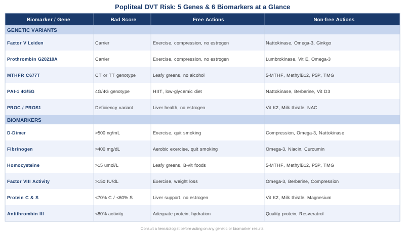
Popliteal vein thrombosis is not simply bad luck, and it is not effectively managed by generic advice alone. Behind every clot is a biological story written in biomarkers and, often, in DNA. The six biomarkers covered here — D-dimer, fibrinogen, homocysteine, Factor VIII, Protein C and S, and Antithrombin III — offer a window into how your coagulation system is functioning right now. The five genetic variants — Factor V Leiden, Prothrombin G20210A, MTHFR C677T, PAI-1 4G/5G, and PROC/PROS1 — tell you what your coagulation system is fundamentally predisposed toward.
These two layers of information together produce something far more useful than a generic risk score: a specific, personal map of where the vulnerabilities lie and where targeted interventions will have the greatest effect. Knowing your homocysteine is elevated because you carry MTHFR C677T and have low methylated B vitamins is not a curiosity — it is directly actionable. Knowing your PAI-1 genotype is 4G/4G explains precisely why fibrinolysis is sluggish and which interventions will help most.
The next smart step is not to implement everything at once, but to start with the most accessible and highest-yield testing: a comprehensive coagulation panel including homocysteine, fibrinogen, D-dimer, Factor VIII activity, Protein C and S, and Antithrombin III, followed by genetic testing for the five variants discussed here if not already completed. Bring those results to a hematologist, vascular medicine specialist, or a physician experienced in thrombophilia workup — and use them to have the kind of specific, evidence-informed conversation that generic risk counseling rarely enables. That conversation, grounded in your own biology, is where meaningful prevention begins.
Cardiovascular Endocrine & Metabolic
Cardiovascular: Blood Vessel Conditions Vascular Conditions
Endocrine & Metabolic: Metabolic Syndrome
Autoimmune: Inflammatory Conditions