This article contains AI generated content.
· UpdatedPopliteus Tendon Tear Genes and Biomarkers: 6 Genes and 7 Biomarkers to Track
Introduction
A popliteus tendon tear is one of those injuries that gets missed, misdiagnosed, or treated too generically. Located at the back of the knee, the popliteus muscle and its tendon play a quiet but critical role in stabilizing the joint during rotation and in unlocking the knee from full extension. When this tendon tears — whether from a traumatic event, chronic overload, or a subtle biomechanical failure — the path to recovery is rarely straightforward.
What makes this injury particularly challenging is how differently people heal. Two people with the same MRI finding can have completely different trajectories. One returns to sport in three months; the other struggles for a year or more. That gap often has less to do with the severity of the initial tear and more to do with underlying biological factors: inflammation levels, collagen quality, tissue repair capacity, and genetic predispositions that no standard consultation addresses.
Generic protocols — rest, ice, compression, and a standardized rehab plan — give most patients a starting point, but not a precise one. They miss the individual biology that drives why some tendons are structurally more vulnerable, why some people mount excessive inflammatory responses, and why certain individuals fail to produce adequate collagen during repair. Without understanding these factors, recovery becomes guesswork.
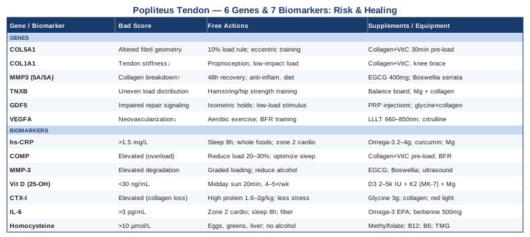
This article takes a more targeted approach. The core section focuses on the most meaningful biomarkers you can actually measure — blood markers that reflect what is happening inside the tendon environment and whether your body has what it needs to heal. A second section covers the genetics layer: key gene variants linked to tendon vulnerability and altered repair biology. Together, these two lenses give you a sharper picture of your individual risk and a more rational basis for the decisions you and your clinician make along the way.
7 Biomarkers That May Reveal How Well Your Popliteus Tendon Can Heal
Biomarkers are measurable signals that reflect biological processes. For a tendon tear, the most relevant ones fall into three broad categories: inflammatory load, collagen metabolism, and nutrient and cofactor status. Each tells a different part of the story, and tracking several together gives a more complete picture than any one marker alone.
1. High-Sensitivity C-Reactive Protein (hs-CRP)
Why it matters: hs-CRP is the most widely available marker of systemic inflammation. After a tendon injury, the body initiates an inflammatory response to clear debris and begin repair — this is necessary and expected. But when inflammation is chronically elevated, even at low levels, it impairs the proliferative phase of healing by disrupting fibroblast activity and collagen synthesis. Elevated hs-CRP before or during rehabilitation is associated with slower recovery timelines and increased risk of re-injury.
What it may reveal: Elevated hs-CRP above 1.0–1.5 mg/L in the context of injury recovery suggests systemic inflammation is elevated beyond what is needed for normal repair. This can come from poor sleep, dietary patterns, gut dysbiosis, obesity, or underlying inflammatory conditions unrelated to the knee.
How to measure it: A standard blood draw, typically included in a basic metabolic or inflammation panel. Cost ranges from $15–$50 USD through direct-to-consumer labs. It is one of the most accessible markers on this list.
If the score is bad, the plan without supplements: The most potent anti-inflammatory interventions that require no supplementation are sleep optimization (7–9 hours per night, consistent timing), elimination of ultra-processed foods and refined seed oils, blood glucose stabilization through lower-glycemic nutrition, and aerobic exercise at low-to-moderate intensity (30 minutes, 5 days per week). Time-restricted eating within a 10–12 hour window has shown meaningful reductions in hs-CRP in several human studies and is a practical starting point that requires no cost.
If the score is bad, the plan with supplements or equipment: Omega-3 fatty acids (EPA+DHA): 2–4g per day, taken with a meal containing fat. Cycle continuously for 3–6 months, then reassess. Common side effects include mild fish breath and GI discomfort at high doses. Curcumin (phytosome or piperine-enhanced form): 500–1000mg twice daily, 8–12 weeks on, then reassess. Avoid at high doses with blood thinners. Magnesium glycinate: 300–400mg at night, continuous. Loose stools can occur at high doses. Cold water immersion or contrast therapy (cold/hot alternating, 10–15 minutes post-exercise) may reduce circulating inflammatory markers with repeated use.
2. COMP (Cartilage Oligomeric Matrix Protein)
Why it matters: Despite its name, COMP is not limited to cartilage. It is a structural glycoprotein found in high concentrations in tendons and ligaments. During active tendon injury or mechanical loading beyond the tissue's capacity, COMP is released into the blood. It is one of the most specific markers of connective tissue stress and breakdown available outside of a biopsy.
What it may reveal: Elevated serum COMP in the context of a known popliteus tendon tear suggests active tissue remodeling or ongoing mechanical stress that may be exceeding the tendon's repair capacity. Researchers have used COMP to track loading responses in Achilles and patellar tendon rehabilitation protocols, making it a useful monitoring tool during progressive tendon loading phases.
How to measure it: Available through specialty labs and some academic medical centers. Cost varies: $80–$200 USD depending on the provider. It is not a routine clinical test, so you may need to request it specifically or work through a sports medicine physician.
If the score is bad, the plan without supplements: Elevated COMP may indicate that loading is progressing too quickly. The first adjustment is reducing training volume and intensity by 20–30% for 2–3 weeks, then reintroducing load using an isometric-to-isotonic progression. Sleep and recovery windows matter significantly — collagen synthesis peaks during sleep, and inadequate rest has been shown to elevate COMP independently of exercise.
If the score is bad, the plan with supplements or equipment: Collagen peptides (Type I/III): 15g taken 30–60 minutes before a loading session, paired with 50mg vitamin C. The vitamin C co-factor is essential for hydroxylation of collagen precursors. Take daily during active rehab; side effects are low, with occasional digestive discomfort. Blood flow restriction (BFR) training: Applied with a cuff to the thigh proximal to the injury site, BFR allows high metabolic stimulus at low mechanical loads — useful when heavy loading remains contraindicated. Administered under physiotherapy supervision initially.
3. MMP-3 (Matrix Metalloproteinase-3)
Why it matters: Matrix metalloproteinases are enzymes responsible for degrading extracellular matrix components, including the collagen fibers that tendons are built from. MMP-3 in particular has been associated with tendon and ligament pathology. When MMP-3 activity is elevated and not adequately balanced by its natural inhibitors (tissue inhibitors of metalloproteinases, or TIMPs), the result is net collagen breakdown — even during attempted healing.
What it may reveal: Elevated serum MMP-3 signals an active matrix degradation environment. In the context of a popliteus tendon tear, persistent elevation suggests that the catabolic processes inside the tissue are not being matched by sufficient anabolic repair activity. MMP-3 is also elevated in inflammatory joint disease, which can coexist with tendon injuries and compound the problem.
How to measure it: MMP-3 can be measured in blood serum. It is more commonly ordered in rheumatoid arthritis monitoring but is accessible through specialty labs. Cost: $50–$120 USD.
If the score is bad, the plan without supplements: Reduce inflammatory triggers: ultra-processed foods, excess alcohol, and smoking all upregulate MMP activity. Introduce consistent graded tendon loading — mechanical load at the right intensity is one of the few interventions that stimulates TIMP production and shifts the MMP/TIMP ratio toward repair. Sleep disruption elevates MMP activity through circadian disruption, making consistent sleep architecture a therapeutic priority.
If the score is bad, the plan with supplements or equipment: Green tea extract (EGCG): EGCG has shown MMP inhibitory activity in both in vitro and some clinical studies. 400–800mg EGCG standardized extract, taken with meals. Cycle 8–10 weeks. Avoid on an empty stomach; caution with liver sensitivity at high doses. Boswellia serrata (AKBA form): 300–400mg twice daily, 8–12 weeks. Generally well tolerated. Therapeutic ultrasound (clinical setting, 1MHz, 0.5–1.5 W/cm²) may stimulate fibroblast activity and help rebalance the catabolic-anabolic environment — typically 2–3 times per week for 4–6 weeks.
4. 25-OH Vitamin D
Why it matters: Vitamin D receptors are present in muscle, bone, tendon, and immune cells. Low vitamin D is linked to impaired muscle function, increased susceptibility to musculoskeletal injury, and slower tendon healing. Several studies in athletic populations have found that vitamin D deficiency correlates with a higher rate of tendon and ligament injuries. The mechanism includes effects on calcium-phosphorus metabolism, anti-inflammatory gene expression, and satellite cell activation.
What it may reveal: Serum 25-OH vitamin D below 30 ng/mL (75 nmol/L) is considered insufficient in most musculoskeletal healing contexts. Levels below 20 ng/mL are frankly deficient. Peter Attia and other performance medicine physicians have argued that optimal healing ranges may sit closer to 40–60 ng/mL, though upper limits matter too — toxicity begins to emerge above 100 ng/mL.
How to measure it: A standard blood test, available nearly everywhere and frequently covered by insurance. Cost: $25–$60 USD. Can be included in a basic annual panel without a specialist referral.
If the score is bad, the plan without supplements: Direct sun exposure: 15–30 minutes of midday sun on large skin surfaces (arms, legs) without sunscreen, 4–5 times per week during summer months. Results are limited in northern latitudes between October and March and in individuals with darker skin tones, who require longer exposure times for equivalent synthesis.
If the score is bad, the plan with supplements or equipment: Vitamin D3: 2,000–5,000 IU daily, paired with vitamin K2 (MK-7 form, 100–200mcg/day) to direct calcium appropriately. Avoid supplementing above 2,000 IU without K2. Retest at 8 and 16 weeks to dial in the dose. Magnesium (required cofactor): Without adequate magnesium, vitamin D conversion to its active form is impaired. 300–400mg magnesium glycinate or malate at night. The risk of over-supplementation includes hypercalcemia — fatigue, nausea, kidney stress — so staying within the 40–70 ng/mL range is the practical target.
5. Collagen Type I C-Telopeptide (CTX-I)
Why it matters: CTX-I is a degradation product of type I collagen — the dominant structural protein in tendons. Elevated CTX-I in blood or urine reflects excessive collagen breakdown, which can outpace synthesis during inadequate recovery, nutritional deficiencies, hormonal imbalance, or excessive mechanical stress.
What it may reveal: High CTX-I alongside slow clinical recovery may point to a catabolic state where the body is breaking down collagen faster than it can rebuild it. This can occur with chronic cortisol elevation, estrogen deficiency (particularly in postmenopausal women), low dietary protein intake, or insufficient collagen co-factors. It is a direct window into the net direction of connective tissue turnover.
How to measure it: Serum or urine CTX-I, best measured in the morning, fasting. Available at major labs. Cost: $50–$100 USD. Often ordered alongside bone turnover markers in metabolic bone workups.
If the score is bad, the plan without supplements: Increase dietary protein toward 1.6–2.0g per kg of body weight daily, with emphasis on glycine-rich foods: bone broth, skin-on chicken, gelatin. Reduce chronic stress — cortisol directly stimulates MMP activity and promotes collagen catabolism. Prioritize consistent sleep over 7.5 hours. Review any corticosteroid use if relevant, as exogenous steroids accelerate CTX-I elevation and actively deplete tendon collagen.
If the score is bad, the plan with supplements or equipment: Glycine: 3–5g before sleep. Glycine is a direct substrate for collagen synthesis and has some evidence for improving sleep quality. Continuous; no known cycling requirement at these doses. Collagen peptides (15g before loading with vitamin C): as described above. DHEA (only if cortisol/DHEA ratio is imbalanced): Only under medical supervision — testing a cortisol awakening response alongside CTX-I provides a better picture of whether adrenal function is contributing. This is not a self-prescribe supplement. Red light therapy panels (660nm/850nm) applied 10–15 minutes daily over the posterior knee show early-stage evidence for fibroblast collagen synthesis stimulation.
6. Interleukin-6 (IL-6)
Why it matters: IL-6 is a pleiotropic cytokine that drives both pro-inflammatory and anti-inflammatory responses depending on context. Acutely after exercise or injury, IL-6 rises and contributes to tissue repair signaling — this is appropriate. But chronically elevated IL-6, particularly when not exercise-driven, reflects an inflammatory state that impairs tendon fibroblast function and promotes collagen degradation via MMP upregulation.
What it may reveal: Elevated fasting IL-6 above 3 pg/mL in someone with a tendon tear and slow recovery may point to persistent low-grade inflammation driven by visceral adiposity, gut permeability, sleep disruption, or prior unresolved systemic stress. It adds depth to the hs-CRP picture — when both are elevated, the inflammatory case is clearer.
How to measure it: Available at most clinical labs. Cost: $40–$90 USD. Should be measured fasted and in the morning, well away from recent exercise sessions, which acutely elevate IL-6 as a normal response.
If the score is bad, the plan without supplements: Reducing visceral fat is the most powerful modifiable driver of chronic IL-6. Structured aerobic activity (zone 2 cardio, 150–200 minutes per week) combined with protein-dominant, lower-carbohydrate nutrition has good evidence for IL-6 reduction. Even partial sleep restriction (6 hours versus 8 hours) elevates IL-6 significantly in controlled studies. Gut health optimization through fiber-rich, diverse diets reduces circulating inflammatory cytokines including IL-6 through microbiome-dependent pathways.
If the score is bad, the plan with supplements or equipment: Omega-3 fatty acids: EPA specifically has the strongest evidence for IL-6 reduction. 2–4g EPA+DHA daily, continuous. Berberine: 500mg twice daily with meals. Activates AMPK pathways that suppress IL-6 and NF-κB signaling. Well studied in metabolic conditions. Cycle 8–12 weeks. GI side effects are possible — start at lower doses. Resveratrol: 250–500mg daily with a fatty meal; some evidence for IL-6 reduction. Cycle 8 weeks on, 4 weeks off. Theoretical interaction with blood thinners at high doses.
7. Homocysteine
Why it matters: Homocysteine is an amino acid byproduct of methionine metabolism. When elevated, it is directly toxic to collagen cross-linking enzymes and inhibits lysyl oxidase — the enzyme responsible for creating strong collagen fiber bonds in tendons. Even if a person is producing sufficient collagen precursors, elevated homocysteine means the resulting fibers may not be structurally sound. This is a specific repair bottleneck that is rarely screened for in standard tendon injury workups.
What it may reveal: Homocysteine above 10–12 µmol/L warrants attention in any connective tissue repair context. Elevation is most commonly driven by low B12, low folate, low B6, or genetic variants in the MTHFR gene that impair methylation — a connection that links this biomarker directly to the genetics section below.
How to measure it: Standard blood test, available at most labs. Often included in cardiovascular risk panels. Cost: $25–$60 USD. One of the most underutilized and inexpensive markers on this list.
If the score is bad, the plan without supplements: Increase dietary consumption of methyl-donor supporting foods: eggs, leafy greens, legumes, and liver. Reduce excess alcohol and smoking — both disrupt B-vitamin status and raise homocysteine independently. Increasing dietary protein from whole animal sources naturally raises methionine cycling and glutathione support.
If the score is bad, the plan with supplements or equipment: Methylfolate (L-5-MTHF): 400–800mcg daily. Preferred over folic acid for those with MTHFR variants. Continuous. Methylcobalamin (B12): 500–1000mcg daily, sublingual form for best absorption. Continuous. Pyridoxal-5-phosphate (B6, active form): 25–50mg daily. Do not exceed 100mg per day over long periods — peripheral neuropathy is a known risk at chronic high doses. TMG (Trimethylglycine/Betaine): 500–1500mg daily. Directly donates methyl groups to convert homocysteine to methionine. Continuous. Generally well tolerated at standard doses.
With a clear picture of the biochemical markers that reflect your healing environment, it is worth turning to the genetic layer — the inherited factors that may predispose some people to popliteus and broader tendon vulnerability before an injury even occurs.
Genetics and Tendon Vulnerability: 6 Key Gene Variants Linked to Injury Risk
Genetics does not determine destiny in tendon health, but it shapes the biological terrain. Understanding your variant status in key tendon-related genes can explain why injury happened, why recovery is slower than expected, and which interventions are most likely to help. The field of tendon genetics is still maturing — most evidence comes from studies on Achilles, ACL, and rotator cuff injuries, which share overlapping biological pathways with popliteus tendon pathology. Where evidence is specifically limited to the popliteus, this is noted clearly.
1. COL5A1 — Collagen Type V, Alpha 1
What it does: COL5A1 encodes a component of type V collagen, which regulates the diameter and mechanical properties of type I collagen fibrils. Narrower, more uniform fibrils created by normal COL5A1 function appear to be protective against excessive deformation under load. A restriction fragment length polymorphism in the 3′ UTR of COL5A1 has been studied in relation to Achilles tendon and ACL injuries in multiple athletic cohorts, with carriers of certain variants showing higher injury rates.
How it may affect you: Individuals carrying specific COL5A1 variants may produce tendons with altered fibril geometry — making them more susceptible to mechanical failure under repetitive load. This may partially explain why some people sustain popliteus and other posterior knee tendon injuries without a clear traumatic event, or why they re-injure on loads that seem manageable.
If the gene is bad, the plan without supplements: Progressive loading with careful attention to volume spikes. The 10% rule — no more than a 10% increase in weekly load per week — is especially important for individuals with COL5A1 variants. Prioritize eccentric and isometric loading protocols, which stimulate collagen remodeling with lower peak mechanical stress. Adequate sleep and recovery windows between training sessions are more important than for average-risk individuals.
If the score is bad, the plan with supplements or equipment: Collagen peptides (15g paired with 50mg vitamin C, taken 30–60 minutes before a loading session) to increase available collagen precursors before mechanical stimulation triggers synthesis. Daily during active rehab phases. Tendons in higher-risk individuals also benefit from longer warm-up protocols — 10–15 minutes of progressive dynamic loading before maximal effort activities reduces the risk of microtear accumulation during peak loading.
2. COL1A1 — Collagen Type I, Alpha 1
What it does: COL1A1 is the primary structural gene for type I collagen, the backbone of tendon architecture. An Sp1 binding site polymorphism in intron 1 of COL1A1 — the so-called "s" allele — has been associated with altered collagen fiber strength. The ss genotype has been linked in some research to increased ACL rupture risk and reduced tendon mechanical stiffness.
How it may affect you: A suboptimal COL1A1 variant may mean your tendons are intrinsically less stiff and more prone to deformation beyond their elastic limit. In the context of a popliteus tear, this may contribute to both the initial injury and slower repair, since new collagen synthesis during healing involves the same gene machinery.
If the gene is bad, the plan without supplements: Emphasis on proprioception and neuromuscular control training — if tendon stiffness is reduced, the nervous system can partially compensate through faster reactive muscle activation. Reduce repetitive high-impact loading (running on hard surfaces, rapid deceleration sport movements) and introduce more controlled loading environments during rehabilitation.
If the score is bad, the plan with supplements or equipment: Collagen peptides and vitamin C as described above. Additionally, consider wearing a posterior knee sleeve or popliteal support brace during return-to-sport activities. Mechanical offloading during the repair phase reduces stress on tendon tissue that is structurally rebuilding itself — particularly important when structural stiffness is a genetic weak point.
3. MMP3 Gene Variants (5A/6A Promoter Polymorphism)
What it does: The MMP3 gene codes for stromelysin-1, a matrix metalloproteinase that degrades several extracellular matrix components including type III collagen and aggrecan. A functional polymorphism (5A/6A) in the promoter region of MMP3 influences how much MMP-3 protein is produced. Individuals with the 5A/5A genotype produce more MMP-3, which creates a more catabolic tendon environment under identical loading conditions.
How it may affect you: Higher MMP-3 expression means faster breakdown of collagen matrix — potentially leading to delayed tendon repair and higher re-injury risk. This genetic variant connects directly to the MMP-3 biomarker discussed above: if you carry the 5A/5A genotype and show elevated serum MMP-3, you have converging evidence of a high-degradation phenotype that warrants specific attention to recovery and anti-inflammatory interventions.
If the gene is bad, the plan without supplements: Prioritize recovery windows between loading sessions — a minimum of 48 hours between heavy tendon-loading sessions is more important for 5A/5A individuals than for the general population. Anti-inflammatory dietary patterns (Mediterranean-style, whole foods emphasis) have been shown to downregulate MMP expression at the epigenetic level over time. Avoid overtraining, which amplifies MMP-3 upregulation through inflammatory cascade.
If the score is bad, the plan with supplements or equipment: EGCG and Boswellia serrata as described in the biomarker section, with the same cycling protocols. Vitamin D3 maintained at optimal levels also has some evidence for TIMP upregulation — the natural inhibitors of MMPs — making vitamin D optimization doubly relevant for individuals with MMP3 5A/5A variants.
4. TNXB — Tenascin-X
What it does: Tenascin-X is an extracellular matrix glycoprotein that plays a key role in collagen fibril organization and mechanical coupling in tendons and ligaments. Complete TNXB deficiency causes a connective tissue disorder closely resembling hypermobile Ehlers-Danlos syndrome. More subtle heterozygous variants, found in the general population, are associated with joint hypermobility and increased susceptibility to tendon and ligament injuries across multiple anatomical sites.
How it may affect you: People with reduced tenascin-X activity may have tendons that distribute mechanical loads less evenly, creating focal stress concentrations where tears are more likely to initiate. The popliteal region is particularly vulnerable during tibial external rotation and rapid knee extension — common movements in running, cutting, and skiing — which are precisely the mechanisms associated with popliteus tendon tears.
If the gene is bad, the plan without supplements: Neuromuscular control training is essential: the surrounding muscles must compensate for tendon tissue that cannot self-organize optimally under load. Focused strength work on the hamstrings, gastrocnemius, and hip external rotators reduces the peak load on the popliteus itself during dynamic activities. Balance and proprioceptive drills help retrain the knee's stabilization reflexes.
If the score is bad, the plan with supplements or equipment: Proprioceptive training tools — balance boards, single-leg loading progressions, perturbation training — to retrain neuromuscular stabilization. Magnesium glycinate and collagen supplementation to support matrix protein function during ongoing tendon maintenance.
5. GDF5 — Growth Differentiation Factor 5
What it does: GDF5 is a member of the TGF-β superfamily and plays a major role in tendon, ligament, and joint development. A common single nucleotide polymorphism (rs143384) in the GDF5 promoter region has been associated with altered tendon cell differentiation and reduced tendon tissue quality in several population studies, with some data suggesting effects in the posterior knee specifically.
How it may affect you: Suboptimal GDF5 variants may impair the activation of tendon-derived progenitor cells during injury, slowing the early repair response. Recovery from a popliteus tear may take longer and be less complete if the growth factor signaling that activates repair cells is blunted from the outset.
If the gene is bad, the plan without supplements: The loading stimulus for tendon progenitor cell activation requires mechanical input. Even during relative rest from sport, gentle isometric holds — submaximal popliteus activation through mild resistance to knee rotation — can maintain a repair-stimulating environment without overloading healing tissue. Sustained, low-load mechanical stimulation has been shown to maintain progenitor cell activity better than complete immobilization.
If the score is bad, the plan with supplements or equipment: Platelet-rich plasma (PRP) injections, administered by a sports medicine physician, deliver concentrated growth factors including signals in the TGF-β family that may partially compensate for reduced endogenous GDF5 activity. Evidence in tendon healing is promising but not yet definitive, and quality of the PRP preparation matters significantly. Glycine and collagen peptide supplementation support matrix assembly even when progenitor cell signaling is suboptimal.
6. VEGFA — Vascular Endothelial Growth Factor A
What it does: Tendons are relatively avascular — they receive limited blood supply, which is a central reason why they heal slowly. VEGFA controls neovascularization, the formation of new blood vessels into healing tissue. Certain VEGFA variants are associated with a reduced angiogenic response to injury, meaning the healing tendon receives less nutrient and oxygen delivery during the critical repair window.
How it may affect you: The popliteus tendon, already marginally vascularized in its mid-substance, is particularly dependent on adequate angiogenesis after tearing. If your VEGFA response is blunted genetically, you may see prolonged healing timelines and reduced tendon strength restoration even with optimal rehabilitation input.
If the gene is bad, the plan without supplements: Aerobic exercise — even non-weight-bearing (cycling, swimming) — stimulates VEGFA expression through hypoxia-inducible factor (HIF) pathways. Maintaining cardiovascular activity during tendon rehab is not just for fitness: it actively supports vascular supply to healing tissue. Blood flow restriction training at the thigh also increases local VEGF signaling — making it doubly useful for individuals with blunted VEGFA genetics.
If the score is bad, the plan with supplements or equipment: Low-level laser therapy (photobiomodulation) has shown ability to stimulate local VEGF production and neovascularization in tendon tissue — 660–850nm wavelengths, 10–20 minutes daily over the posterior knee. Nitric oxide support through citrulline malate (3–6g daily) and dietary nitrates (beet root, leafy greens) supports vasodilation and nutrient delivery to healing tissue with a favorable side effect profile.
The genetic and biomarker picture together give you a much more complete understanding of the individual terrain you are working with. What follows is a set of practical insights from the research and clinical communities that apply regardless of your specific genetic or biomarker status.
What the Tendon Biology Research Has Changed: 10 Things That May Shift Your Recovery
Over the past decade, the field of tendon biology has been reshaped by researchers like Keith Baar (UC Davis), Jill Cook (La Trobe University), and Karim Khan, with their work gaining broader reach through performance medicine practitioners like Peter Attia. Much of what follows contradicts conventional wisdom — and it is the part that is often not communicated in a standard physiotherapy consultation.
1. Tendons Need Load, Not Rest
Complete rest after a tendon tear causes collagen disorganization and weakens the surrounding tissue. The question is not whether to load, but how much and what type. Isometric contractions — sustained holds at specific joint angles — are currently the best-supported first-line mechanical intervention. They reduce pain, stimulate collagen synthesis, and do not cause the microtrauma that dynamic loading does in the acute phase. For the popliteus, isometric tibial rotation resistance holds in a supported seated position are an early safe option.
2. The Collagen-Vitamin C Timing Window Is Real
Research by Keith Baar and colleagues demonstrated that consuming collagen peptides with vitamin C approximately 60 minutes before a loading session increases collagen synthesis in the tendon itself. The mechanism is that the resulting amino acid spike in blood — particularly glycine and proline — coincides with the anabolic window that mechanical loading opens. This is a specific, time-dependent intervention, not a general supplement strategy, and the timing detail is what makes it meaningful.
3. Tendons Respond Slowly and Nonlinearly
Tendon collagen turnover is measured in weeks to months, not days. Imaging and clinical symptoms frequently improve before the tendon has adequate mechanical strength. Many re-injuries occur during the "false recovery" window when symptoms have resolved but structural restoration is incomplete. Programs that progress based on objective strength testing — not pain alone — are more protective, and returning to sport after only 6–8 weeks should be approached with significant caution.
4. Sleep Is a Collagen Synthesis Window
Growth hormone, released primarily during deep sleep, is one of the most powerful stimulators of collagen synthesis in the body. For tendon injuries, sleep quality is a genuine therapeutic variable. Chronic partial sleep restriction — even losing 90 minutes per night — substantially impairs collagen turnover markers over time. Addressing sleep is not peripheral to recovery; it may determine whether the anabolic environment needed for repair is available at all.
5. Chronic Cortisol Actively Degrades Tendons
Glucocorticoids — whether endogenous from chronic psychological stress or exogenous from corticosteroid injections — increase MMP activity, suppress collagen synthesis, and impair tendon cell viability. Multiple long-term studies have found that repeated corticosteroid injections into tendons provide short-term pain relief at the cost of higher rupture rates over the following months. Managing cortisol through stress reduction, sleep, and nutrition is mechanistically central to tendon recovery, not a lifestyle nicety.
6. Eccentric Loading Has a Specific Evidence Base
Eccentric protocols — loading during the lengthening phase of muscle contraction — have been shown in multiple randomized controlled trials to remodel abnormal tendon tissue and improve mechanical properties. For posterior knee rehabilitation, this translates to controlled exercises emphasizing the lengthening phase of the hamstring and popliteal musculature under resistance — applied gradually and progressively as pain and function allow.
7. Tendon Stiffness, Not Size, Predicts Function
Imaging studies show that tendon cross-sectional area correlates poorly with function or re-injury risk. Stiffness — measured via ultrasound elastography or force-displacement testing — is what predicts performance. This means a hypertrophied-looking tendon on imaging can still be mechanically compromised, and standard MRI alone is insufficient for return-to-sport decisions in individuals who want genuine risk assessment.
8. The Posterior Knee Is a Biomechanical Chain
The popliteus does not work in isolation. It is part of a posterolateral corner complex involving the fibular collateral ligament, popliteofibular ligament, and posterolateral capsule. Injury to any one component increases load on the others. Rehabilitation that addresses the entire posterior knee kinetic chain — including hip external rotation strength, hamstring force production, and foot and ankle mechanics — consistently produces better long-term outcomes than popliteus-specific protocols alone.
9. Blood Flow Restriction Changes the Early Rehab Calculus
BFR training allows significant hypertrophic and strength stimulus at loads as low as 20–30% of one-repetition maximum — compared to the 70–85% typically required for the same effect without restriction. For early tendon rehabilitation, where high mechanical load is contraindicated but muscle atrophy and tendon stimulus deprivation are ongoing risks, BFR represents a meaningful middle path that is increasingly supported by clinical evidence.
10. Return-to-Sport Decisions Should Integrate Biology
Traditional return-to-sport criteria rely primarily on time and subjective symptom resolution. Emerging practice in sports medicine incorporates objective biomarker tracking — COMP, hs-CRP — alongside imaging (ultrasound elastography) and strength symmetry testing. Returning to sport with unresolved biological markers of ongoing breakdown substantially increases re-injury risk, regardless of how the knee feels subjectively.
These principles provide a framework that is genuinely different from what most people receive at a standard sports medicine or physiotherapy consultation. Beyond this research framework, a handful of complementary approaches also have meaningful clinical support for tendon injury recovery specifically.
Complementary Approaches With Clinical Evidence
The following modalities have the most relevant evidence base for tendon injury. None replaces medical management or rehabilitation, but each adds a different dimension of support when applied appropriately.
Low-Level Laser Therapy (Photobiomodulation)
Low-level laser therapy uses specific wavelengths of light — typically 660nm red and 808–850nm near-infrared — to stimulate cellular energy production in mitochondria. For tendons, this has two relevant effects: increased ATP production in tenocytes (tendon cells) and stimulation of local growth factors including VEGF and TGF-β, which drive repair and neovascularization. This makes it particularly relevant for individuals with blunted VEGFA genetics or those with a persistently elevated COMP, suggesting inadequate repair stimulus.
A meta-analysis examining LLLT for Achilles tendinopathy published in Lasers in Medical Science found significant reductions in pain and improvements in function compared to placebo across multiple randomized trials. While the popliteus tendon specifically has not been studied in isolation in this modality, the cellular mechanisms are shared across tendon types and posterior knee application is both feasible and safe.
A practical protocol involves a clinical-grade device delivering 50–100 joules per session at 660–850nm wavelengths, applied to the posterior knee in a grid pattern over 10–15 minutes, 3–5 times per week during active healing phases. Home devices (panels and wands) offer cost-effective options, though clinical devices have more documented dosing protocols. Avoid application over active hematoma in the first 72 hours. Always use appropriate eye protection when using laser-based devices.
Massage Therapy
Massage applied to the posterior knee musculature — particularly the popliteus, medial gastrocnemius, and semimembranosus — can reduce muscle guarding, improve local circulation, and restore neuromuscular coordination that is disrupted after a tendon injury. Transverse friction massage applied to the tendon by a skilled therapist has been used clinically to address adhesion formation and to stimulate mechanotransduction in healing tissue.
The Cyriax methodology of deep transverse friction massage has been studied in lateral knee tendinopathy, with randomized controlled trials finding significant improvements in pain and function compared to standard physiotherapy alone. Popliteus-specific evidence is limited to case reports and clinical experience rather than large RCTs, but the anatomical rationale is sound and the intervention carries minimal risk when applied correctly.
Practically, 2–3 sessions per week of posterior knee massage during the subacute healing phase — weeks 2–8 post-injury — is a reasonable frequency. Patients can learn self-massage techniques using a massage ball applied to the posterior knee, though deep transverse friction to the tendon itself should be performed by a trained therapist. Avoid deep pressure directly over the neurovascular bundle running through the popliteal fossa, which contains the popliteal artery and tibial nerve.
Breathing-Based Therapies
Diaphragmatic breathing and controlled respiratory protocols — particularly slow breathing at 5–6 breaths per minute, or extended exhale patterns — activate the parasympathetic nervous system, reduce cortisol output, lower circulating IL-6, and improve tissue oxygenation. These effects directly address two of the most important biological barriers to tendon recovery: chronic stress-driven cortisol elevation and inflammatory cytokine burden.
A randomized crossover study published in Psychoneuroendocrinology demonstrated that slow-paced breathing significantly reduced salivary cortisol and pro-inflammatory cytokines in healthy adults and those with musculoskeletal pain conditions. Effects were maintained with consistent daily practice over 8 weeks, suggesting cumulative benefit rather than acute-only response.
A practical protocol for tendon recovery: 10 minutes of slow breathing (inhale 4 seconds, exhale 6–8 seconds) twice daily — once upon waking and once before sleep. The pre-sleep session pairs well with progressive muscle relaxation of the posterior knee musculature, which reduces nocturnal muscle tension and improves the quality of deep sleep. This is the window during which growth hormone and collagen synthesis peak — making anything that deepens sleep a genuine recovery intervention, not just a comfort measure.
Yoga
Yoga's relevance in tendon injury recovery operates through three mechanisms: improving tissue flexibility to reduce compensatory loading on injured tendons, strengthening adjacent stabilizing muscles through body-weight loading, and reducing systemic cortisol through documented effects on the hypothalamic-pituitary-adrenal axis. Lower cortisol means less MMP upregulation and better collagen preservation — connecting directly to the biomarker and genetic picture described earlier.
A randomized trial published in the International Journal of Yoga found that 12 weeks of regular yoga practice significantly reduced serum cortisol and hs-CRP in participants compared to control groups — both biomarkers directly relevant to tendon recovery. While no study has examined yoga specifically for popliteus tendon tears, the systemic effects on inflammation and local effects on posterior knee flexibility make it a meaningful and low-risk adjunct.
For this condition, the safest entry point is a restorative or Hatha yoga practice that avoids deep squat positions and full knee flexion in the early healing phase. Poses such as supported reclining butterfly, gentle prone hip extension, and low lunge variants that do not compress the posterior knee are appropriate starting points. Advance to more active poses — warrior series, single-leg balance postures — as strength and pain resolution progress. Always work within a pain-free range and communicate clearly with any yoga instructor about the injury.
Conclusion
A popliteus tendon tear sits at the intersection of biomechanics, inflammation biology, and individual genetic variation. Standard care gives most people a foundation, but it rarely accounts for the biological factors that most directly determine speed and quality of recovery — and those factors are now measurable.
Tracking the seven biomarkers outlined here gives you real data on your inflammatory load, collagen turnover, and nutrient status. Understanding your genetic predispositions tells you where your terrain is naturally more challenging and which interventions are most likely to move the needle. The complementary approaches add layered support that works alongside, not instead of, standard rehabilitation.
The next smart step is not overhauling everything at once. Pick one or two biomarkers that are most accessible to you, measure them, and use the results to guide one or two specific changes — in nutrition, supplementation, or recovery habits. Review the results in 8–12 weeks and build from there. Work with a sports medicine physician or clinician comfortable interpreting these markers in the context of your injury. Better information leads to better decisions, and in tendon recovery, better decisions make a measurable difference.
Musculoskeletal: Joint Conditions Tendon & Ligament Conditions Sports Injuries
Autoimmune: Inflammatory Conditions Connective Tissue Conditions