This article contains AI generated content.
· UpdatedPost-Polio Syndrome Genes and Biomarkers: 5 Genes and 7 Biomarkers to Track
Introduction
If you are living with post-polio syndrome, you already know how little the standard clinical conversation offers. The fatigue, the new muscle weakness, the cold intolerance arriving decades after the original infection — these are real, measurable, and biologically explicable, yet they are still met in many clinics with the same limited response: rest more, pace yourself, accept the progression. That advice is not wrong, but it is profoundly incomplete.
What makes PPS so difficult to manage well is that it operates across several intersecting biological systems simultaneously — neuroinflammation, motor neuron survival, hormonal signaling, mitochondrial function, and immune regulation. The degree to which each of these is disrupted varies considerably from one person to the next. Two people with very similar polio histories can have radically different biomarker profiles driving their current symptoms, and they will benefit from very different interventions. Generic protocols cannot account for this. Precision tracking can.
This article is built around a straightforward premise: better data leads to better decisions. Knowing where your inflammatory markers stand, how your IGF-1 compares to age-adjusted norms, whether your thyroid is contributing to your fatigue, and whether your motor neurons are showing signs of ongoing stress does not require a research center. It requires targeted testing, informed interpretation, and a willingness to act on what the numbers reveal.
Two complementary strategies anchor the pages ahead. The first is a practical biomarker tracking plan — seven measurements that illuminate the core mechanisms of PPS and guide interventions that are far more targeted than anything a general recommendation can offer. The second is a genetics overview revealing how five inherited variants may be shaping the biological terrain of your condition. A book summary challenges the conventional management approach in ways that many clinicians still have not absorbed. And a final section covers complementary modalities with genuine human clinical evidence. None of this is a cure. All of it is information that can move the needle.
7 Biomarkers to Track if You Have Post-Polio Syndrome
Biomarker tracking in PPS is not yet standard care in most clinics, but the science increasingly supports it. Several measurable blood and serum values correlate directly with the core mechanisms driving the condition — neuroinflammation, motor neuron stress, hormonal decline, and mitochondrial function. Tracking them over time does not just reveal where problems exist; it reveals which interventions are working and which are not. That feedback loop is what makes precision management possible.
1. Neurofilament Light Chain (NfL)
Why it matters: Neurofilament light chain is a structural protein released into the bloodstream and cerebrospinal fluid when axons and motor neurons are physically damaged or under significant metabolic stress. In post-polio syndrome, the surviving enlarged motor units — those that sprouted new connections after the original poliovirus destroyed a large portion of the motor neuron pool — are under chronic mechanical and metabolic strain. When they begin to fail, NfL rises. It is arguably the most direct available proxy for active motor neuron stress in PPS, and it has become a standard monitoring tool in motor neuron disease research more broadly.
How to measure it: Serum NfL testing is increasingly available through reference laboratories and specialty neurology centers. Cost typically ranges from $150 to $400 depending on the country and lab. A neurologist familiar with motor neuron diseases is the appropriate person to order and interpret this test. Cerebrospinal fluid measurement is more accurate but requires lumbar puncture and is rarely warranted outside of diagnostic workups. For monitoring purposes, serum NfL drawn every 6–12 months provides meaningful trend data.
If the score is bad, the plan without supplements: The single most important free intervention is structured energy conservation and pacing. Overuse of damaged motor units accelerates their loss — this is not a metaphor but a measurable biological reality. Implementing mandatory rest periods throughout the day (not just when forced by fatigue), reducing both physical and cognitive load, and addressing sleep quality are the highest-leverage free interventions. Sleep is when motor neuron repair and clearance processes peak; sleep apnea, which is disproportionately common in PPS due to brainstem damage from the original infection, can dramatically elevate NfL. Identifying and treating sleep-disordered breathing can meaningfully reduce motor neuron stress over time. An anti-inflammatory dietary pattern — Mediterranean or similar, centered on vegetables, olive oil, oily fish, and legumes — is free and directly supports neuronal survival.
If the score is bad, the plan with supplements or equipment: Omega-3 fatty acids at 2–4 g EPA+DHA daily (with food, using a high-quality triglyceride-form product to prevent oxidation) have well-documented anti-neuroinflammatory effects and should be considered foundational. Magnesium glycinate at 200–400 mg nightly supports neuronal function and significantly improves slow-wave sleep depth, both of which matter for motor neuron health. A sleep study (polysomnography, cost $500–$2,000 depending on setting) is a worthwhile diagnostic investment if NfL is elevated and sleep quality is poor — CPAP or BiPAP therapy in PPS patients with sleep-disordered breathing can produce measurable downstream improvements in neurological markers. For those working with a neurologist, intravenous immunoglobulin (IVIG) has been explored in PPS with mixed but occasionally encouraging results in pilot studies; this is a clinical intervention, not self-treatment, but worth raising in the right clinical context.
2. Interleukin-6 and High-Sensitivity C-Reactive Protein (IL-6 / hs-CRP)
Why it matters: Chronic neuroinflammation is now considered a central mechanism in PPS rather than a peripheral consequence. Research by Dr. Marinos Dalakas and colleagues has shown elevated pro-inflammatory cytokines — particularly IL-6 and TNF-α — in the cerebrospinal fluid and blood of PPS patients. These are not merely correlates; they likely drive fatigue, pain, and progressive motor unit dysfunction through direct effects on surviving neurons. High-sensitivity CRP is an accessible, inexpensive proxy for systemic inflammation and one of the most useful screening tools available for tracking inflammatory burden over time.
How to measure it: Hs-CRP is available at virtually any clinical laboratory for $10–$40 and is frequently covered by insurance. IL-6 is less routinely ordered but available at most reference labs for $30–$100. Both should be drawn fasting, in the morning, at least 48 hours away from acute illness, heavy exertion, or significant physical injury. Tracking quarterly for the first year, then biannually once stable, is a reasonable monitoring protocol.
If the score is bad, the plan without supplements: Hs-CRP above 1.0 mg/L in a resting, non-infected PPS patient warrants attention; above 3.0 mg/L suggests meaningful systemic inflammation driving symptom burden. Time-restricted eating — a 12-hour overnight fast as a consistent baseline — consistently lowers CRP across multiple randomized trials without cost. Cold exposure, even brief cold shower endings or cold water face immersion, activates norepinephrine-driven anti-inflammatory pathways. Removing dietary ultra-processed foods, seed oils high in omega-6 polyunsaturated fat, and alcohol addresses the three most common dietary drivers of elevated hs-CRP. Adequate sleep (7–9 hours, consistent timing) directly suppresses IL-6 production through well-characterized circadian mechanisms.
If the score is bad, the plan with supplements or equipment: Fish oil at 2–4 g EPA+DHA per day (with meals, refrigerated to prevent oxidation) is among the best-supported anti-inflammatory interventions available, with effects on both IL-6 and hs-CRP documented in multiple meta-analyses. Curcumin with piperine (500–1000 mg curcuminoids daily, taken 5 days on / 2 days off to limit GI sensitivity) has clinically meaningful anti-IL-6 effects documented in inflammatory and metabolic conditions. Low-dose naltrexone (LDN) at 1.5–4.5 mg nightly is gaining attention in neuroinflammatory conditions including PPS; it works primarily by modulating microglial and astrocyte activation, the same glial cells that sustain the neuroinflammation identified in PPS research. LDN requires a prescription and compounding pharmacy but is widely available and relatively low-cost ($30–$60/month) in many countries. If combining fish oil and curcumin, cycle curcumin 5/7 days to reduce cumulative GI load.
3. IGF-1 (Insulin-Like Growth Factor 1)
Why it matters: IGF-1 is a trophic hormone essential for motor neuron survival, skeletal muscle fiber maintenance, and peripheral nerve regeneration. Research published in Neurology and Journal of the Neurological Sciences has found that PPS patients tend to have lower IGF-1 levels compared to age-matched controls — independent of activity level or body composition. This matters enormously because IGF-1 supports the enlarged, overworked motor units that are doing extra work in PPS. When it falls, those units lose a critical trophic signal. Low IGF-1 also accelerates sarcopenia at exactly the time when muscle reserve is most precious.
How to measure it: A standard IGF-1 blood test is available at most clinical laboratories for $50–$150. It should be drawn in the morning, fasting. Age- and sex-specific reference ranges are essential for interpretation — values that appear within the broad normal range may still be in the lower quartile for the individual's demographic, which carries clinical significance. Peter Attia consistently recommends targeting the upper quartile of the age-adjusted normal range for anyone prioritizing neuromuscular longevity and muscle preservation.
If the score is bad, the plan without supplements: Resistance exercise — even gentle isometric or low-load resistance training — is the most powerful natural IGF-1 stimulant available. In PPS, the critical constraint is staying within the energy envelope: brief, non-exhausting resistance work (10–20 minutes, every other day) guided by a physical therapist familiar with neuromuscular conditions. Post-exertional fatigue persisting beyond 30 minutes after a session is a signal to reduce load immediately. Adequate protein intake (1.2–1.6 g/kg body weight daily) directly supports IGF-1 signaling in muscle tissue. Sleep is equally critical: 70–80% of growth hormone — the upstream driver of IGF-1 — is released during slow-wave sleep. Fixing sleep quality often produces measurable IGF-1 improvement within 8–12 weeks.
If the score is bad, the plan with supplements or equipment: Zinc (15–30 mg daily with food, cycling 5 days on / 2 days off to prevent long-term copper displacement) supports growth hormone axis function and is one of the most commonly deficient minerals in aging adults. Vitamin D (addressed below) also positively influences IGF-1 signaling. A pulse protein approach — concentrating 35–40 g of complete protein in a single meal rather than spreading small amounts across the day — has shown stronger IGF-1 stimulation than equal total protein distributed in small doses. For those with persistently low IGF-1 and significant symptoms, an endocrinology consultation to evaluate growth hormone secretion is clinically warranted.
4. 25-OH Vitamin D
Why it matters: Vitamin D is not simply a bone mineral regulator — it functions as a pleiotropic hormone with direct roles in immune modulation, muscle fiber composition (particularly type II fast-twitch fibers), neuronal survival, and regulation of inflammatory cytokine production. In PPS, its relevance spans at least three domains simultaneously: reducing neuroinflammation, supporting the remaining muscle fiber function, and modulating the immune activity that may perpetuate residual antigen-driven responses. Low vitamin D is extraordinarily common in the general population, and PPS patients — many of whom have reduced mobility and limited sun exposure — are at compounded risk.
How to measure it: A 25-OH vitamin D blood test costs $30–$80 at most laboratories and is frequently covered by insurance when ordered alongside fatigue or musculoskeletal complaints. Testing twice per year — in fall and spring — captures clinically meaningful seasonal variation. Many physicians order only total vitamin D; specifying 25-hydroxyvitamin D (25-OH D) ensures the most useful form is measured.
If the score is bad, the plan without supplements: Levels below 30 ng/mL in a PPS patient should prompt immediate action; most functional medicine practitioners — including those working within Peter Attia's longevity framework — target 40–60 ng/mL as the optimization range rather than the conventional lab floor of 20 ng/mL. Midday sun exposure with significant skin area exposed for 15–30 minutes (adjusted for skin tone, latitude, and season) generates 1,000–4,000 IU of vitamin D daily at peak summer. Fatty fish (salmon, sardines, mackerel) consumed 3–4 times weekly provides modest but consistent dietary vitamin D alongside beneficial EPA+DHA simultaneously.
If the score is bad, the plan with supplements or equipment: Vitamin D3 combined with vitamin K2 (MK-7 form) is the standard supplementation approach — D3 at 2,000–5,000 IU daily with K2 at 100–200 mcg ensures calcium metabolism is properly directed. Retest after 3 months to confirm the target range has been reached and titrate accordingly. Toxicity at these doses is rare but possible with sustained very high intakes above 10,000 IU; testing rather than guessing is essential. Magnesium glycinate at 200–400 mg daily is required for the enzymatic activation of vitamin D in the liver and kidney — many people supplementing D3 without magnesium see limited response; co-supplementation meaningfully improves D3 conversion efficiency.
5. Ferritin and Full Iron Panel
Why it matters: Iron is essential for mitochondrial oxidative metabolism, neurotransmitter synthesis, and proper neurological function. Ferritin — the primary storage form of iron — is one of the most clinically useful single markers a PPS patient can track, but it tells an incomplete story. The full panel (serum iron, total iron-binding capacity, transferrin saturation, and ferritin) is considerably more informative. In a condition defined by debilitating fatigue, iron deficiency — even without overt anemia — is among the most commonly overlooked and most readily treatable contributors. Conversely, elevated ferritin acting as an acute-phase reactant can mask actual iron status and signal systemic inflammation, making it a dual-purpose diagnostic marker.
How to measure it: A full iron panel with ferritin costs $40–$100 at standard labs. It should be drawn fasting and in the morning, as ferritin has meaningful diurnal variation. Thomas Dayspring, among the most respected lipidologists and metabolic clinicians in the field, consistently includes ferritin in his standard annual laboratory panel given its dual utility as both a storage marker and an inflammatory signal. Annual testing is a reasonable baseline; quarterly tracking during active correction is more useful.
If the score is bad, the plan without supplements: Ferritin below 50 ng/mL in a symptomatic person, or transferrin saturation below 20%, warrants active correction. Increasing heme iron sources (red meat, liver, shellfish) and pairing plant-based iron foods with vitamin C to improve non-heme absorption are the most direct dietary interventions. Avoiding tea, coffee, and calcium-rich foods within one hour of iron-containing meals significantly improves absorption. For elevated ferritin with no identified cause, addressing inflammatory root causes (diet, sleep, alcohol) is primary; regular blood donation has been studied as a practical approach to reducing iron stores in those with elevated ferritin without hemochromatosis.
If the score is bad, the plan with supplements or equipment: For iron deficiency: ferrous bisglycinate is significantly better tolerated than ferrous sulfate (markedly less GI irritation and constipation) at 25–50 mg elemental iron daily. Recent research on hepcidin regulation suggests that taking iron on alternating days rather than daily improves net absorption and reduces GI burden. Retest after 8–12 weeks. For elevated ferritin without diagnosed hemochromatosis: avoid all supplemental iron entirely, address inflammatory root causes, and discuss therapeutic phlebotomy with a physician in confirmed cases of iron overload.
6. Creatine Kinase (CK)
Why it matters: Creatine kinase is an enzyme released from muscle fibers when they are physically damaged or metabolically stressed. In PPS, elevated CK at rest — measured after at least 48 hours of minimal physical activity — is a direct indicator that the existing overloaded motor units are under excessive mechanical strain. This is not a subtle signal. It means that current activity levels are actively damaging the muscle fibers that PPS patients can least afford to lose. Monitoring CK over time is one of the most concrete ways to determine whether an individual's physical activity program is appropriate or destructive.
How to measure it: CK is a standard metabolic marker available at any laboratory for $30–$60. The critical detail is the timing of the draw: no strenuous physical activity for 48 hours beforehand, and ideally 72 hours for individuals with muscle damage history. Testing every 3–6 months during any exercise modification provides meaningful trend data. Genova Diagnostics and standard hospital labs all include CK as a routine measurement.
If the score is bad, the plan without supplements: The primary intervention for elevated resting CK is aggressive activity modification. This means reducing total daily movement load, eliminating exercises that produce post-exertional fatigue, and implementing structured rest breaks. Physical therapy with a specialist experienced in energy conservation and neuromuscular conditions is the most effective approach — an occupational therapist can also assess daily activity patterns for modifiable inefficiencies. Reducing body weight, where elevated, reduces the mechanical load on compromised motor units significantly and can lower CK without any other change.
If the score is bad, the plan with supplements or equipment: Creatine monohydrate at 3–5 g daily (continuous, no loading phase necessary) has been shown across multiple neuromuscular trials to reduce muscle damage markers and support phosphocreatine resynthesis; its safety profile in motor neuron conditions is well-established and broadly favorable. Magnesium glycinate supports muscle relaxation and reduces the background muscle tension that increases CK at rest. A heart rate monitor ($30–$100 for a basic wrist or chest strap model) used during daily activities helps patients identify and stay within safe cardiovascular effort zones, preventing the inadvertent overexertion that most frequently causes CK spikes. Orthotic device assessment — ankle-foot orthoses, canes, rollators — with a physical therapist can redistribute mechanical load and measurably reduce the motor unit strain reflected in CK readings.
7. Thyroid Panel: TSH and Free T3
Why it matters: Hypothyroidism and post-polio syndrome share an almost identical symptom profile: profound fatigue, cold intolerance, muscle weakness, slow reflexes, and cognitive sluggishness. In clinical practice, undiagnosed or undertreated thyroid dysfunction is one of the most common conditions that worsens apparent PPS while remaining entirely treatable. Even subclinical hypothyroidism — TSH above 2.5 with symptoms — can significantly add to the fatigue burden in a nervous system already operating with reduced reserve. Free T3, the biologically active thyroid hormone at the tissue level, provides more clinically useful information than TSH alone, yet the majority of clinicians order only TSH.
How to measure it: A full thyroid panel including TSH, free T4, and free T3 costs $50–$150 at most laboratories. Requesting all three — not TSH alone — is worth the additional cost. Allan Sniderman and clinicians focused on comprehensive cardiometabolic health consistently emphasize the diagnostic value of free T3 as a functional marker of thyroid activity at the tissue level. Testing annually is appropriate for PPS patients; quarterly during any thyroid optimization intervention.
If the score is bad, the plan without supplements: TSH above 2.5–3.0 mIU/L with symptoms warrants discussion with a physician. Free T3 below 3.0 pg/mL (lab ranges vary, but consistently in the lower third of range) suggests suboptimal tissue-level thyroid activity even when TSH appears acceptable. Selenium is the most critical dietary mineral for thyroid function — found in Brazil nuts (1–2 daily consistently meets selenium requirements), seafood, and organ meats. Iodine-containing foods (seaweed, dairy, eggs) support T4 synthesis. Reducing exposure to raw cruciferous vegetables in large quantities (cooking deactivates thyroid-disrupting glucosinolates) is a no-cost adjustment. Chronic stress and disrupted circadian rhythm are among the strongest suppressors of thyroid axis function — both of which are correctable through lifestyle.
If the score is bad, the plan with supplements or equipment: Selenium supplementation at 200 mcg selenomethionine daily (continuous; this is both the best-tolerated form and the best-studied for thyroid applications) reduces thyroid antibody levels and supports T4-to-T3 conversion through selenoprotein-dependent deiodinase enzymes — the same enzymes that activate the majority of circulating thyroid hormone. Iodine supplementation is more nuanced: helpful when deficient, potentially damaging in autoimmune thyroid conditions (Hashimoto's); thyroid antibody testing (TPO-Ab, TG-Ab) should precede any iodine supplementation. Confirmed hypothyroidism requires medical management with thyroid hormone replacement; discussion with an endocrinologist open to measuring free T3 as well as TSH is worth pursuing when fatigue remains disproportionate.
Taken together, these seven biomarkers form a metabolic and neurological monitoring panel tailored to the specific mechanisms driving PPS. None of them alone explains the full picture; together, they reveal patterns that can direct action with precision that generic advice cannot approach.
5 Genes That May Shape Your Post-Polio Syndrome Experience
Genetic variants do not determine outcomes in post-polio syndrome, but they shape the biological terrain on which the condition develops and progresses. Understanding key inherited polymorphisms — particularly those governing inflammation, neuroplasticity, mitochondrial defense, and neurodegeneration risk — helps explain why identical activity levels or identical diets produce very different results in different people. It also identifies which interventions are likely to yield the most return for a specific individual. The evidence base for most of these variants in PPS specifically is mechanistic and early-stage rather than established through large-scale PPS cohort studies; the connections are scientifically grounded but should be considered directional rather than definitive.
HLA-DRB1: The Immune Identity Gene
Human leukocyte antigen genes encode the proteins that allow immune cells to identify self from non-self and to present foreign antigens for destruction. HLA-DRB1 alleles — particularly those in haplotypes associated with vigorous immune responses (including some HLA-DR3, DR4, and DR6 variants) — have been linked in immunological research to more intense post-infectious immune activation and to prolonged immune responses against viral antigens. In the context of PPS, the hypothesis — supported by Dalakas and others — is that poliovirus leaves behind persistent antigenic material in the spinal cord, and individuals with certain HLA profiles may mount a more sustained, more damaging immune response against it, contributing to the neuroinflammation measured in cerebrospinal fluid studies.
If the gene is unfavorable, the plan without supplements: Anti-inflammatory dietary patterns (Mediterranean, whole food, low-glycemic) directly modulate the downstream inflammatory output of activated immune cells regardless of HLA genotype. Chronic psychological stress increases HLA-driven immune gene expression through glucocorticoid and sympathetic pathways — reducing stress load is not optional for those with high-inflammatory HLA profiles. Avoiding overexertion, infection, and prolonged emotional strain — all known immune triggers — is particularly important for those with genetically heightened immune reactivity.
If the gene is unfavorable, the plan with supplements or equipment: HLA typing is available through specialty immunology labs and some direct-to-consumer genomics panels for $150–$300; it provides lifetime reference data and can inform clinical immune modulation discussions. For known high-inflammatory HLA profiles, the supplement stack overlaps substantially with the IL-6 biomarker interventions: fish oil continuous at 2–4 g EPA+DHA daily, curcumin with piperine at 500 mg on 5/7 days, and LDN with medical supervision as a microglial modulator. Side effects to monitor: curcumin at high doses may impair iron absorption — cycle off during iron repletion periods.
IL-6 Gene Polymorphism (rs1800795): The Baseline Inflammation Dial
The rs1800795 polymorphism in the IL-6 gene promoter region influences how much IL-6 the body produces in response to stress, infection, and physical exertion. The GG genotype is associated with higher IL-6 output compared to GC and CC genotypes. In PPS — where IL-6 elevation in cerebrospinal fluid and blood is a documented feature — this variant can clarify whether observed inflammatory elevation is primarily genetic in origin, lifestyle-driven, or both. Combined with serum IL-6 measurement, it provides a more complete mechanistic picture.
If the gene is unfavorable, the plan without supplements: GG carriers have a higher inflammatory baseline that free interventions can meaningfully modulate: time-restricted eating (12-hour overnight minimum), cold exposure, removal of pro-inflammatory foods (refined carbohydrates, linoleic-acid-rich seed oils, alcohol), and regular low-intensity aerobic movement within the PPS energy envelope all reduce IL-6 gene expression through NF-κB pathway suppression. Consistent sleep timing — the single variable most consistently correlated with lower IL-6 in population studies — is among the highest-leverage free targets.
If the gene is unfavorable, the plan with supplements or equipment: The intervention here mirrors the IL-6 biomarker section: omega-3 supplementation, curcumin cycling, and potentially LDN under medical supervision. Consumer genetic tests (23andMe, AncestryDNA) include rs1800795 in their raw data and can be interpreted through free tools like Promethease; full pharmacogenomic panels from labs like Genomind run $200–$400 and provide clinical-grade interpretation.
BDNF Val66Met (rs6265): The Neuroplasticity Gene
Brain-derived neurotrophic factor is a critical survival signal for motor neurons and other neurons throughout the nervous system. The Val66Met substitution at position 66 of the BDNF gene reduces activity-dependent secretion of BDNF — the form released in response to exercise, learning, and cognitive engagement. Met allele carriers (particularly Met/Met homozygotes) show measurably lower BDNF release in response to physical activity compared to Val/Val individuals. In PPS, where the remaining enlarged motor units depend heavily on trophic support to maintain function, this reduction in neuroplasticity signaling has direct clinical relevance. Research on BDNF in aging and motor neuron disease consistently shows that lower BDNF correlates with faster functional decline.
If the gene is unfavorable, the plan without supplements: Aerobic exercise is the most powerful BDNF stimulator available regardless of genotype — Met allele carriers produce less BDNF per session but still produce significantly more than at rest. In PPS, the critical parameter is exercise dose: brief, moderate-intensity aerobic sessions (10–20 minutes at conversation pace, swimming or cycling preferred) every other day, monitored for post-exertional fatigue. Cold water immersion and sauna (alternating heat-cold protocols) stimulate BDNF through separate norepinephrine-dependent pathways. Cognitive engagement — learning new skills, social conversation, creative work — activates BDNF expression through pathways completely independent of physical activity, making it a particularly important complementary strategy for PPS patients with limited physical capacity.
If the gene is unfavorable, the plan with supplements or equipment: Lion's mane mushroom extract (500–1000 mg standardized hericenones and erinacines daily, cycling 4 weeks on / 1 week off) has the strongest clinical support among natural compounds for nerve growth factor and BDNF-pathway stimulation. Magnesium threonate (1,500 mg daily in the evening) has shown neuroplasticity-related benefits in several clinical studies and crosses the blood-brain barrier more efficiently than other magnesium forms. Side effects of lion's mane are generally mild — occasional GI sensitivity; rare reports of breathing discomfort in mushroom-allergic individuals.
APOE: The Neurodegeneration Risk Gene
The APOE gene encodes apolipoprotein E, a protein responsible for cholesterol transport in the brain and for clearing protein aggregates including amyloid. The ε4 allele — carried by approximately 25% of the general population — impairs neuronal membrane repair, increases baseline neuroinflammation, and reduces the efficiency of lipid transport to neurons under stress. While APOE ε4 is best known as a major risk factor for Alzheimer's disease, its downstream effects on neuronal repair capacity and inflammatory signaling are also relevant in any condition featuring chronic motor neuron stress — including PPS. APOE ε4 carriers may have meaningfully reduced capacity to repair the sprout-and-reinnervation cycles that surviving motor neurons attempt to sustain over decades.
If the gene is unfavorable, the plan without supplements: The evidence-based lifestyle framework for APOE ε4 carriers — developed through the work of Dale Bredesen and the broader functional neurology community — is highly applicable here: high-quality dietary fat sources (avocado, olive oil, small fatty fish, nuts), deliberate avoidance of refined carbohydrates and trans fats, time-restricted eating (extending overnight fasting to 12–14 hours), high aerobic fitness within the PPS energy envelope, and aggressive prioritization of deep sleep quality. These are free or food-cost interventions with strong mechanistic rationale for APOE ε4 carriers specifically. They reduce neuroinflammation, improve cerebrovascular function, and support lipid clearance pathways that are genetically compromised.
If the gene is unfavorable, the plan with supplements or equipment: DHA-rich omega-3 supplementation is particularly important for APOE ε4 carriers — DHA preferentially over EPA (1–2 g DHA daily from algae-based or high-DHA triglyceride-form fish oil) supports neuronal membrane integrity in a population with impaired lipid transport. Genetic testing for APOE is available through 23andMe and physician-ordered genetic panels. A note of care: APOE ε4 status carries Alzheimer's disease risk implications that some individuals prefer not to know — this is a personal decision and there is no clinical or ethical obligation to test.
SOD2 Ala16Val (rs4880): The Mitochondrial Defense Gene
Superoxide dismutase 2 (MnSOD) is the primary antioxidant enzyme protecting the mitochondrial matrix from oxidative damage. The rs4880 variant introduces a valine-to-alanine substitution in the mitochondrial targeting sequence, with the TT genotype reducing efficient import of the SOD2 protein into the mitochondrial matrix. The result is lower antioxidant protection within the mitochondria — the organelles that power every motor neuron and muscle fiber. In PPS, where the enlarged surviving motor units have extraordinarily high metabolic demands and generate proportionally large quantities of reactive oxygen species, this variant represents a meaningful reduction in the mitochondrial stress tolerance that the remaining neuromuscular system depends upon.
If the gene is unfavorable, the plan without supplements: Manganese-rich foods — whole grains, legumes, nuts, leafy greens — support SOD2 substrate availability. A dietary pattern high in plant polyphenols (dark berries, green tea, dark chocolate, cruciferous vegetables) activates Nrf2, a transcription factor that upregulates SOD2 gene expression and compensates partially for the import inefficiency. Minimizing exogenous oxidative stressors — tobacco smoke, excessive alcohol, ultra-processed food oxidative products, excessive supplemental iron without deficiency — reduces the reactive oxygen species burden that SOD2 must neutralize.
If the gene is unfavorable, the plan with supplements or equipment: CoQ10 in ubiquinol form (100–200 mg daily with food) supports the mitochondrial electron transport chain and reduces the oxidative burden on SOD2 — the active ubiquinol form is particularly important for individuals over 40, as conversion from ubiquinone declines with age. MitoQ (10 mg daily) is a targeted mitochondrial antioxidant with superior penetration into the mitochondrial matrix compared to standard CoQ10, directly addressing the site of SOD2 deficiency in TT carriers; cost is higher ($50–$80/month). Alpha lipoic acid (300–600 mg, cycling 5 days on / 2 days off — avoid long-term continuous daily use due to potential biotin depletion with sustained high doses) regenerates multiple antioxidants including CoQ10, vitamin C, and vitamin E within the cell. Genetic testing for SOD2 rs4880 is included in most functional genomics panels through Genova Diagnostics, Genomind, and 3X4 Genetics.
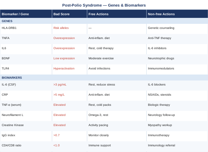
Understanding which of these genetic variants you carry adds a meaningful layer of context to the biomarker data — it helps explain why certain interventions may require more effort from some individuals than others, and it identifies where targeted supplementation is most likely to produce return.
The Polio Paradox: 10 Things That May Change How You Think About PPS
The Polio Paradox by Richard L. Bruno, PhD, is among the most substantive and direct books written specifically for polio survivors and the clinicians managing their care. Bruno — who himself survived non-paralytic polio — spent decades at the Harvest Center for Post-Polio Care in New Jersey synthesizing clinical experience with emerging neuroscience. His central argument challenges the prevailing clinical instinct with precision: the standard "push through fatigue gently" model applied to PPS is not just unhelpful — it is biologically counterproductive. What follows are the ten most impactful principles from his work, each of which challenges something the conventional medical approach still gets wrong.
PPS Is a Neuronal Problem, Not a Muscular One
The poliovirus targeted and destroyed anterior horn cells in the spinal cord — the motor neurons that directly drive muscle contraction. The surviving neurons compensated by sprouting new axonal connections, absorbing the orphaned muscle fibers of dead neighbors. These enlarged motor units — some managing 5 to 10 times the normal fiber count — sustained function for decades through heroic over-performance. When they begin to fail, the primary site of failure is neuronal, not muscular. Muscle weakness in PPS is downstream of neuronal exhaustion, not upstream of it. This distinction completely changes what interventions make sense.
Cognitive Work Draws from the Same Depleted Budget as Physical Work
The brainstem reticular activating system — responsible for regulating arousal, attention, motor preparation, and cognitive sustenance — was a frequent target of poliovirus, even in cases that appeared clinically mild or non-paralytic. This means that sustained mental effort: prolonged concentration, emotional processing, professional cognitive demands, and even social stimulation draws from the same neurological energy reserve as physical movement. A PPS patient who rests physically while remaining cognitively overloaded has not actually recovered. This insight reframes rest as neurological rest, not merely physical inactivity.
The "Polio Personality" Drives Overuse
Bruno identified a remarkably consistent psychological profile across PPS patients: high achievers who adapted to disability in childhood by performing at or above able-bodied peers, who learned to suppress fatigue signals as a survival strategy, and who built entire adult identities on productivity and self-sufficiency. This personality profile — deeply adaptive in early life — becomes a direct biological threat in PPS. The reflex to push through, minimize symptoms, and maintain appearances accelerates motor neuron overuse. Recognizing and actively working against this pattern is not a psychological luxury; it is part of the medical management of the condition.
Non-Paralytic Polio Left Hidden Damage
Up to 40% of people who had poliovirus infection without documented paralysis still showed significant motor neuron loss on autopsy and advanced imaging studies conducted decades later. The dismissal that many PPS patients encounter — "your polio was so mild, this can't be related" — is medically inaccurate and clinically harmful. The threshold for late-onset motor neuron failure does not require historical visible paralysis. This misunderstanding leads to years of systematic underdiagnosis.
Fatigue Is the Master Symptom — Treat It First
Muscle weakness and pain in PPS are predominantly fatigue-dependent: they worsen sharply when the neurological fatigue threshold is exceeded and improve partially when fatigue is adequately managed. Conventional clinical approaches often target weakness and pain directly with exercise and analgesics while treating fatigue as a secondary complaint. Bruno argues that this sequence is inverted. Fatigue management is the master lever — reduce it, and weakness and pain follow. This is not intuitive, but it is consistently supported by clinical observation across PPS cohorts.
Sleep-Disordered Breathing Is a Hidden Amplifier
The poliovirus frequently damaged the brainstem respiratory centers that regulate automatic breathing during sleep. Bruno's clinical research found elevated rates of obstructive and central sleep apnea, as well as nocturnal hypoventilation, in PPS patients relative to general population controls. These breathing disorders during sleep produce chronic sleep fragmentation, morning fatigue, cognitive impairment, and elevated inflammatory markers — all of which directly worsen PPS symptom load. Identifying and treating sleep-disordered breathing (polysomnography, CPAP or BiPAP) can produce improvements in PPS that months of other interventions cannot match.
Graded Exercise Therapy Can Worsen PPS — Unlike Most Chronic Fatigue Conditions
Graded exercise therapy — the graduated, progressive increase in physical activity commonly recommended for chronic fatigue syndrome and similar conditions — has produced documented neurological worsening in a subset of PPS patients. This is mechanistically coherent: unlike conditions where deconditioning is the primary driver of fatigue, PPS involves irreversible structural motor neuron loss. Progressively loading an already compromised and shrinking motor neuron pool accelerates the very damage it is intended to reverse. Energy conservation, pacing, and selective strengthening only for muscles not yet affected by PPS is Bruno's supported alternative.
Cold Sensitivity Is Neurological, Not Just Circulatory
Cold intolerance in PPS is frequently explained away as poor circulation or aging. Bruno's framework reveals the underlying mechanism: poliovirus damaged the hypothalamic and brainstem centers responsible for thermoregulation, leaving PPS patients with impaired ability to detect and compensate for cold exposure. This is not a peripheral vascular problem that can be improved with exercise; it reflects structural damage to the thermoregulatory system itself. Understanding this helps patients stop self-blaming and instead take practical structural steps: thermal layers, electric blankets, heated environments, and explicit avoidance of prolonged cold exposure that reliably triggers fatigue cascades.
Assistive Technology Protects Neurological Reserve
Bruno is unambiguous on this point: using a cane, orthotic brace, scooter, or power wheelchair is not conceding defeat. It is protecting the remaining motor neuron capacity that a patient has from being unnecessarily spent on locomotion. Every motor neuron activation that can be replaced by assistive technology preserves that neuron for activities that matter more — work, social connection, family life. PPS patients who resist assistive devices to appear more capable are spending their most finite resource on appearances. This is the reframe that many patients and clinicians most need to hear.
Psychological Adaptation Is Part of the Medical Treatment
Bruno's research consistently found that psychological support — peer groups, trauma-informed therapy, formal cognitive-behavioral work on illness acceptance — produced measurable improvements in functional outcomes alongside physical management. This is not incidental. Many PPS patients spent childhood concealing disability, overperforming, and absorbing messages that their limitations were personal failures. The psychological residue of this history actively interferes with the rest, pacing, and acceptance of assistance that effective PPS management requires. Treating the psychological dimension is not separate from treating the neuromuscular dimension; for many patients, it is the bottleneck.
Complementary Approaches with Clinical Evidence
The following modalities were selected specifically for their mechanistic relevance to post-polio syndrome and the availability of human clinical evidence — not simply for general wellness applications. None is a substitute for the biomarker-guided and neurological management strategies outlined above, but each offers a complementary pathway with meaningful evidence behind it.
Mindfulness-Based Stress Reduction (MBSR)
MBSR is an 8-week structured program developed at the University of Massachusetts Medical School, combining body scan meditation, sitting and walking mindfulness, and gentle movement. In PPS, its primary relevance lies in the neuroinflammatory and autonomic dimensions of the condition — not simply as stress reduction, but as a direct intervention on the chronic sympathetic nervous system activation that elevates IL-6, degrades sleep quality, and depletes neurological energy reserves. Multiple randomized controlled trials across chronic fatigue and chronic pain populations have shown MBSR reduces perceived fatigue severity, lowers inflammatory cytokine production, and improves sleep architecture. A landmark 2012 study by Creswell and colleagues, published in Brain, Behavior, and Immunity, demonstrated that an MBSR intervention reduced loneliness-associated increases in IL-6 gene expression — directly relevant to the neuroinflammatory mechanism operative in PPS.
The specific technique most applicable to PPS is body scan meditation — a systematic, non-effortful practice of focused attention to body sensations, typically conducted lying down, lasting 20–45 minutes. For PPS patients with limited tolerance for seated practices, the reclining format eliminates postural fatigue as a barrier to practice. The standard MBSR curriculum (8 weekly 2.5-hour sessions plus daily home practice) is available in-person and through validated online platforms developed by UMass Center for Mindfulness.
Begin with 10 minutes of breath-focused practice daily, increasing by 5 minutes per week as tolerance builds. The cognitive demands of MBSR practice should be factored into daily neurological energy budgeting — MBSR is not a free lunch for the nervous system, particularly initially. Most practitioners report significant fatigue and pain reduction benefits emerging between weeks 4 and 8 of consistent practice. Monthly continuation practice sustains benefits beyond the initial course; occasional weekend intensives help recalibrate practice when it has drifted.
Tai Chi
Tai chi is a slow, deliberate movement art combining coordinated posture sequences, weight shifting, breath control, and sustained attention. For PPS patients managing muscle weakness, postural instability, and elevated fall risk — all common complications — tai chi offers a uniquely appropriate movement modality. It is low-impact, easily modified, builds proprioception and postural stability through mechanisms distinct from conventional strengthening, and has been studied in neurological and aging populations with consistently favorable results. A 2016 Cochrane review examining tai chi in neurological conditions found consistent improvements in dynamic balance and functional mobility. Critically, the paced, non-fatiguing character of tai chi aligns naturally with the energy conservation and pacing principles central to PPS management — unlike most exercise modalities, it can be practiced at a stimulus level that stays within the neurological energy envelope when properly modified.
Yang-style tai chi — the most widely studied form, involving a 24-movement short form — is typically practiced for 30–45 minutes, 3–5 days per week in population studies. For PPS, seated tai chi adaptations practiced entirely in a chair or wheelchair preserve the proprioceptive, coordinated movement, and parasympathetic nervous system benefits while eliminating standing fatigue and fall risk entirely. An instructor experienced with neurological populations or older adults is strongly preferable for the initial 4–8 weeks of practice.
Begin with twice-weekly 20-minute sessions, monitoring explicitly for post-exertional fatigue — if any fatigue persists beyond 30–40 minutes after a session, session duration should be reduced, not pushed through. Free resources (YouTube instructional channels for tai chi for seniors or chair tai chi) offer accessible starting points, though initial in-person guidance substantially improves technique and reduces the risk of compensatory movement patterns that could elevate CK in weakened muscles. Measurable balance improvements typically emerge after 8–12 weeks of consistent practice.
Biofeedback
Biofeedback involves real-time measurement of physiological signals displayed back to the patient, who then learns through practice to consciously influence those signals. In PPS, the most clinically relevant application is heart rate variability (HRV) biofeedback — a technique that trains the autonomic nervous system toward parasympathetic dominance by guiding breathing at an individual's cardiac resonance frequency (typically 5–6 breaths per minute). This parasympathetic shift directly reduces sympathetically-driven IL-6 production, lowers baseline inflammatory markers, improves sleep quality, and conserves the neurological energy otherwise spent on chronic threat-response activation. A secondary application is EMG biofeedback for specific affected muscle groups, which helps PPS patients learn to reduce unnecessary co-contraction and resting muscle tension — directly lowering the metabolic demand on overloaded motor units and reducing resting CK.
HRV biofeedback involves 20-minute daily practice sessions with a device providing real-time breath-pacing guidance and heart rate feedback. The HeartMath Inner Balance sensor (compatible with iOS/Android) and the Polar H10 chest strap paired with appropriate apps are commonly used home options costing $80–$200. Clinical biofeedback sessions with a certified practitioner ($80–$150 per session) are most valuable in the initial phase to establish correct technique; 6–12 sessions followed by independent home practice is a reasonable protocol.
HRV biofeedback is exceptionally well-suited to PPS because it requires no physical exertion whatsoever, can be performed reclined, and directly targets the autonomic and inflammatory mechanisms operative in the condition. It is one of the few interventions that simultaneously addresses fatigue, inflammation, sleep, and pain through a single mechanism. Begin with 10-minute daily sessions and increase gradually to 20 minutes over 2–4 weeks. Measurable resting HRV improvement typically appears within 4–6 weeks of consistent daily practice, and this improvement correlates with reductions in reported fatigue and inflammatory markers in chronic condition populations.
Photobiomodulation (Low-Level Laser Therapy)
Photobiomodulation uses specific wavelengths of red and near-infrared light to stimulate cytochrome c oxidase — the terminal enzyme of the mitochondrial electron transport chain — reducing oxidative stress, improving mitochondrial ATP production, and dampening local and systemic neuroinflammation. In PPS, its theoretical and emerging practical relevance spans the two domains most consistently implicated in the condition: mitochondrial function (directly relevant to the SOD2 variant discussed above and to the high metabolic demands of enlarged motor units) and neuroinflammation (relevant to IL-6, NfL, and HLA-driven immune activity). While large PPS-specific randomized trials do not yet exist, PBM has demonstrated measurable effects in ALS pilot studies, peripheral neuropathy, and muscle damage recovery in multiple published human trials. Its safety profile is favorable when appropriate dosing is maintained.
The evidence-based wavelength range for neurological and musculoskeletal PBM applications is red light at 630–660 nm and near-infrared at 808–850 nm, with power density in the range of 10–100 mW/cm². For PPS, both paraspinal application (targeting the cervical and lumbar motor neuron entry zones affected in PPS) and direct application to affected muscle groups have mechanistic rationale. Clinical PBM practitioners use more powerful devices with broader coverage; home devices in the $150–$1,500 range vary enormously in actual output, and power density verification is essential before investing — many consumer "red light" panels use sub-therapeutic doses.
Approach PBM as a complementary adjunct to the biomarker-guided and lifestyle strategies above, not as a standalone intervention. Start with 5–10 minutes per treatment area, 3–4 times per week, monitoring for post-treatment fatigue responses — some people with neurological conditions report transient fatigue in the first few sessions, which typically resolves within 2–3 weeks of continued use. The key practical step is confirming device specifications before purchase: wavelength and power output specifications should be provided by the manufacturer, not estimated from general marketing language. Consistent application over 8–12 weeks is typically required to observe measurable effects.
Conclusion
Post-polio syndrome does not respond well to generic management, and it never has. The specific mechanisms driving fatigue, weakness, and neurological decline in PPS — neuroinflammation, motor neuron stress, trophic hormone deficiency, mitochondrial compromise, and autonomic dysregulation — are measurable, and the ability to measure them is the foundation of meaningful intervention. The seven biomarkers covered here give a clear biological picture; the five genetic variants add a layer of context that explains individual variation in response; and the clinical approaches and book insights provide a framework for action that challenges the passive "rest and accept" model still too common in clinical practice.
The most useful next step is not to overhaul everything simultaneously. It is to start with two or three of the most accessible and informative biomarkers — hs-CRP and 25-OH vitamin D are both inexpensive and universally available — and use what they reveal to prioritize one or two interventions. Track what changes. Add layers as the picture becomes clearer. Work with a clinician who is willing to engage with this level of specificity. The decisions that come from better data are almost always better than the ones made without it.
Neurological Endocrine & Metabolic
Musculoskeletal: Muscle Conditions
Neurological: Nerve Conditions Spinal Cord Conditions
Respiratory: Sleep & Breathing Disorders
Endocrine & Metabolic: Thyroid Conditions
Autoimmune: Inflammatory Conditions