This article contains AI generated content.
· UpdatedPosterior Knee Capsule Tear — 5 Genes And 7 Biomarkers To Track
Introduction
A posterior knee capsule tear places you in a particular kind of limbo. The injury is painful enough to stop daily activity, but the standard care pathway — rest, some ice, a physiotherapy referral, maybe an MRI — rarely explains why it happened or why recovery is slower for some people than others. The back of the knee is anatomically intricate, and capsular tears there receive far less clinical nuance than ACL or meniscus injuries, despite involving the same biological repair mechanisms.
What standard rehabilitation almost never accounts for is the biological individuality underneath every soft tissue injury. The posterior knee capsule is composed primarily of collagen. Whether that collagen repairs cleanly depends on factors that do not appear on a standard orthopaedic assessment: your baseline inflammatory status, how aggressively your matrix-degrading enzymes are operating, whether your genetic profile supports collagen tensile strength, and whether key micronutrients are present in adequate concentrations.
Generic recovery protocols assume your biology is average. For some people it is; for others, a chronically elevated hs-CRP, a COL5A1 risk variant, or a vitamin D level below 30 ng/mL means the healing environment is fundamentally compromised — and no amount of standard physiotherapy fully compensates for that.
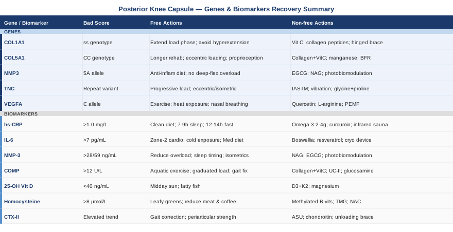
Better data leads to better decisions. Tracking seven specific blood and urinary biomarkers gives you a measurable window into the molecular state of your recovery. Understanding five key genetic factors helps explain your baseline vulnerabilities and exactly where to apply targeted compensation. Together, these two layers transform a frustrating wait-and-see process into an informed, adjustable protocol.
Summary
This article covers seven biomarkers — hs-CRP, IL-6, MMP-3, COMP, 25-OH Vitamin D, homocysteine, and CTX-II — and explains in practical terms what each reveals about whether your posterior knee capsule is healing or stalling. For each marker, you will find the measurement method, cost range, and specific action plans with and without supplements. The article then examines five genes — COL1A1, COL5A1, MMP3, TNC, and VEGFA — that shape your baseline soft tissue repair capacity, along with targeted protocols for each unfavorable genotype. Beyond the molecular picture, Andrew Huberman's connective tissue healing framework offers 10 evidence-drawn principles that most physiotherapy protocols quietly ignore. Finally, four complementary modalities with meaningful clinical backing round out the recovery toolkit. If your knee is not responding the way it should, the explanation is likely in one of these pages.
7 Biomarkers to Track for Posterior Knee Capsule Recovery
Biomarkers give you something imaging cannot: a moving picture of the biological processes underneath your injury. An MRI shows structure; biomarkers show function. The seven below were selected because they directly relate to collagen integrity, inflammatory activity, matrix degradation, and tissue remodeling — the four core processes that determine whether a posterior knee capsule heals well or remains compromised.
Biomarker 1: High-Sensitivity C-Reactive Protein (hs-CRP)
Why it matters
hs-CRP is a liver-produced acute-phase protein that rises in response to systemic inflammation. In the context of a posterior knee capsule tear, some early elevation is normal and necessary — it is part of the repair signal. The concern arises when hs-CRP remains chronically elevated, typically above 1.0 mg/L, which indicates a systemic inflammatory state that slows tissue maturation, increases collagen catabolism, and impairs fibroblast function.
Peter Attia considers hs-CRP among the most accessible and informative routine markers available precisely because it reflects metabolic and inflammatory biology across multiple systems simultaneously. In soft tissue injury recovery, a high baseline or failure to normalize within 4-6 weeks post-injury is a meaningful clinical signal that the healing environment needs direct intervention.
How to measure it
Standard blood draw. Available at virtually every commercial laboratory. Cost: $10–30, often bundled in inflammation or cardiovascular panels. Optimal for healing: below 1.0 mg/L. Above 3.0 mg/L warrants immediate attention. Most informative as a trend: test at baseline, then at weeks 6 and 12.
If the score is bad: the plan without supplements
- Remove the primary dietary drivers of CRP elevation: ultra-processed foods, seed oils, refined carbohydrates, and alcohol. Even two weeks of dietary clean-up produces measurable CRP reduction. - Sleep 7-9 hours per night. Sleep deprivation is one of the most potent independent drivers of hs-CRP elevation, operating entirely separately from diet. - Practice time-restricted eating: a 12–14 hour overnight fast is associated with meaningful reductions in inflammatory markers in human trials without any caloric restriction. - Reduce psychological stress through structured daily downregulation (even 10 minutes of quiet activity). Chronic stress elevates both IL-6 and CRP via HPA-axis activation.
If the score is bad: the plan with supplements or equipment
- Omega-3 fatty acids (EPA+DHA): 2–4g daily. Multiple randomized trials demonstrate significant hs-CRP reduction with sustained omega-3 supplementation. Use a triglyceride-form product. Cycling: continuous use is appropriate; reassess at 3 months. Side effects: mild fishy aftertaste; possible mild blood thinning at high doses. - Curcumin (95% curcuminoid extract) + piperine: 500–1000mg curcumin + 5mg piperine daily. Reduces NF-κB signaling, the transcription factor driving CRP synthesis. Evidence from human RCTs in joint inflammation. Cycling: 8 weeks on, 2 weeks off. Side effects: GI upset at high doses; may potentiate anticoagulants. - Magnesium glycinate: 300–400mg at bedtime. Reduces CRP and supports sleep quality simultaneously — two relevant mechanisms. Safe for continuous use. - Infrared sauna: 20–30 minutes, 3–4 sessions per week. Human studies including in inflammatory conditions show reduced CRP and IL-6 with repeated heat exposure. Reassess at 8 weeks. Caution: hydrate adequately; avoid if cardiovascular instability is present.
Biomarker 2: Interleukin-6 (IL-6)
Why it matters
IL-6 is a pleiotropic cytokine released by immune cells, fibroblasts, and injured tissue. In the immediate aftermath of a posterior capsule tear, IL-6 rises appropriately as part of the acute repair signal. The problem is persistent elevation — chronically high IL-6 drives fibroblast dysfunction, inhibits anabolic signaling, promotes catabolic matrix activity, and prevents the transition from proliferation to remodeling that marks healthy tissue healing.
Measuring IL-6 alongside hs-CRP gives a more granular picture. CRP signals that inflammation exists; IL-6 helps locate whether it is primarily a systemic metabolic issue or an ongoing local tissue-level response.
How to measure it
Serum IL-6 requires a standard blood draw but is not available at all routine labs. Quest Diagnostics and LabCorp both offer it. Cost: $50–150. Reference: below 7 pg/mL. A single snapshot is less useful than trending every 6-8 weeks during active recovery.
If the score is bad: the plan without supplements
- Reduce visceral fat, the primary driver of chronically elevated IL-6: zone-2 cardiovascular work at 150+ minutes per week, adapted to avoid posterior knee loading. Upper-body ergometers or swimming are appropriate alternatives during recovery. - Cold exposure: brief cold showers or partial limb immersion for 3–5 minutes daily suppresses IL-6 acutely and has systemic anti-inflammatory effects with regular use. - Mediterranean dietary pattern: associated with meaningful IL-6 reductions in multiple controlled dietary intervention trials.
If the score is bad: the plan with supplements or equipment
- Boswellia serrata (AKBA standardized extract): 200–400mg daily. Inhibits 5-lipoxygenase (5-LOX), reducing leukotriene production and IL-6 synthesis. Human evidence for joint inflammation. Cycling: 8 weeks on, 2 weeks off. Side effects: rare mild GI irritation. - Resveratrol: 250–500mg daily with fat. Inhibits NF-κB and reduces IL-6 in human supplementation studies. Cycling: 12 weeks on, 4 weeks off to avoid receptor adaptation. Side effects: minimal at standard doses; possible interaction with anticoagulants. - Game Ready or Normatec compression cryotherapy device: reduces local IL-6 production at the injury site mechanically. Apply to posterior knee 2x daily for 15–20 minutes during the first 8 weeks post-injury. Practical and well-tolerated.
Biomarker 3: Matrix Metalloproteinase-3 (MMP-3)
Why it matters
MMP-3, or stromelysin-1, is an enzyme that degrades collagen, fibronectin, laminin, and other extracellular matrix components. It plays a necessary role in tissue remodeling — the problem is when its activity becomes dysregulated. In the posterior knee capsule specifically, elevated MMP-3 means the body is dismantling its connective tissue matrix faster than it can rebuild it.
Elevated serum and synovial fluid MMP-3 has been associated with accelerated ligamentous degradation, persistent capsular laxity, and delayed soft tissue healing in knee conditions. This biomarker is often elevated in people who experience ongoing joint irritation months after an injury that should have resolved. The MMP-3 promoter gene variant (discussed in the genetics section) explains why some individuals have a structurally high MMP-3 baseline regardless of lifestyle.
How to measure it
Serum MMP-3 via blood draw. Available through specialty labs and some academic medical centers. Cost: $150–300. Normal range: typically below 28 ng/mL in women, below 59 ng/mL in men (lab-dependent). Most useful when measured at injury baseline and again at 8-12 weeks of recovery.
If the score is bad: the plan without supplements
- Eliminate repetitive mechanical overload on the healing posterior capsule: deep squats, aggressive stretching into hyperextension, and running on hard surfaces all sustain elevated MMP-3 by maintaining a mechanical stress signal in the tissue. - Protect circadian alignment: MMP activity follows circadian rhythms and is significantly upregulated by sleep disruption. Consistent sleep timing reduces MMP-3 more reliably than many dietary interventions. - Introduce isometric loading over excessive end-range stretching: submaximal isometric holds in mid-range positions have tissue-protective effects in connective tissue research, whereas aggressive passive stretching at end-range can paradoxically increase MMP expression.
If the score is bad: the plan with supplements or equipment
- N-acetyl glucosamine (NAG): 1500mg daily. Inhibits MMP-3 expression in human chondrocyte and synoviocyte research. Cycling: continuous; reassess at 3 months. Side effects: minimal. - EGCG (green tea extract): 400–800mg daily standardized extract. Reduces MMP-3 expression in multiple in vitro and some human studies via AP-1 inhibition. Cycling: 8 weeks on, 2 weeks off. Side effects: possible GI irritation on empty stomach; rare hepatotoxicity at very high doses; do not exceed 800mg. - Photobiomodulation (660–850nm LED or laser panel): reduces local MMP activity in soft tissue via mitochondrial signaling. Apply to posterior knee for 10–15 minutes, 3–4x weekly. See the complementary approaches section for a full protocol. No significant side effects at standard therapeutic doses.
Biomarker 4: Cartilage Oligomeric Matrix Protein (COMP)
Why it matters
COMP is a structural glycoprotein found in the extracellular matrix of cartilage, tendons, and ligaments. When connective tissue is mechanically stressed or damaged, COMP fragments are shed into the bloodstream, making serum COMP a sensitive marker of active connective tissue turnover. For a posterior knee capsule tear, elevated COMP signals ongoing matrix breakdown — and elevated COMP that persists beyond 8-12 weeks suggests tissue remodeling is stalled or that excessive loading is maintaining a catabolic state.
In longitudinal studies of knee joint injuries, higher early-timepoint serum COMP predicted worse structural outcomes at one year. It is one of the most informative yet underused injury biomarkers available outside of academic research settings.
How to measure it
Serum COMP via blood draw. Available through specialty and functional medicine labs. Cost: $200–400. Normal adult range is typically below 12 U/L, though lab-specific reference ranges vary. Timing: measure at injury baseline and at 8-week follow-up; the trend matters more than any single value.
If the score is bad: the plan without supplements
- Reduce repetitive impact: running, jumping, and forceful knee flexion sustain elevated COMP by keeping the capsular tissue in a state of repeated micro-stress. Substituting aquatic exercise or cycling maintains fitness while dramatically reducing joint load. - Graduated weight-bearing protocol: systematic, progressive increases in load stimulate matrix remodeling without overwhelming the catabolic threshold. The goal is not avoidance but calibrated loading. - Gait optimization: altered gait mechanics from pain compensation increase joint load asymmetrically and keep COMP elevated. Early physiotherapy focused on gait normalization matters more than people realize.
If the score is bad: the plan with supplements or equipment
- Hydrolyzed collagen peptides (15–20g) + vitamin C (50–200mg): taken 45–60 minutes before rehabilitation exercise. This timing-specific protocol, studied by Keith Baar's group and Shaw et al., significantly increases collagen synthesis markers in healing connective tissue. Daily during active healing (12–16 weeks). Side effects: very low; occasional mild digestive sensitivity. - Undenatured type II collagen (UC-II): 40mg daily. Uses the oral tolerance pathway to modulate immune-mediated matrix degradation. Effects build over 4-8 weeks. Cycling: 12 weeks continuous then reassess. Side effects: minimal. - Glucosamine sulfate: 1500mg daily. Modulates proteoglycan synthesis and may reduce COMP release from stressed connective tissue. Evidence is mixed but biologically plausible. Cycling: 6-month courses acceptable.
Biomarker 5: 25-OH Vitamin D
Why it matters
Vitamin D deficiency is widespread and has consequences that extend well beyond bone health. Vitamin D receptors are expressed on tenocytes, fibroblasts, and chondrocytes — exactly the cells responsible for repairing the posterior knee capsule. Deficiency impairs collagen synthesis rate, reduces the muscle strength that dynamically offloads the capsule during movement, and is independently associated with increased soft tissue injury risk and slower functional recovery.
Thomas Dayspring, one of the most cited specialists in clinical lipidology and preventive medicine, has discussed at length the non-cardiovascular dimensions of vitamin D biology, including its direct effects on musculoskeletal tissue repair and immune regulation. For anyone recovering from a soft tissue injury, this is a non-negotiable baseline measurement.
How to measure it
Serum 25-hydroxyvitamin D, standard blood draw. One of the most widely available and affordable panels available. Cost: $30–80. Optimal range for active healing: 40–60 ng/mL. Many people fall below 30 ng/mL without any symptoms. Test at baseline; if supplementing, retest at 8-12 weeks.
If the score is bad: the plan without supplements
- Midday sun exposure: 20–30 minutes of direct sunlight on arms and legs when season and latitude permit. Darker skin pigmentation significantly reduces synthesis efficiency and may require longer exposure or supplementation regardless of sun access. - Dietary vitamin D sources: fatty fish (salmon, mackerel, sardines), egg yolks, and fortified dairy. Practical but rarely sufficient to correct genuine deficiency — dietary correction alone typically raises levels by only 5-10 ng/mL.
If the score is bad: the plan with supplements or equipment
- Vitamin D3: 2000–5000 IU daily for maintenance; up to 10,000 IU short-term (8 weeks) under clinical guidance for significant deficiency. Always co-administer with vitamin K2 (MK-7, 100–200mcg) to support appropriate calcium distribution and prevent arterial calcification. Side effects: hypercalcemia at excessive long-term doses above 10,000 IU — periodic testing is the safeguard. Cycling: dose-to-target, then maintenance. - Magnesium (glycinate or malate): 300–400mg daily. Critical cofactor for vitamin D activation via the 25-hydroxylase and 1-alpha-hydroxylase enzymes. Without adequate magnesium, supplemental D3 is poorly converted. Safe for continuous use.
Biomarker 6: Homocysteine
Why it matters
Homocysteine is an amino acid intermediate in the methionine metabolism cycle. Elevated homocysteine — above 10 µmol/L — interferes with lysyl oxidase activity, the enzyme responsible for forming the crosslinks that give collagen its tensile strength. For a posterior knee capsule that must withstand repeated shear and tensile loads during recovery and return to function, impaired collagen crosslinking means structurally weaker tissue even when everything else about the healing process proceeds normally.
Gary Brecka, a human biologist widely known for his work on genetic methylation pathways, has identified chronically elevated homocysteine as one of the most underappreciated drivers of connective tissue fragility and re-injury risk. The MTHFR gene variant, which affects roughly 40% of the population, is one of the most common reasons homocysteine remains elevated despite adequate dietary protein intake.
How to measure it
Serum homocysteine via standard blood draw. Cost: $20–60. Available at most commercial labs. Optimal range: below 8 µmol/L. Values above 15 µmol/L are clinically significant. Retest at 8-12 weeks if supplementing.
If the score is bad: the plan without supplements
- Moderate methionine-heavy foods: red meat in excess, when methylation capacity is limited, raises homocysteine disproportionately. Reducing portion frequency is a simple first step. - Reduce heavy coffee consumption: consistent high coffee intake impairs folate metabolism and raises homocysteine in susceptible individuals. - Emphasize folate-rich whole foods: leafy greens (spinach, kale), legumes, eggs, and meat provide the B-vitamin cofactors involved in homocysteine clearance. For mild elevations, dietary adjustment alone can produce meaningful normalization within 8-12 weeks.
If the score is bad: the plan with supplements or equipment
- Methylated B-vitamin combination: 5-MTHF (methylfolate) 400–800mcg + methylcobalamin (B12) 1000mcg + pyridoxal-5-phosphate (P5P, active B6) 25–50mg daily. This stack bypasses the MTHFR enzyme defect — the most common reason homocysteine remains elevated despite adequate dietary intake. Multiple controlled trials confirm homocysteine lowering with this combination. Cycling: continuous with periodic homocysteine monitoring every 8-12 weeks. Side effects: very low at standard doses; high-dose B6 (>200mg) over extended periods can cause peripheral neuropathy — stay at therapeutic range. - Betaine (trimethylglycine, TMG): 1500–3000mg daily. Provides an alternative methyl group donor that lowers homocysteine through a MTHFR-independent pathway. Well tolerated and effective even in MTHFR-affected individuals. Cycling: continuous. - NAC (N-acetylcysteine): 600mg daily. Reduces oxidative stress associated with elevated homocysteine, which compounds its collagen-crosslinking interference. Side effects: rare nausea; start low and titrate.
Biomarker 7: CTX-II (C-Terminal Crosslinked Telopeptide of Type II Collagen)
Why it matters
CTX-II is a urinary marker of type II collagen degradation, primarily reflecting catabolism of cartilaginous and fibrocartilaginous structures. The posterior knee capsule inserts into and mechanically interacts with structures containing type II collagen, and when the capsule tears, secondary articular stress and joint irritation frequently elevate CTX-II beyond the direct capsular contribution.
Pierre Garnero, a pioneer in bone and cartilage biomarker research, identified elevated urinary CTX-II as a sensitive predictor of progressive joint deterioration. In the context of a posterior capsule tear, persistent CTX-II elevation at 2-3 months post-injury may indicate that secondary articular damage is more substantial than imaging suggests — a signal to reduce loading and focus on the matrix-protective interventions below.
How to measure it
First-morning void urine, corrected for creatinine concentration. Measured via ELISA at specialty functional medicine labs. Cost: $100–250. Most valuable as a trend marker — lower over time indicates improving matrix protection. Available through specialty ordering services in most countries.
If the score is bad: the plan without supplements
- Address gait mechanics: uncontrolled limping and altered joint kinematics generate disproportionate peak cartilage stress and drive CTX-II up. Early gait correction through physiotherapy is among the highest-leverage interventions available. - Modest weight reduction if indicated: each additional kilogram increases effective knee joint load by 3-4 kilograms during walking. Even a 5kg reduction produces measurable CTX-II improvement in individuals with elevated body mass. - Progressive periarticular strengthening: a well-conditioned quadriceps and hamstring complex absorbs peak loads before they reach the capsule and articular cartilage. Strengthening these muscles is matrix-protective, not just functionally important.
If the score is bad: the plan with supplements or equipment
- Avocado-soybean unsaponifiables (ASU): 300mg daily. Reduces cartilage catabolism and has demonstrated CTX-II reduction in randomized controlled trials for knee osteoarthritis. Mechanism is direct inhibition of IL-1β-mediated matrix degradation. Cycling: 6-month courses with 4-week intervals. Side effects: minimal. - Chondroitin sulfate: 1200mg daily. Inhibits type II collagen degradation enzymes in human trials. Effects build over 3-4 months; most relevant for the subacute and chronic recovery phases. Cycling: 6 months continuous. - Hydrolyzed type I/II collagen (15–20g) + vitamin C (as described under COMP): the collagen synthesis support protocol directly reduces net CTX-II by increasing the anabolic side of the collagen balance equation. - Unloading knee brace during high-load activities: specifically for individuals with compromised articular cartilage under the posterior capsule, an unloading brace during the return-to-activity phase reduces CTX-II by redistributing joint contact stress. Consult with a sports medicine physician for fitting.
The Genetic Layer: 5 Genes That Shape Your Capsule's Healing Capacity
Biomarkers tell you what is happening in real time. Genetics tell you why your tissue responds the way it does, and where your structural vulnerabilities originate. The five genes below have the strongest human evidence for influencing soft tissue repair, collagen architecture, matrix remodeling, and vascularization in musculoskeletal injuries. None of them represent a fixed destiny — but each one tells you specifically where to compensate, and how.
Gene 1: COL1A1 — The Blueprint for Type I Collagen Strength
COL1A1 encodes the alpha-1 chain of type I collagen — the primary structural protein in the posterior knee capsule, tendons, and ligaments throughout the body. The most studied functional variant is the Sp1 binding site polymorphism (rs1800012), located in intron 1 of the gene. The ss genotype (homozygous T allele) is consistently associated with reduced collagen structural integrity, lower tensile strength in fibrous connective tissues, and elevated risk of soft tissue injury across multiple independent cohorts.
Garnero and colleagues were among the first to link this COL1A1 polymorphism to musculoskeletal vulnerability, and the association has been replicated across ACL tears, rotator cuff injuries, and Achilles tendinopathy. For someone recovering from a posterior capsule tear, carrying the risk genotype means the starting point for collagen tensile integrity is lower than average — not unmovable, but requiring explicit compensation.
If the gene is unfavorable: the plan without supplements
- Extend the conservative loading phase by 2-4 weeks: with reduced baseline collagen tensile strength, aggressive mechanical loading before adequate fibroblast proliferation risks re-tear or chronic instability. - Train hyperextension avoidance: the posterior capsule is maximally stressed at end-range extension. Proprioceptive drills that specifically reinforce avoiding this position — single-leg landing mechanics, neuromuscular control exercises — are particularly important for COL1A1 risk carriers. - High-quality protein: 1.6–2.0g per kilogram body weight daily to maximize substrate availability for collagen fibroblasts. Animal proteins provide the complete amino acid profile required. - Eccentric loading specificity: slow, controlled eccentric exercises (reverse wall slides, hamstring curls at controlled tempo) stimulate fibroblast activity and collagen organization without applying the peak mechanical loads that risk re-injury in weaker tissue.
If the gene is unfavorable: the plan with supplements or equipment
- Vitamin C: 500–1000mg taken 30–60 minutes before any loading exercise. Essential cofactor for prolyl and lysyl hydroxylase enzymes that form stable collagen crosslinks. Particularly relevant for COL1A1 risk carriers because it maximizes the stability of whatever collagen is being synthesized. Continuous during healing. Side effects: low; loose stool at very high doses (>2000mg). - Collagen peptides (15–20g hydrolyzed) + vitamin C before loading exercise: as above. COL1A1 risk carriers get the most benefit from timing this supplementation to the pre-exercise window when fibroblast activity is highest. - Orthosilicic acid: 10–25mg daily. Emerging evidence for collagen crosslink stabilization through silicon incorporation into the collagen matrix. Cycling: safe for long-term use. Side effects: very low. - Functional hinged knee brace or proprioceptive sleeve: for COL1A1 risk carriers returning to activity, a properly fitted functional brace reduces peak loads on the healing capsule and provides real-time proprioceptive feedback. Recommended during all loading activities for the first 6 months post-injury, then reassessed.
Gene 2: COL5A1 — Collagen Fibril Architecture
COL5A1 encodes the alpha-1 chain of type V collagen, a quantitatively minor but architecturally critical component that regulates fibril diameter in type I collagen tissues. Disrupted fibril diameter control produces weaker, mechanically less efficient ligamentous tissue despite apparently normal type I collagen abundance. The 3' UTR variant rs12722 is the most studied functional polymorphism.
Posthumus et al. (2009) in the American Journal of Sports Medicine demonstrated that the CC genotype at rs12722 was significantly associated with ACL rupture in a case-control study. Subsequent studies in cricket players, rugby athletes, and endurance sports populations have replicated COL5A1 associations across multiple soft tissue injury phenotypes, establishing this as one of the most robust genetic risk variants in musculoskeletal medicine.
If the gene is unfavorable: the plan without supplements
- Expect and plan for a longer rehabilitation timeline: COL5A1 risk carriers should anticipate a recovery course 25-30% longer than standard clinical guidelines. Building this expectation into the plan avoids the temptation to return to loading too early. - Prioritize eccentric strengthening: eccentric loading specifically stimulates collagen fibril reorganization and maturation. Nordic curl progressions, slow hamstring curls on a machine, and eccentric calf raises are directly relevant to posterior knee capsule recovery. - Lifelong proprioception maintenance: single-leg balance, perturbation training, and unstable surface exercises should be introduced early and maintained indefinitely in COL5A1 risk carriers. Dynamic joint stabilization reduces the mechanical demand on capsular tissue across all activities.
If the gene is unfavorable: the plan with supplements or equipment
- Gelatin/collagen + vitamin C protocol (as above): directly relevant for COL5A1 risk because fibril diameter organization depends on the same collagen synthesis substrates. - Manganese: 5–10mg daily as manganese gluconate or glycinate. Required cofactor for glycosaminoglycan synthesis and collagen fibril organization. Cycling: 8 weeks on, 4 weeks off. Side effects: do not exceed 11mg daily long-term — high chronic manganese intake has neurological consequences. - Blood flow restriction (BFR) training: BFR with light loads (20-30% 1RM) stimulates collagen synthesis and muscle hypertrophy without generating the high peak mechanical loads that threaten fragile fibril architecture. Use a validated cuff system with professional instruction. Frequency: 3-4 sessions per week. BFR offers a meaningful rehabilitation bridge for COL5A1 carriers who cannot tolerate standard loading progression.
Gene 3: MMP3 — The Tissue Remodeling Regulator
The MMP3 gene encodes matrix metalloproteinase-3 and contains a well-characterized promoter polymorphism (rs3025058, also written as the 5A/6A variant) that meaningfully affects transcription rate. The 5A allele drives substantially higher MMP-3 production — meaning 5A carriers have a structurally more aggressive matrix-degrading environment throughout their lives.
In the context of a posterior knee capsule tear, a 5A carrier enters the injury with elevated baseline catabolic activity. Their MMP-3 biomarker will trend high, their tissue turnover will be more aggressive, and they are more likely to experience ongoing capsular laxity or persistent joint irritation even after formally completing standard rehabilitation. This is not a rare variant; the 5A allele is present in a substantial portion of the population.
If the gene is unfavorable: the plan without supplements
- Minimize lifestyle factors that upregulate MMP expression: ultra-processed food, alcohol, and sleep deprivation all activate NF-κB and AP-1 transcription factors that drive MMP3 gene expression. For 5A carriers, these exposures have disproportionate downstream consequences on soft tissue integrity. - Extend the offloading phase in deep knee flexion: avoid compressive loading with deep flexion for 4-6 weeks longer than standard guidelines. Deep flexion generates posterior capsular stress precisely where healing tissue is weakest. - Anti-inflammatory eating with time restriction: combining a 12-14 hour overnight fast with an anti-inflammatory food pattern reduces NF-κB activation, directly lowering MMP3 promoter activity.
If the gene is unfavorable: the plan with supplements or equipment
- EGCG (green tea extract): 400–800mg daily: inhibits AP-1 transcriptional activity that drives the MMP3 5A promoter. More mechanistically targeted in 5A carriers than in baseline-normal individuals. Cycling: 8 weeks on, 2 weeks off. Side effects: GI irritation on empty stomach; do not exceed 800mg. - N-acetyl glucosamine: 1500mg daily: reduces MMP-3 induction in synoviocyte tissue studies. Safe for long-term continuous use. - Photobiomodulation (660/850nm, 10–15 min, 4x weekly): reduces local MMP activity at treatment sites via mitochondrial signaling. Directly relevant for both the MMP-3 biomarker level and the 5A genotype. See the complementary approaches section for device and protocol specifics. - Low-dose doxycycline (prescription only, physician supervision required): sub-antimicrobial doses of tetracycline antibiotics are established MMP inhibitors discussed in academic connective tissue literature. This is not a standard clinical indication — raise with a sports medicine physician if other interventions are insufficient.
Gene 4: TNC — Tenascin-C and Scaffold Organization
Tenascin-C (TNC) is an extracellular matrix glycoprotein expressed abundantly in tendons, ligaments, and joint capsules. It acts as a mechanical signal transducer — its expression increases under tensile load and it organizes the collagen scaffold during injury repair, essentially serving as scaffolding around which healing collagen fibrils organize. A pentanucleotide repeat polymorphism in the TNC gene has been associated with Achilles tendinopathy and other ligamentous injuries in human studies.
Mokone et al. (2006) demonstrated that TNC repeat variants were significantly associated with Achilles tendinopathy in South African athletes, consistent with TNC's mechanobiological role in fibrous connective tissue repair broadly, including capsular structures.
If the gene is unfavorable: the plan without supplements
- Progressive mechanical loading is not optional — it is the treatment: TNC expression is load-dependent. Without appropriate tensile loading during rehabilitation, TNC cannot organize the collagen scaffold effectively. The therapeutic goal is controlled, progressive loading — not avoidance. - Favor eccentric and isometric protocols: these loading types most effectively stimulate tenascin-C expression in healing ligamentous tissue relative to purely concentric work. - Avoid complete immobilization: total rest dramatically reduces TNC expression and leads to disorganized collagen architecture. Even gentle active range-of-motion exercises within pain-free limits maintain the mechanical TNC expression signals critical to scaffold formation.
If the gene is unfavorable: the plan with supplements or equipment
- Instrument-assisted soft tissue mobilization (IASTM) by a trained therapist: when performed correctly, IASTM provides tensile stimulation to healing connective tissue and may substitute for some of the mechanical signaling that drives TNC expression in tissue where direct loading is still limited. 1-2 sessions per week from week 6-8 onward. - Localized vibration therapy: 30–50Hz applied to the posterior knee during isometric holds, 10–15 minutes per session, 3x weekly. Stimulates mechanoreceptors and may support TNC-mediated scaffold organization. Consumer-grade vibration devices are sufficient for this application. - Glycine + proline supplementation or bone broth: daily during the healing phase. TNC, like all extracellular matrix proteins, requires proline and glycine as synthesis substrates. Bone broth (1-2 cups daily) or direct amino acid supplementation supplies both. Side effects: negligible.
Gene 5: VEGFA — Vascularization and Healing Capacity
VEGFA (Vascular Endothelial Growth Factor A) drives angiogenesis — the formation of new blood vessels essential to tissue repair. The posterior knee capsule is relatively hypovascular at baseline, like most fibrous connective tissue. After a tear, healing depends substantially on VEGF-mediated capillary growth to supply oxygen, nutrients, and paracrine growth factors to fibroblasts at the repair site. The rs2010963 (+405G>C) promoter variant affects VEGF production levels and has been associated with differential healing trajectories in musculoskeletal injuries.
Gary Brecka has emphasized VEGF-pathway genetics as part of his broader framework for understanding why certain individuals struggle to heal soft tissue injuries, arguing that reduced vascularization capacity keeps the repair environment chronically substrate-limited regardless of other optimizations.
If the gene is unfavorable: the plan without supplements
- Maintain exercise-induced VEGF signaling: even low-intensity exercise powerfully stimulates VEGF release from active muscle. Upper-body and contralateral-limb exercise during posterior knee recovery maintains systemic VEGF signaling even when the injured knee cannot be loaded fully. - Regular heat exposure: heat stress reliably upregulates VEGF via heat shock protein pathways. A 15-20 minute sauna or hot bath 3-4x weekly provides meaningful angiogenic stimulus with no mechanical stress on the healing knee. Avoid applying intense heat directly to the injured site during the acute inflammatory phase (first 72 hours). - Nasal breathing during rehabilitation: nasal-only breathing during low-intensity exercise induces mild hypoxia, which is the strongest physiological VEGF stimulus. Incorporate nasal breathing in all light rehab activities.
If the gene is unfavorable: the plan with supplements or equipment
- Quercetin: 500–1000mg daily: upregulates HIF-1α/VEGF signaling pathway in multiple human and cell culture studies. Cycling: 8 weeks on, 3 weeks off. Side effects: minimal; possible interaction with thyroid medications and some antibiotics. - L-arginine: 3–6g daily: precursor to nitric oxide, which acts synergistically with VEGF in angiogenesis. Cycling: use during active healing phase (12-16 weeks). Side effects: GI upset at high doses; caution in individuals with herpes simplex history. - PEMF (Pulsed Electromagnetic Field Therapy): home devices applying therapeutic electromagnetic pulses have demonstrated VEGF upregulation in soft tissue healing research. Apply to posterior knee for 20-30 minutes daily. Consumer devices (multiple brands available in the $200-600 range) are appropriate for this application. Side effects: generally safe; contraindicated with cardiac pacemakers or implanted electronic devices. - Iron optimization if deficient: iron deficiency blunts VEGF-mediated angiogenesis and is common in active individuals. Ensure ferritin above 50 ng/mL. Supplement only if confirmed deficient; excess iron is harmful and pro-inflammatory.
What Andrew Huberman's Connective Tissue Protocol Gets Right
Andrew Huberman, a neurobiologist and professor at Stanford, dedicated a full episode of the Huberman Lab podcast to the science of connective tissue healing, synthesizing peer-reviewed literature on collagen biology, tendon mechanobiology, and injury recovery. The discussion draws heavily from the research of Keith Baar at UC Davis — who has conducted some of the most practically actionable connective tissue research in the past decade — and challenges several assumptions that remain embedded in standard rehabilitation.
What makes this episode particularly relevant to posterior knee capsule recovery is that Huberman presents the healing process not as passive and time-dependent, but as highly responsive to specific nutritional and mechanical inputs that most recovery programs miss entirely. Below are the ten most practically impactful insights from that framework.
1. The Pre-Exercise Collagen Synthesis Window
Collagen production is not a background steady-state process — it is stimulated acutely by the combination of available substrate and mechanical loading. Ingesting 15g hydrolyzed gelatin with 50-200mg vitamin C approximately 60 minutes before rehabilitation exercise significantly increases collagen synthesis in healing connective tissue compared to loading alone. This was demonstrated by Shaw et al. in human subjects and is mechanistically explained by the acute rise in circulating collagen precursor amino acids coinciding with fibroblast activation during loading. The timing is specific and matters.
2. Brief, Frequent Loading Outperforms Long, Infrequent Sessions
Connective tissue biology responds better to brief (5-10 minute), frequent (2-3 times daily) loading sessions at moderate intensity than to a single long session three times per week. This reflects the short stimulation window of collagen synthesis and the tissue remodeling biology described by Baar's lab. For the posterior capsule, this could mean three short daily sessions of isometric holds rather than a single extended physiotherapy appointment. It also means recovery is more distributed across the day and less likely to create sustained catabolic loading windows.
3. Sleep Is the Primary Healing Mechanism, Not Recovery from It
Growth hormone surges during slow-wave sleep are the principal anabolic signal for connective tissue repair. Huberman makes the case that 7-9 hours of high-quality sleep is not optional — it is the mechanism through which the tissue actually heals. Disrupted sleep (extremely common in people with pain) substantially reduces GH pulse amplitude, slows tissue maturation, and extends the recovery timeline independently of all other interventions. Practical sleep optimization — consistent timing, low-light evenings, cool room temperature, zero alcohol — is the highest-leverage intervention available.
4. Vitamin D Is Directly Connected to Collagen Production Rate
Vitamin D receptors on fibroblasts directly regulate collagen synthesis rate. Baar's research, cited by Huberman, confirms that deficiency meaningfully impairs healing even when all other nutritional factors are optimized. This aligns with the biomarker section above. The target is 40-60 ng/mL serum 25-OH vitamin D during the active recovery phase — not just "not deficient."
5. NSAIDs Suppress Both Pain and Healing Simultaneously
One of the most clinically important points in Huberman's discussion: NSAIDs (ibuprofen, naproxen, diclofenac) block prostaglandin synthesis via COX inhibition, which reduces pain but simultaneously inhibits the prostaglandin-mediated signaling that is required for tendon and ligament fibroblast proliferation. Long-term or aggressive NSAID use during soft tissue recovery likely trades short-term pain relief for a slower tissue maturation process. This is supported by multiple animal studies and some human data on tendon healing. Warden's 2007 review in Sports Medicine raised this concern directly. The implication is not to suffer through pain without any intervention — but to limit NSAID duration to the acute phase and switch to alternative pain management approaches as quickly as possible.
6. Moist Heat Before Loading Is More Healing-Supportive Than Chronic Icing
While ice has a legitimate role in acute pain management during the first 24-48 hours, chronic icing beyond that window reduces blood flow and IGF-1 signaling at the repair site without meaningfully accelerating tissue healing. Huberman's synthesis of the current literature suggests that moist heat applied for 10-15 minutes before loading sessions is more appropriate for the subacute and remodeling phases: it increases local circulation, promotes fibroblast activity, and prepares the tissue mechanically for loading. This does not mean avoiding ice for comfort — it means not treating ice as a therapeutic healing tool beyond the acute phase.
7. Glycine Is Likely Rate-Limiting in Most Western Diets
Collagen is one-third glycine by amino acid composition. Dietary glycine intake from typical Western food patterns is consistently below the level required for optimal collagen synthesis, particularly during active tissue repair. Huberman highlights that supplemental glycine — 3-8g daily from gelatin or collagen protein — elevates the glycine availability inside fibroblasts and is likely a meaningful bottleneck that most people never address. Bone broth is the most food-based way to obtain high glycine; collagen peptide supplements are the most quantitatively reliable approach.
8. Motor Imagery Prevents Disuse Atrophy Without Mechanical Loading
Huberman draws from motor neuroscience research to explain that complete rest accelerates disuse atrophy beginning within 72 hours. More importantly, the reduction in neural drive to muscles supporting the knee increases mechanical stress on the healing capsule by removing its primary dynamic protection. Motor imagery — mentally rehearsing movement patterns without physical execution — and contralateral limb training both maintain cortical motor maps and neural drive during periods where the injured knee cannot be loaded. This is practical during the first 2-4 weeks when loading is most restricted.
9. Omega-3 Fatty Acids Support Pro-Resolution Rather Than Suppression
Unlike NSAIDs, omega-3 fatty acids modulate prostaglandin and resolvin biosynthesis to promote resolution of inflammation — meaning the inflammatory process completes efficiently and transitions to repair, rather than being blocked mid-process. 2-4g EPA+DHA daily supports this pro-resolution state. The distinction between suppression (NSAIDs) and resolution (omega-3s) is biologically meaningful and clinically important for connective tissue healing timelines.
10. Complete Alcohol Abstinence During the Healing Phase
Alcohol reduces growth hormone secretion, impairs hepatic processing of the systemic inflammatory response, elevates IL-6, disrupts sleep architecture (particularly slow-wave sleep where GH surges occur), and reduces protein synthesis rate. The combined effect on the healing anabolic environment is substantial. Huberman recommends complete abstinence during the first 8-12 weeks post-injury based on the aggregate evidence — not as a moralizing point, but as a direct protocol optimization. Even moderate evening consumption (1-2 drinks) measurably impairs the slow-wave sleep quality that drives connective tissue repair.
Complementary Approaches with Clinical Evidence for Posterior Knee Recovery
The protocols above address the molecular and nutritional dimensions of posterior capsule healing. The following approaches address pain, tissue perfusion, and neuromuscular control through different mechanisms — and each carries meaningful human clinical evidence for relevance in knee soft tissue conditions.
Low-Level Laser Therapy / Photobiomodulation
Photobiomodulation applies red and near-infrared wavelengths (typically 630-850nm) to injured tissue to stimulate mitochondrial cytochrome c oxidase, increasing ATP production, reducing oxidative stress, and promoting fibroblast proliferation and collagen synthesis. For a posterior knee capsule tear, LLLT offers a non-invasive method of enhancing the local biological healing environment without any additional mechanical tissue stress. It is particularly relevant for individuals with MMP-3 5A gene variants, where local MMP activity is structurally elevated.
Systematic reviews of LLLT for knee pain and soft tissue conditions have found clinically meaningful outcomes when appropriate doses are applied. Bjordal and colleagues in their assessment of low-level laser for joint conditions found significant pain reduction and functional improvement compared to sham protocols. Studies on Achilles tendinopathy and lateral epicondylosis have demonstrated accelerated collagen organization and reduced pro-inflammatory cytokine levels following photobiomodulation at 3-5 joules/cm² — mechanisms directly applicable to capsular healing biology.
Application: use a class 3B or class 4 clinical device, or a quality home device (660/850nm, minimum 50mW per diode). Apply to the posterior knee for 10-15 minutes per session. Frequency: 4-5 sessions per week during the first 8 weeks, reducing to 3x weekly in the remodeling phase. Move the device slowly across the posterior joint line, covering both the medial and lateral aspects. Caution: avoid over skin with active infection or open wounds. Evidence for LLLT specifically in posterior capsule tears is extrapolated from the broader ligament and tendon literature — but the mechanistic relevance is strong.
Massage Therapy
Therapeutic massage — particularly myofascial release and cross-fiber friction techniques — is directly applicable to posterior knee capsule recovery. The gastrocnemius, semimembranosus, biceps femoris, and popliteus muscles all attach in or near the posterior capsule region. Restriction or hypertonicity in these muscles increases posterior capsular stress, perpetuates pain, and limits the range of motion needed for normal rehabilitation progression. Manual therapy that systematically addresses posterior chain myofascial tension reduces this mechanical load on healing tissue.
Brosseau and colleagues' Cochrane Database review of deep transverse friction massage for tendon and ligament conditions found short-term pain and function improvements. More mechanistically, cross-fiber friction applied to the posterior capsular region at 6-12 weeks post-injury promotes collagen fiber realignment parallel to lines of functional stress — the same mechanism demonstrated in tendon and ligament healing research on scar tissue remodeling and adhesion prevention.
Realistic application: work with a licensed massage therapist experienced in orthopaedic or sports massage. During weeks 1-6, focus should be on the posterior leg musculature — hamstrings and gastrocnemius — to reduce the indirect capsular loading. Direct posterior capsular work via cross-fiber friction is appropriate from weeks 6-12 onward, when tissue is in the proliferative-to-remodeling transition. Frequency: 1-2 sessions per week. Important caution: direct manipulation of a capsule in the acute inflammatory phase (first 4-6 weeks) can worsen damage and inhibit organized healing. Timing and palpation-based clinical judgment by a qualified therapist are essential.
Biofeedback
EMG biofeedback provides real-time visual or auditory feedback on muscle activation patterns, enabling neuromuscular retraining that cannot be achieved through voluntary effort alone. Following a posterior knee capsule tear, compromised proprioception and altered muscle firing — particularly in the hamstrings, popliteus, and medial gastrocnemius — are nearly universal findings. These dysfunctional patterns increase passive mechanical load on the healing capsular tissue by removing the dynamic joint protection these muscles normally provide.
Clinical evidence for EMG biofeedback in knee rehabilitation has been demonstrated in multiple controlled studies. Research following ACL reconstruction — structurally analogous to posterior capsule recovery in neuromuscular retraining requirements — found that biofeedback-guided training consistently accelerated the return to normal muscle activation patterns compared to standard exercise protocols alone. Christanell et al. (2008) demonstrated measurably improved functional outcomes with EMG biofeedback after ACL reconstruction, establishing the mechanism's clinical validity for posterior capsule recovery by direct analogy.
Application: begin at 4-6 weeks post-injury, once the acute phase has resolved. Initial sessions focus on activating the biceps femoris and medial gastrocnemius in non-weight-bearing positions to establish baseline neuromuscular control before introducing standing and functional tasks. Sessions can be conducted with a physiotherapist using clinical equipment, or supported at home with wearable EMG devices. Frequency: 3 sessions per week, 20-30 minutes per session, for a minimum of 8 weeks. Progress is typically measurable within 4-6 sessions.
Tai Chi
Tai chi is a slow, controlled, weight-shifting movement practice that develops proprioception, balance, lower-extremity muscle endurance, and joint kinesthetic awareness without generating the high peak joint forces of conventional exercise. For posterior knee capsule recovery, this occupies a therapeutic loading window that most other modalities cannot: sufficient stimulus to maintain proprioceptive signaling and muscle activity, without the mechanical demands that exceed healing tissue capacity.
Wang C et al. (2009) published a randomized controlled trial in Arthritis & Rheumatism demonstrating that 12 weeks of tai chi significantly reduced knee pain and improved physical function and proprioception in patients with knee osteoarthritis compared to control. Although the population differs from a capsule tear, the therapeutic mechanism is identical: slow, weight-shifting, low-impact loading improves neuromuscular joint control while permitting tissue healing to proceed without mechanical overload. Multiple subsequent RCTs have confirmed these findings across diverse knee conditions.
Application: join a beginner tai chi class or follow a structured online program — Yang-style is most extensively studied. Begin at 4-6 weeks post-injury, adapting sequences that involve deep knee flexion to stay within pain-free range. Frequency: 3 sessions per week, 30-45 minutes each, for a minimum of 12 weeks to observe meaningful effect. An instructor familiar with knee injury modifications can guide appropriate adaptations. Caution: some sequences involve positions that stress the posterior capsule — work within pain-free range throughout and communicate clearly with the instructor about the specific injury location.
Conclusion
A posterior knee capsule tear is not simply a structural problem that time will eventually solve. The biology underneath it — your inflammatory baseline, the rate at which your matrix-degrading enzymes operate, your collagen synthesis capacity, your vascularization genetics — determines whether recovery is clean and predictable or protracted and unpredictable. The seven biomarkers in this article offer measurable, repeatable windows into that biology. The five genetic factors explain the vulnerabilities that shaped your starting point and point toward targeted, evidence-based compensations.
None of this replaces a good physiotherapist or orthopaedic physician. But better information changes what questions you ask, what you supplement with, what you monitor, and what you realistically expect. The immediate next step is concrete: have your hs-CRP, 25-OH vitamin D, and homocysteine measured through your primary care provider or a direct-access lab. If any are out of optimal range, address them — the interventions are well-tolerated, affordable, and mechanistically sound. If you have access to genetic testing through a sports genetics panel, COL1A1 and COL5A1 genotyping are particularly worth including.
Recovery from a posterior knee capsule tear is entirely achievable. The cleaner and more intentional the biological environment you build around it, the more predictable that outcome becomes.
Musculoskeletal: Joint Conditions Tendon & Ligament Conditions Sports Injuries
Autoimmune: Inflammatory Conditions Connective Tissue Conditions