This article contains AI generated content.
· UpdatedPosterior Knee Impingement Syndrome — 7 Biomarkers and 5 Genes to Track
Introduction
There is a particular frustration in feeling pain at the back of your knee, especially when you cannot fully bend or extend without that deep, aching pinch. You may have been told to rest, apply ice, and do some generic stretching. Maybe you have tried those things, and the relief was partial or short-lived. What nobody often explains is that posterior knee impingement syndrome sits at the intersection of mechanics, tissue quality, and biology — and ignoring any of these layers is why so many people keep cycling through flare-ups.
Posterior knee impingement syndrome occurs when soft tissue structures at the back of the knee — the posterior capsule, the popliteal fat pad, the posterior horns of the menisci, or neurovascular tissue — get compressed between the femur and tibia during movement, especially hyperextension or extreme flexion. It is common in athletes, dancers, cyclists, and anyone with subtle joint hypermobility. But the same anatomical mechanics play out differently from person to person, and that difference often comes down to individual biology.
Generic rehabilitation protocols are designed around average anatomy and average inflammatory responses. They do not account for whether your connective tissue is structurally predisposed to laxity, whether your body is running a low-grade inflammatory environment that slows repair, or whether your cartilage is degrading faster than it should be under load. That is not a flaw in the protocols themselves — it is simply the limit of one-size-fits-all medicine when applied to a condition with genuinely individual roots.
This article takes a more targeted approach. The first section covers seven blood and urine biomarkers you can track to understand the biological environment around your knee right now — from inflammation markers to cartilage stress signals. The second section explores five key genetic variants that may shape your connective tissue quality and inflammatory response, and what you can do when a variant works against you. You will also find a summary of one of the most research-grounded podcasts on connective tissue recovery, and a curated set of complementary approaches backed by human evidence. Better information does not guarantee a cure, but it consistently leads to better decisions.
7 Biomarkers to Track for Posterior Knee Impingement Syndrome
Most people with posterior knee impingement never look beyond imaging and clinical examination. That makes sense for diagnosis, but it misses the ongoing biological story — why recovery stalls for some, why reinjury happens, and why inflammation persists long after the mechanical insult seems resolved. The following seven biomarkers give you a window into that story.
1. High-Sensitivity C-Reactive Protein (hs-CRP)
hs-CRP is a protein produced by the liver in response to systemic inflammation. While it is not joint-specific, chronically elevated hs-CRP indicates that the body is running a background inflammatory state that impairs healing. In the context of posterior knee impingement, synovitis — inflammation of the synovial lining — can sustain elevated hs-CRP even when the original mechanical stress has been addressed. Studies published in PubMed have consistently linked elevated systemic CRP to delayed musculoskeletal recovery outcomes.
How to measure it
A standard blood draw at any laboratory. Request high-sensitivity CRP, not standard CRP, as it detects lower-level chronic inflammation. Cost ranges from $20 to $50 in most countries, often covered by insurance with a musculoskeletal diagnosis. Target range: below 1.0 mg/L is optimal; 1.0–3.0 mg/L is borderline; above 3.0 mg/L is elevated and warrants investigation.
If the score is bad, the plan without supplements
The most impactful non-supplement interventions are dietary and behavioral. Removing ultra-processed foods, refined seed oils, and added sugars has shown meaningful CRP reductions in randomized trials. Daily brisk walking (30 minutes) is anti-inflammatory at modest intensity but pro-inflammatory if overdone — keep sessions at a pace where you can hold a conversation. Prioritize 7 to 9 hours of sleep: sleep deprivation is a consistent driver of elevated CRP. Stress management matters too: chronic psychological stress elevates cortisol which feeds systemic inflammation; breath-based practices (slow 4-count inhale, 6-count exhale) can help regulate this.
If the score is bad, the plan with supplements or equipment
Omega-3 fatty acids (EPA+DHA): 2–4g per day of combined EPA+DHA from fish oil or algal oil. Cycle after 3 months with re-testing. Side effects are minimal at this dose; higher doses may affect clotting. Curcumin with piperine: 500–1000mg twice daily with meals. Piperine (black pepper extract) increases bioavailability 20-fold. Run for 8 weeks, then re-test. Some individuals experience mild GI discomfort. A 2016 meta-analysis on PubMed found significant CRP reductions with curcumin supplementation in inflammatory conditions.
2. COMP — Cartilage Oligomeric Matrix Protein
COMP is a non-collagenous protein secreted by chondrocytes and tenocytes during cartilage and tendon matrix synthesis. Serum COMP rises when these tissues are under acute mechanical stress or undergoing accelerated degradation. In posterior knee impingement, the repeated compression of posterior cartilage surfaces — particularly during hyperextension — can drive COMP upward even before visible cartilage changes appear on MRI. COMP is therefore a leading indicator of cartilage stress, not just a lagging one. Research on COMP as a joint biomarker has been building since the late 1990s, with multiple studies linking elevated serum COMP to progression of joint damage.
How to measure it
COMP is measured via a blood test, but it is not available at standard labs — you need a specialty lab or a sports medicine clinic with access to research panels. Cost typically ranges from $100 to $300 depending on the provider. Some functional medicine practitioners can order it through specialty panels. It is rarely covered by standard insurance but is a worthwhile investment if cartilage health is a primary concern.
If the score is bad, the plan without supplements
High COMP signals cartilage matrix under stress. The most important non-supplement action is load management and movement quality correction. Identify and eliminate the specific movements driving hyperextension — this may require a biomechanical assessment. Cyclic, low-load joint movement (like cycling or swimming without kicks) keeps COMP lower than either immobilization or high-impact loading. Proprioceptive exercises — single-leg balance work, closed-chain movements on unstable surfaces — restore joint position sense and reduce uncontrolled loading at the posterior capsule.
If the score is bad, the plan with supplements or equipment
Hydrolyzed collagen peptides: 10–15g per day, ideally 30–60 minutes before joint-loading exercise alongside 50mg vitamin C. Research from studies on collagen supplementation shows this timing increases collagen synthesis markers in cartilage tissue. Cycle for 12 weeks, then reassess. Undenatured type II collagen (UC-II): 40mg per day, taken on an empty stomach. This works via oral tolerization rather than substrate supply, and is distinct from regular collagen. Side effects are minimal. A compression sleeve during loading can also reduce peak forces on the posterior compartment — not as a permanent fix, but to reduce COMP elevation during the remodeling phase.
3. CTX-II — C-Terminal Cross-Linking Telopeptide of Type II Collagen
CTX-II is a urine marker of type II collagen degradation — the specific collagen type that forms the structural scaffold of articular cartilage. Unlike COMP which reflects mechanical stress, CTX-II directly quantifies the breakdown rate of cartilage collagen. If CTX-II is elevated, the posterior cartilage surfaces are losing structural integrity faster than they are rebuilding. This is a critical distinction because the treatment for elevated COMP (reduce mechanical stress) partially overlaps with elevated CTX-II but the supplement protocol differs significantly. Studies by Lohmander and colleagues established CTX-II as one of the most sensitive urinary biomarkers for cartilage collagen turnover.
How to measure it
First-morning urine collection, creatinine-corrected. Specialty lab required — not standard. Cost ranges from $100 to $250. Some functional medicine panels include it. Interpret with caution in isolation: physical activity in the 24 hours prior inflates CTX-II, so standardize collection by avoiding vigorous exercise the day before.
If the score is bad, the plan without supplements
Intermittent mechanical loading — not rest — is the evidence-based approach for cartilage that is catabolically active. Complete immobilization accelerates cartilage degradation. Short bouts of low-impact, controlled loading (e.g., 10-minute cycling sessions, 3 times per day) stimulate anabolic cartilage signaling. Temperature also matters: cold plunges or ice massage after loading can blunt excessive catabolic cytokine release in the posterior joint space.
If the score is bad, the plan with supplements or equipment
Glucosamine sulfate: 1500mg per day in divided doses. The sulfate form (not HCl) has the strongest evidence for reducing CTX-II. Effect timeline is 3–6 months. Chondroitin sulfate: 800–1200mg per day alongside glucosamine — the combination shows better CTX-II reduction than either alone in some trials. Avocado soybean unsaponifiables (ASU): 300mg per day, shown to reduce cartilage degradation markers in OA populations. Cycle these over 6 months with re-testing. Side effects are generally mild; glucosamine has a theoretical interaction with blood thinners.
4. Vitamin D (25-OH)
Vitamin D is far more than a bone mineral. Its receptor is expressed in chondrocytes, synoviocytes, and immune cells — all directly relevant to posterior knee impingement. Deficiency below 30 ng/mL is associated with increased synovial inflammation, reduced cartilage proteoglycan content, and impaired neuromuscular function that can worsen joint mechanics. Many people in northern latitudes or with limited sun exposure are chronically deficient without knowing it. Multiple studies link vitamin D insufficiency to worse outcomes in inflammatory joint conditions.
How to measure it
Standard blood test (25-hydroxyvitamin D), available at any laboratory. Cost: $30 to $80, often covered by insurance. Optimal range for musculoskeletal health is typically 50–80 ng/mL, which is higher than the clinical minimum of 20 ng/mL. Test in early autumn to capture the post-summer peak — the lowest values appear in late winter.
If the score is bad, the plan without supplements
Direct midday sun exposure on arms, legs, and torso for 20–30 minutes daily. Avoid sunscreen during this window if skin tolerance allows. This alone can raise 25-OH D by 5–15 ng/mL over 8–12 weeks in the right season and latitude.
If the score is bad, the plan with supplements or equipment
Vitamin D3 + K2: Start at 4000–5000 IU of D3 daily, always paired with 100–200mcg MK-7 (menaquinone-7, a form of K2) to direct calcium appropriately. Re-test at 90 days. Do not exceed 10,000 IU without close monitoring, as toxicity is possible though rare below that threshold. Magnesium (glycinate or malate form, 300–400mg before bed) supports D3 metabolism and is frequently depleted alongside vitamin D deficiency.
5. Interleukin-6 (IL-6)
IL-6 is a cytokine with a complex dual role: acutely, it signals tissue repair; chronically, it drives destructive joint inflammation. In the posterior knee, synovial tissue produces IL-6 in response to mechanical irritation and cartilage stress. Chronically elevated serum IL-6 can indicate active synovitis in the posterior compartment even when CRP looks borderline. Thomas Dayspring and other precision medicine practitioners have highlighted IL-6 as an underappreciated driver of subacute inflammation that standard panels miss. Research on synovial IL-6 has established its role in cartilage matrix degradation pathways.
How to measure it
Blood test available through specialty labs and some functional medicine panels. Cost: $50 to $150. It is not routinely ordered but increasingly available through comprehensive inflammation panels. Ideal serum IL-6 is below 3 pg/mL; levels above 7 pg/mL suggest active inflammatory activity.
If the score is bad, the plan without supplements
Visceral fat is one of the most potent drivers of elevated IL-6. Reducing waist circumference through a whole-food, lower-carbohydrate diet and regular moderate-intensity exercise (particularly zone 2 cardio) is the most evidence-supported non-pharmacological approach. Sleep quality matters substantially here: IL-6 elevation is directly correlated with poor sleep architecture and short sleep duration.
If the score is bad, the plan with supplements or equipment
Boswellia serrata extract (AKBA): 100–250mg of the standardized AKBA fraction twice daily. This resin extract inhibits 5-LOX, a key enzyme in the IL-6 pathway, and has specific evidence for joint inflammation. Run for 8–12 weeks. Mild GI side effects are possible. Omega-3 EPA+DHA: as noted above, EPA specifically competes with arachidonic acid and reduces IL-6 downstream synthesis. Combine with dietary shifts for best results. Re-test IL-6 after 90 days.
6. Omega-3 Index
The omega-3 index measures the percentage of EPA and DHA in red blood cell membranes, reflecting a 3-month average of omega-3 status. It is the most reliable way to assess your long-term anti-inflammatory fatty acid environment — more accurate than a single plasma lipid panel. Below 4% is considered high risk; above 8% is optimal. Peter Attia and Thomas Dayspring both cite the omega-3 index as one of their routine cardiovascular and inflammation panel markers. For joint tissue specifically, EPA and DHA are incorporated into synovial cell membranes and reduce the production of pro-inflammatory prostaglandins and leukotrienes.
How to measure it
A finger-prick blood spot card, mailed to a specialty lab (such as OmegaQuant). Cost: $50 to $100. Results in 2–3 weeks. This is not available through most standard labs and requires proactive ordering. Re-test every 4–6 months when actively supplementing.
If the score is bad, the plan without supplements
Increase fatty fish consumption to 3–4 servings per week (wild salmon, sardines, mackerel, herring). Simultaneously reduce linoleic acid (LA) intake by cutting refined vegetable oils — the omega-6 to omega-3 ratio matters as much as absolute omega-3 intake. Dietary change alone can raise the index from 4% to 6–7% over 6 months.
If the score is bad, the plan with supplements or equipment
EPA+DHA fish oil or algal oil: 2–4g combined daily. For rapid index improvement, use 4g daily for the first 3 months, then re-test and reduce to a maintenance dose if target is reached. Studies show that reaching above 8% on the index corresponds to significantly reduced inflammatory cytokine expression. Take with a fat-containing meal to improve absorption. Side effects at these doses are minimal; rare fishy burp, mitigated by enteric-coated formulations.
7. MMP-3 — Matrix Metalloproteinase-3
MMP-3 (stromelysin-1) is an enzyme that degrades multiple extracellular matrix components including collagen types II, III, IV, IX, and X, as well as aggrecan. In the posterior knee, elevated MMP-3 signals active breakdown of the structural proteins holding the posterior capsule, synovium, and meniscal tissue together. It is elevated in both inflammatory arthritis and in post-traumatic joint conditions. Critically, MMP-3 has both systemic and local joint origins — synovial fibroblasts are major producers in response to inflammatory cytokines like IL-1β and TNF-alpha. Research has linked serum MMP-3 levels to the rate of joint space narrowing and soft tissue degradation.
How to measure it
Blood serum test, available through specialty labs and some rheumatology panels. Cost: $100 to $200. Not routinely ordered; you may need to request it specifically through a functional medicine or sports medicine practitioner. Reference ranges vary by lab, but below 60 ng/mL is generally considered normal.
If the score is bad, the plan without supplements
Mechanical unloading protocols — reducing compressive and shear forces on the posterior compartment — are the primary non-supplement tool. This means modifying exercise selection, correcting any posterior pelvic tilt that drives knee hyperextension, and prioritizing pool-based or cycling-based aerobic conditioning during the recovery window. Adequate sleep and stress reduction reduce IL-1β and TNF-alpha, which drive MMP-3 transcription.
If the score is bad, the plan with supplements or equipment
EGCG (epigallocatechin gallate) from green tea extract: 400–500mg per day standardized to EGCG. This polyphenol has demonstrated MMP-3 inhibitory effects in joint tissue studies. Cycle 8 weeks on, 4 weeks off. Avoid taking on empty stomach. Curcumin (as above) also directly suppresses MMP-3 transcription via NF-κB inhibition. A quality joint compression brace worn during loading activities reduces peak intra-articular pressure, limiting the mechanical trigger for MMP-3 upregulation during the remodeling phase.
What Your Genes May Reveal About Posterior Knee Vulnerability
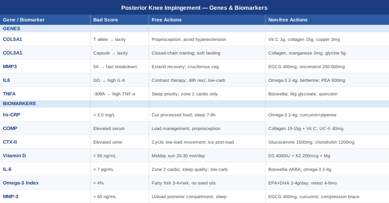
Biomarkers tell you what is happening right now. Genetic variants tell you why certain tissues may be structurally predisposed to respond poorly to load — and what you can do to compensate. This section covers five key genes relevant to posterior knee impingement syndrome. Note that single-gene effects are modest; what matters is the cumulative picture and what it suggests about personalized prevention.
COL5A1 — The Connective Tissue Laxity Gene
COL5A1 encodes collagen type V, which regulates the fibril diameter of collagen type I in tendons, ligaments, and joint capsules. The variant rs12722 (C/T) is the most studied — the T allele is associated with smaller, irregular collagen fibrils, increased joint laxity, and significantly elevated risk of ligament and tendon injury. Multiple studies across athletic populations have linked this variant to Achilles tendon rupture, ACL injury, and generalized joint hypermobility — all conditions that share a posterior knee impingement mechanism.
If the gene is bad, the plan without supplements
The structural deficit in COL5A1 variants means the posterior capsule may be more prone to stretching under load. The compensatory strategy is neuromuscular control: proprioceptive training (single-leg balance, perturbation training, wobble board), eccentric hamstring strengthening, and targeted posterior chain activation (glute-ham raises, Nordic curls). Frequency: 4 sessions per week, 20–30 minutes focused work. Taping (dynamic tape or Kinesio tape) can reduce hyperextension range of motion during sports temporarily. Avoid end-range hyperextension in all training contexts.
If the score is bad, the plan with supplements or equipment
Vitamin C (ascorbic acid): 500–1000mg per day, essential for prolyl and lysyl hydroxylase enzymes that cross-link collagen fibers. In a COL5A1-variant individual, maximizing the quality of whatever collagen is produced becomes the target. Hydrolyzed collagen peptides: 15g before exercise, combined with vitamin C (see COMP section). Copper: 2–4mg per day (as copper glycinate or bisglycinate) — copper is a cofactor for lysyl oxidase, the enzyme that cross-links collagen fibrils. Deficiency is more common than appreciated, especially when zinc supplementation is not balanced. Cycle: 12 weeks on, 4 weeks off, re-assess.
COL3A1 — Joint Capsule Integrity
COL3A1 encodes collagen type III, the dominant collagen in blood vessels, skin, and joint capsules including the posterior knee capsule. Variants in this gene are associated with Ehlers-Danlos syndrome at the extreme end, but subclinical variants are much more common and contribute to posterior capsule laxity without meeting clinical syndrome criteria. Research suggests these variants impair the posterior capsule's ability to resist hyperextension forces, directly predisposing to impingement.
If the gene is bad, the plan without supplements
Stability-first training: prioritize closed-chain exercises (leg press, step-downs, Bulgarian split squats) over open-chain work that stresses the posterior capsule. Learn and consistently apply soft landing mechanics — land with slight knee flexion, never lock out. Compression sleeves worn during activities can partially substitute for reduced capsular stiffness. Physical therapy to address any posterior pelvic tilt, which loads the posterior capsule under passive tension.
If the score is bad, the plan with supplements or equipment
The supplement approach mirrors COL5A1: collagen peptides, vitamin C, and copper. Manganese: 2–5mg per day — a cofactor for glycosyltransferases involved in collagen glycosylation. Often overlooked but relevant in connective tissue synthesis. Proline and glycine amino acids (available in glycine powder and bone broth): glycine at 3–5g before bed supports collagen synthesis during the overnight recovery window. These are low-risk, long-cycle interventions — 3 months minimum before expecting connective tissue remodeling.
MMP3 — The Matrix Degradation Gene
The 5A/6A polymorphism in the MMP3 promoter region directly regulates how much MMP-3 enzyme is produced. The 5A allele is associated with higher MMP-3 transcription — meaning individuals carrying this variant degrade their extracellular matrix faster under the same mechanical or inflammatory stimulus. This is clinically relevant because two people with the same knee impingement injury may recover at dramatically different rates depending on their MMP3 genotype. Research has confirmed the 5A variant as a risk factor for accelerated soft tissue degradation.
If the gene is bad, the plan without supplements
For 5A carriers, the priority is avoiding prolonged mechanical overload — this genotype responds more destructively to repetitive impingement than the general population. Recovery windows should be extended. Sleep quality is particularly important: matrix metalloproteinase activity follows a circadian rhythm and is modulated by cortisol — sleep deprivation directly amplifies MMP-3 activity. Regular but non-excessive antioxidant intake from vegetables (cruciferous vegetables, berries) reduces the oxidative stress that upregulates MMP3 expression.
If the score is bad, the plan with supplements or equipment
EGCG (as above) is especially relevant for 5A MMP3 carriers given its direct MMP-3 inhibitory effect. Resveratrol: 250–500mg per day — this polyphenol suppresses NF-κB, which drives MMP-3 gene expression. Cycle 8 weeks on, 4 off. Take with fat for better absorption. Side effects are rare. These interventions do not change the underlying genotype, but they can meaningfully shift the expression level of a genotype that is otherwise running hot.
IL6 — Inflammatory Cytokine Regulation
The rs1800795 variant (G/C) in the IL6 gene promoter affects baseline IL-6 production. The GG genotype is associated with higher baseline and stimulus-triggered IL-6 levels, translating directly to a more intense local inflammatory response after posterior knee mechanical irritation. Gary Brecka and other functional medicine practitioners highlight the IL6 variant as a key modulator of recovery speed, noting that GG carriers frequently show prolonged inflammatory responses to the same injury stimulus. Studies have linked the GG genotype to higher post-exercise IL-6 levels and longer recovery times.
If the gene is bad, the plan without supplements
For GG carriers, active inflammation management between training sessions is the priority. This includes contrast therapy (alternating cold and warm water immersion), dietary anti-inflammatory patterns (Mediterranean-style, low refined carbohydrate), and strategic rest periods — GG carriers likely need 48 hours between high-load sessions rather than 24. Reducing visceral fat is especially impactful: adipose tissue in GG carriers produces disproportionately more IL-6 than in other genotypes.
If the score is bad, the plan with supplements or equipment
Omega-3 EPA+DHA (2–4g daily) reduces the downstream conversion of IL-6 stimuli. Berberine: 500mg twice daily with meals — this alkaloid activates AMPK and has demonstrated IL-6 suppression in inflammatory models. Cycle 8 weeks on, 4 weeks off; it is not intended for permanent daily use. Palmitoylethanolamide (PEA): 600mg twice daily — an endogenous fatty acid amide with a strong joint-specific anti-inflammatory track record and excellent safety profile. PEA studies show particularly good results for persistent joint inflammatory pain.
TNFA — The Inflammatory Amplifier
TNFA encodes tumor necrosis factor alpha, a master inflammatory cytokine. The variant rs1800629 (G/A, often called -308 G>A) is associated with higher TNF-alpha production. TNF-alpha is one of the earliest signals that turns on both MMP-3 expression and IL-6 production — making TNFA variants a kind of upstream amplifier of the entire inflammatory cascade. In the posterior knee, elevated TNF-alpha accelerates synovial inflammation and posterior capsule degradation under conditions that would be tolerable for someone without this variant. Research has linked the A allele to increased inflammatory burden in joint injury scenarios.
If the gene is bad, the plan without supplements
Sleep architecture is the most underutilized tool for TNFA modulation: TNF-alpha is tightly regulated by sleep, and even one night of poor sleep upregulates it significantly. TNFA A-allele carriers should treat sleep as a therapeutic priority — blackout curtains, cool room temperature (65–68°F), consistent sleep and wake times. Moderate aerobic exercise consistently downregulates TNF-alpha over time; high-intensity exercise acutely spikes it, which is a problem in already-inflamed joints.
If the score is bad, the plan with supplements or equipment
Boswellia serrata (AKBA): 100–250mg twice daily — directly inhibits TNF-alpha downstream pathways. Magnesium glycinate: 300–400mg before bed — magnesium deficiency amplifies TNF-alpha production, and many people with elevated TNFA expression are also marginally deficient. Quercetin: 500–1000mg per day with bromelain for absorption — has documented TNF-alpha inhibitory effects in human studies and pairs well with omega-3s for a synergistic anti-inflammatory approach. Cycle: 12 weeks on, 4 weeks off.
The Huberman Lab Podcast on Connective Tissue Recovery That May Change Your Approach
The Huberman Lab episode featuring Dr. Keith Baar — a leading researcher in connective tissue adaptation at UC Davis — contains some of the most evidence-grounded and clinically actionable insights on tendons, ligaments, and joint capsule recovery available in podcast format. Baar's research directly challenges the common clinical advice of passive rest for connective tissue injuries. Here are the ten most impactful takeaways.
1. Tendons and Ligaments Are Not Just Passive Cables
They actively adapt to mechanical loading, producing more collagen when loaded correctly. Passive rest does not stimulate this adaptation — it often leads to disorganized collagen deposition. Posterior capsule healing requires strategic loading, not immobilization.
2. Collagen Synthesis Has a Narrow Daily Window
Connective tissue collagen synthesis peaks in the 6 to 8 hours after loading, then returns to baseline. Loading every 6 hours (rather than once daily) maximizes cumulative synthesis within the tissue.
3. Vitamin C Is Non-Negotiable
Collagen synthesis is enzymically dependent on vitamin C. Without adequate levels, proline and lysine cannot be hydroxylated, producing structurally weak collagen. 50mg of vitamin C taken 30–60 minutes before loading, alongside gelatin or collagen peptides, is the specific protocol Baar recommends.
4. Gelatin Is Better Than Plain Collagen Protein for Joint Tissue
Gelatin (hydrolyzed from whole collagen) contains the full amino acid composition including glycine, proline, and hydroxyproline that connective tissue preferentially uses. 15g of gelatin or hydrolyzed collagen 45 minutes pre-loading, with vitamin C, is the studied protocol.
5. Blood Supply to Connective Tissue Is Extremely Limited
Unlike muscle, tendons and ligaments have minimal vascularity. This is why recovery takes months, not weeks. Warming the joint before loading (warm bath, cycling) increases local blood flow and enhances nutrient delivery during the critical window.
6. Isometric Exercise Is the Entry Point for Injured Connective Tissue
Isometrics produce high tendon force without joint movement — making them ideal for irritated posterior knee structures. 5 sets of 45-second isometric holds at moderate intensity, twice daily, is the starting protocol for most connective tissue injuries before transitioning to eccentric work.
7. Eccentric Loading Remodels Collagen Architecture
Eccentric (lengthening under load) contractions are uniquely effective at organizing collagen fibers along lines of force. For the posterior knee, controlled eccentric hamstring work — particularly the Nordic curl — is the evidence-supported progression once pain has reduced.
8. The Maturation Phase Takes 12 to 18 Months
Even when pain is resolved, newly synthesized connective tissue takes 12 to 18 months to fully mature and achieve near-normal tensile strength. Returning to full sport before this window is complete is the primary driver of re-injury — not a failure of rehabilitation.
9. Cold Therapy Timing Matters
Ice applied immediately after loading blocks the anabolic signaling cascade in connective tissue. Baar recommends delaying cold application by at least 2 hours post-loading if the goal is tissue synthesis, not just pain management.
10. mTOR in Connective Tissue Is Mechanically Driven, Not Nutritionally Driven
Unlike muscle, where protein/leucine intake spikes mTOR, connective tissue mTOR activation is driven almost entirely by mechanical loading. This means supplements alone — without the right loading stimulus — will not produce connective tissue adaptation. The combination of correct loading AND targeted nutrition is what produces results.
Complementary Approaches With Clinical Support
The following approaches have meaningful human clinical evidence in musculoskeletal conditions and are relevant to posterior knee impingement syndrome. They are not replacements for structured rehabilitation, but they have a genuine adjunctive role.
Low-Level Laser Therapy and Photobiomodulation
Photobiomodulation (PBM) uses near-infrared or red light to penetrate soft tissue and stimulate mitochondrial activity in cells, reducing local inflammation and promoting collagen synthesis. In the context of posterior knee impingement, PBM is relevant because it targets exactly the tissue types involved — synovial cells, tenocytes, and chondrocytes — without adding mechanical load to an already irritated posterior compartment.
A 2017 systematic review and meta-analysis in PubMed-indexed journals found statistically significant pain reduction and functional improvement with PBM in knee joint conditions compared to sham treatment. Wavelengths between 810nm and 1064nm appear most effective for deeper tissue penetration.
For posterior knee impingement, apply a class 3B or class 4 device (or consumer near-infrared device of comparable power) to the posterior knee for 10–15 minutes, 4–5 times per week. Position the device directly over the posterior fossa. Use during the subacute and chronic phases — avoid during acute flares with significant swelling. Consumer-grade devices (660nm + 850nm combined panels) are available in the $200–500 range and can be used at home. Avoid direct eye exposure.
Massage Therapy
Manual soft tissue therapy — particularly targeted to the popliteal fossa, posterior capsule area, and hamstring-to-gastrocnemius transition zone — addresses the muscular tension and fascial adhesions that compound posterior knee impingement. When the posterior musculature is chronically tight, it increases resting compression in the posterior compartment. Manual therapy directly reduces this mechanical load.
A randomized trial published in manual therapy literature found meaningful reduction in knee pain scores and improved range of motion with targeted massage compared to exercise alone. The effect size was moderate, but the combination with active rehabilitation was more effective than either alone.
For posterior knee impingement, seek a sports massage therapist or physiotherapist experienced in posterior chain work. Sessions of 45–60 minutes, once or twice per week during the acute phase, transitioning to once per week for maintenance, are realistic. Techniques include longitudinal stripping of the hamstrings, cross-fiber friction at the posterior capsule insertion points, and myofascial release of the popliteal region. Avoid aggressive deep tissue work during active inflammation.
Yoga
Yoga — particularly styles that emphasize controlled posterior chain lengthening and proprioceptive joint loading (Iyengar, Hatha, Restorative) — is relevant for posterior knee impingement because it simultaneously addresses flexibility, neuromuscular control, and body awareness. One of the primary drivers of posterior knee impingement is uncontrolled hyperextension, and yoga trains individuals to feel and manage knee positioning during movement.
A meta-analysis of yoga for musculoskeletal knee conditions found significant improvements in pain, function, and range of motion compared to usual care. Iyengar yoga, which uses props to achieve correct joint alignment, was specifically noted as appropriate for those with structural knee vulnerabilities.
For posterior knee impingement, avoid end-range hyperextension in any pose — this is the exact mechanism of injury. Micro-bend the standing knee in Warrior poses. Use blocks in forward folds to prevent forced knee lockout. Begin with restorative sessions 2–3 times per week; transition to active flow classes only when pain is consistently below 3/10. A qualified Iyengar teacher can modify any pose to protect the posterior knee.
Biofeedback
Biofeedback trains individuals to consciously modify neuromuscular patterns — in this case, the unconscious tendency to hyperextend the knee. EMG-based biofeedback applied to the hamstrings and quadriceps can identify imbalances in posterior chain activation and teach corrective firing patterns. For posterior knee impingement specifically, restoring hamstring pre-activation before ground contact is one of the most biomechanically important corrections.
A study on EMG biofeedback in knee rehabilitation found that biofeedback-augmented training produced faster improvement in dynamic valgus and hyperextension control compared to standard exercise alone — consistent with the mechanism of impingement reduction.
For practical application, seek a sports physiotherapist or rehabilitation specialist who offers real-time EMG biofeedback during gait and sport-specific movement analysis. Sessions typically run 45 minutes, 1–2 times per week, for 6–8 weeks. Some home biofeedback devices are available but clinical guidance significantly improves outcomes. The goal is habituation — once the correct pattern is learned, biofeedback is no longer needed.
Mindfulness Meditation and MBSR
Mindfulness-Based Stress Reduction (MBSR) may seem distant from a mechanical joint problem, but the evidence connecting chronic pain, central sensitization, and stress-mediated inflammatory amplification is robust. Posterior knee impingement often develops a central sensitization component — where the nervous system becomes sensitized to pain signals independent of ongoing tissue damage. MBSR directly targets this component.
A meta-analysis of MBSR for chronic musculoskeletal pain found significant and durable reductions in pain intensity, pain catastrophizing, and inflammatory markers compared to waitlist controls. The effect on IL-6 specifically has been documented in controlled trials.
MBSR is an 8-week structured program, with sessions typically running 2.5 hours weekly plus daily 20–45 minute home practice. Look for certified MBSR instructors through established medical centers or use validated online programs. The body scan meditation — where attention is systematically moved through body regions — is particularly useful for building awareness of pain without amplifying it. This is not a distraction technique; it is a retraining of pain response at the neural level.
Conclusion
Posterior knee impingement syndrome does not resolve predictably with rest and generic advice because it is not a generic condition. The biomarkers covered here — from hs-CRP and omega-3 index to COMP and CTX-II — give you a measurable picture of the biological environment your knee is healing in. The genetic variants, particularly COL5A1, MMP3, and TNFA, help explain why your connective tissue behaves the way it does under load, and what compensatory strategies are most relevant for your individual biology.
The most important next step is not to act on all of this simultaneously. Start by requesting hs-CRP, 25-OH vitamin D, and an omega-3 index — three accessible, affordable tests that give you immediate, actionable information. Bring the results to a sports medicine physician, a functional medicine practitioner, or a physiotherapist who works with biomarkers. From there, the interventions become targeted rather than speculative. Better data leads to better decisions, and that is what sustained recovery — not just symptom management — requires.
Musculoskeletal: Joint Conditions Tendon & Ligament Conditions Sports Injuries
Autoimmune: Inflammatory Conditions Connective Tissue Conditions