This article contains AI generated content.
· UpdatedPrader-Willi Syndrome Genes and Biomarkers — 6 Genes And 7 Biomarkers To Track
Introduction
Living with Prader-Willi syndrome — whether as a parent, caregiver, or the person directly affected — often means navigating a system where explanations feel incomplete and advice feels generic. You hear about growth hormone therapy and caloric restriction. You hear about behavioral management and "structure." What you rarely hear, even in specialized clinics, is a precise account of which genes are driving which symptoms, or which blood markers could tell you whether what you are doing is actually working.
Most guides to PWS treat it as one condition with one approach. In biological terms, it is closer to six overlapping conditions unfolding simultaneously — each driven by a different gene region, each affecting a different system, from appetite circuits in the hypothalamus to serotonin receptor function to the neurons that produce oxytocin. The hyperphagia in PWS is not simply a behavioral problem. It emerges from a specific molecular architecture: elevated ghrelin, disrupted melanocortin signaling, absent SNORD115-mediated serotonin receptor editing, and a shortage of oxytocin-producing neurons. These are separate targets with separate solutions.
This is not a cure article. The genetics of Prader-Willi syndrome are fixed at birth, and no intervention reverses the underlying imprinting deficit. But "fixed genotype" does not mean "fixed outcome." The research of the past decade — including trials on intranasal oxytocin, melanocortin pathway drugs, and growth hormone optimization — has demonstrated that meaningful functional improvements are possible when interventions are matched to the specific molecular pathway being disrupted. The difference between managing PWS generically and managing it with precision is increasingly large.
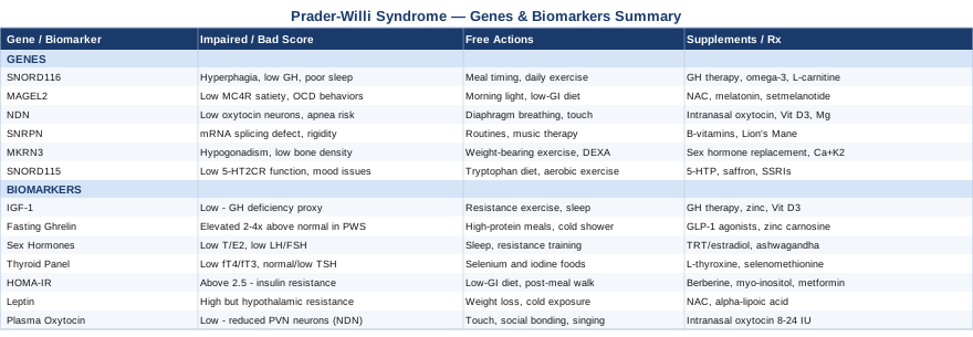
This article maps the six most clinically relevant gene regions, explaining what each one does, what happens when it is silenced, and what concrete steps — with and without supplements or medical interventions — may help compensate. It then covers seven biomarkers that offer a real-time window into how well those systems are functioning. The goal is not more information for its own sake. It is better questions for specialists, smarter lab requests, and a clearer sense of where the levers actually are.
Summary
This article covers the six key gene regions silenced in Prader-Willi syndrome — including SNORD116, MAGEL2, NDN, SNRPN, MKRN3, and SNORD115 — explaining what each one controls, which symptoms it drives, and what interventions (lifestyle, nutritional, and medical) are supported by current evidence. It then presents seven high-value biomarkers — from IGF-1 and fasting ghrelin to oxytocin and HOMA-IR — with guidance on how to measure them, what target ranges to aim for, and what to do when scores fall outside those ranges. Beyond the genetics and biomarkers, you will find a summary of the most compelling research insights from a leading scientist in appetite biology, plus five evidence-supported complementary approaches including music therapy, microbiome interventions, and breathing-based therapies. Each section is designed to translate science into something you can actually use in conversation with your care team.
The 6 Gene Regions That Define Prader-Willi Syndrome — And What To Do About Each
Prader-Willi syndrome is caused by the loss of expression of paternally inherited genes on chromosome 15q11.2-q13. In about 65–75% of cases, this results from a deletion of the paternal region. In 20–25% of cases, both copies of chromosome 15 come from the mother (maternal uniparental disomy), leaving no paternal alleles expressed. A small percentage (1–3%) results from defects in the imprinting center itself. In all three cases, the same set of genes goes silent. The clinical consequence is not a single deficit — it is a cascade across six partially independent gene systems.
The comprehensive genetic overview of this condition is documented in the GeneReviews entry for Prader-Willi Syndrome, maintained at the National Institutes of Health.
Gene 1: SNORD116 Cluster — The Hypothalamic Hunger Regulator
What it controls: SNORD116 is a cluster of small nucleolar RNAs (snoRNAs) that regulate the processing and expression of other RNA molecules, particularly in the hypothalamus. This region appears to be the most critical single contributor to the PWS phenotype. Mice with isolated deletion of SNORD116 develop hyperphagia, growth hormone deficiency, and metabolic dysregulation nearly identical to full PWS deletion models.
What goes wrong when it is silenced: The hypothalamus loses part of its capacity to process satiety signals correctly. POMC (pro-opiomelanocortin) processing is disrupted, growth hormone pulsatility decreases, and circadian-linked gene expression in the hypothalamus is blunted. The result is persistent hunger even after adequate caloric intake, low muscle mass, elevated body fat, and disrupted sleep architecture.
If the gene region is affected — the plan without supplements
The first line of management does not require any pharmacological intervention. Structured meal timing — three meals and one planned snack at fixed daily intervals — reduces the anticipatory anxiety that amplifies food-seeking behavior. Visual countdown timers for meals help manage the time distortion that many individuals with PWS experience around food. Physical access to food should be secured (locked refrigerators and pantries are a legitimate medical recommendation, not a punishment). Regular aerobic exercise at moderate intensity, performed daily for 30–45 minutes, supports endogenous growth hormone release and improves hypothalamic insulin sensitivity over time.
If the gene region is affected — the plan with supplements or equipment
Growth hormone therapy (recombinant human GH, 0.5–1 mg/day by subcutaneous injection) is FDA-approved for PWS and remains the most evidence-supported pharmacological intervention. It improves body composition, muscle tone, bone density, and cognitive function. It must be initiated and monitored by a pediatric or adult endocrinologist. Key side effects to monitor: scoliosis progression, sleep apnea worsening (particularly in the first months of therapy), and glucose tolerance changes. It is not cycled — it is maintained continuously with regular IGF-1 monitoring.
Omega-3 fatty acids (2–4 g/day EPA+DHA from fish oil) reduce hypothalamic neuroinflammation that worsens when SNORD116-dependent processing is disrupted. Take with meals to minimize GI side effects. No mandatory cycling required at these doses. L-carnitine (50–100 mg/kg/day, divided doses) supports mitochondrial fatty acid oxidation, which is commonly impaired in PWS due to low GH and reduced muscle mass. Monitor for GI symptoms; dose can be split morning and afternoon.
Gene 2: MAGEL2 — The Melanocortin Pathway and Social Behavior Gene
What it controls: MAGEL2 encodes a protein that stabilizes the WASH regulatory complex in endosomes, which in turn supports recycling of melanocortin-4 receptors (MC4R) to the cell surface. MC4R is the primary post-synaptic receptor for POMC-derived satiety signals. Beyond appetite, MAGEL2 plays a role in circadian rhythm entrainment, dopamine system maturation, and — critically — social behavior. Isolated MAGEL2 mutations cause Schaaf-Yang syndrome, a PWS-spectrum condition with similar features.
What goes wrong when it is silenced: MC4R signaling is blunted, meaning the brain receives a weaker satiety signal even when nutrients are present. Dopamine reward circuitry is dysregulated, contributing to rigidity, obsessive-compulsive behaviors, and social difficulties. Circadian alignment is impaired, worsening sleep and metabolic timing.
If the gene is affected — the plan without supplements
Morning light exposure (bright natural light or a 10,000-lux lamp for 20–30 minutes within the first hour of waking) is the single most effective free intervention for MAGEL2-related circadian dysfunction. It resets the suprachiasmatic nucleus independently of the MAGEL2-mediated pathway. A low-glycemic diet reduces the demand placed on MC4R signaling by limiting post-meal glucose spikes that require strong hypothalamic compensation. Social engagement in structured, predictable contexts supports dopamine system health without overwhelming the impaired regulation capacity.
If the gene is affected — the plan with supplements or equipment
N-acetylcysteine (NAC) at 600–1200 mg/day (taken in divided doses with meals) reduces glutamate dysregulation that worsens when dopamine circuits are impaired. A 2009 pilot trial found NAC reduced repetitive and compulsive behaviors in adults with OCD-spectrum disorders; smaller reports suggest similar benefit in PWS-related rigidity. Side effects: mild nausea at higher doses. Cycle 8 weeks on, 2 weeks off to prevent glutathione system adaptation. Melatonin (0.5–3 mg, 30–60 minutes before target sleep time) supports the circadian deficit. Low-dose is preferred. Cycle 5 nights on, 2 nights off to preserve receptor sensitivity.
Setmelanotide (Imcivree), a melanocortin-4 receptor agonist, has received FDA approval for obesity associated with POMC deficiency and LEPR mutations. Clinical trials in PWS are ongoing. This is a prescription-only injectable medication and requires specialist enrollment. Side effects include injection site reactions, hyperpigmentation, and nausea.
Gene 3: NDN (Necdin) — The Neuronal Survival and Oxytocin Gene
What it controls: Necdin is a nuclear protein expressed highly in post-mitotic neurons. It promotes neuronal differentiation and survival by suppressing apoptosis, and plays a specific role in the development of oxytocin-expressing neurons in the paraventricular nucleus (PVN) of the hypothalamus.
What goes wrong when it is silenced: PVN oxytocin neuron count is reduced by approximately 50% in PWS (Swaab et al. 1995). This explains several features that do not fit neatly into pure appetite dysregulation: impaired bonding behaviors, anxiety, social reciprocity deficits, and — counterintuitively — aspects of the hyperphagia itself, since oxytocin acts as a satiety signal and an appetite suppressant. Breathing control (the hypotonia and respiratory depression in newborns with PWS) is also partly NDN-dependent.
If the gene is affected — the plan without supplements
Sleep apnea evaluation is non-negotiable given NDN's role in brainstem respiratory control. Polysomnography should be performed before starting GH therapy and monitored annually thereafter. Diaphragmatic breathing practice (10 minutes twice daily: inhale 4 counts, hold 2, exhale 6) strengthens respiratory musculature and improves brainstem CO2 sensitivity. Physical touch — massage, deep pressure input, weighted blankets — stimulates oxytocin release through peripheral sensory pathways, partially compensating for central oxytocin insufficiency.
If the gene is affected — the plan with supplements or equipment
Intranasal oxytocin (8–40 IU administered nasally, 30–60 minutes before social or mealtime interactions) is the most actively investigated intervention for NDN-related PWS features. A 2017 randomized trial by Tauber et al. (PMID 28278518) showed improvements in hyperphagia, social behavior, and temper outbursts in children with PWS. Effects are modest and variable. This is not yet FDA-approved for PWS; use requires physician oversight. Side effects: headache, nausea. Vitamin D3 (2000–5000 IU/day, tested against 25-OH vitamin D levels targeting 50–80 ng/mL) provides neuroprotective support for remaining oxytocin neurons. Take with vitamin K2 (MK-7, 100 mcg/day) to ensure calcium routing. Magnesium glycinate (200–400 mg at night) supports neuronal function and reduces the anxiety amplified by oxytocin insufficiency.
Gene 4: SNRPN — The RNA Splicing Coordinator and Imprinting Anchor
What it controls: SNRPN encodes SmB', a core spliceosome protein required for accurate pre-mRNA splicing across thousands of transcripts. Its promoter region also overlaps the primary imprinting center (IC1) for the entire 15q11-q13 domain. Loss of SNRPN therefore disrupts both direct splicing function and the imprinting control mechanism for the entire gene cluster.
What goes wrong when it is silenced: Brain-specific mRNA splicing is dysregulated, affecting synaptic plasticity genes, neurexins, and contactin-associated proteins — all of which contribute to cognitive flexibility and learning. The imprinting center defect that occurs in approximately 1–3% of PWS cases is directly traceable to this region.
If the gene is affected — the plan without supplements
Consistent environmental structure reduces the cognitive load imposed by impaired neural flexibility. Predictable daily routines, advance preparation for transitions, and visual schedules all reduce the behavioral meltdowns that emerge when cognitive flexibility circuits are taxed. Music-based cognitive training (rhythmic auditory stimulation, melodic intonation therapy) engages neuroplasticity pathways through the auditory and motor systems, partially bypassing disrupted synaptic mechanisms.
If the gene is affected — the plan with supplements or equipment
B vitamins complex (B6 as P5P 25–50 mg, methylfolate 400–800 mcg, methylcobalamin 1000 mcg) support RNA methylation and one-carbon metabolism critical to splicing regulation. Avoid high-dose B6 (>200 mg/day) due to peripheral neuropathy risk. Lion's Mane mushroom extract (Hericium erinaceus, 500–1000 mg/day standardized to hericenones/erinacines) stimulates nerve growth factor (NGF) production, supporting synaptic remodeling. A 2009 randomized trial in humans (Mori et al.) showed cognitive improvements in older adults; smaller studies in neurodevelopmental conditions are emerging. Cycle 3 months on, 1 month off. Phosphatidylcholine (500–1000 mg/day from lecithin or as pure supplement) supports synaptic membrane integrity.
Gene 5: MKRN3 — The Puberty Gate and Bone Health Regulator
What it controls: MKRN3 is an E3 ubiquitin ligase that normally suppresses GnRH pulse generation during childhood, preventing premature activation of the hypothalamic-pituitary-gonadal (HPG) axis. It is expressed exclusively from the paternal allele, making it a classic imprinted gene.
What goes wrong when it is silenced: The HPG axis may be inadequately suppressed during childhood (contributing to rare precocious adrenarche in some PWS cases) but more commonly, the central hypogonadism that defines PWS — with low LH, FSH, and sex hormones — leads to absent or incomplete puberty, reduced bone density, low muscle mass, and (in adults) significant metabolic consequences.
If the gene is affected — the plan without supplements
Weight-bearing exercise (walking, resistance bands, adapted strength training) stimulates bone formation independently of sex hormones through mechanotransduction pathways. DEXA scan from early adolescence is recommended to establish a bone density baseline. Calcium-rich foods (dairy or fortified plant alternatives) should be consistently included. Regular endocrinology review for pubertal staging from age 8–10 in both sexes.
If the gene is affected — the plan with supplements or equipment
Sex hormone replacement therapy — testosterone in males (testosterone undecanoate or cypionate, dose and route per endocrinologist) and estradiol in females — is the standard intervention for hypogonadism in adolescent and adult PWS. It improves bone density, muscle mass, mood, and energy. This requires specialist initiation and monitoring. Calcium (1000–1200 mg/day, from food or calcium citrate supplement split across meals) and vitamin K2 MK-7 (100–200 mcg/day) support bone mineralization. Collagen peptides (5–10 g/day, type I/III) provide substrate for connective tissue health, which is frequently poor in PWS due to hypotonia and hormonal deficiency.
Gene 6: SNORD115 Cluster — The Serotonin Receptor Editor
What it controls: SNORD115 (also called HBII-52) directs A-to-I RNA editing of the serotonin 2C receptor (5-HT2CR) pre-mRNA at a specific adenosine site. This editing step changes the receptor's G-protein coupling efficiency. Without SNORD115, the unedited 5-HT2CR isoform predominates — a form with significantly reduced serotonin sensitivity.
What goes wrong when it is silenced: The 5-HT2CR becomes less responsive to serotonin. Since this receptor mediates satiety signaling (drugs that activate 5-HT2CR, such as lorcaserin, reduce food intake), its hypofunction contributes to hyperphagia through a mechanism entirely separate from SNORD116. This also explains the mood dysregulation, impulsivity, and compulsive behaviors characteristic of PWS — all of which are influenced by serotonin tone.
If the gene is affected — the plan without supplements
Tryptophan-rich diet (turkey, eggs, pumpkin seeds, quinoa) supports serotonin synthesis upstream of the receptor. Regular aerobic exercise (30–45 minutes at 65–75% maximum heart rate) consistently increases serotonin release in the raphe nuclei — the effect is measurable after a single session and cumulative with weeks of practice. Morning light exposure augments serotonin production through the retino-hypothalamic pathway. Consistent sleep protects the serotonin-to-melatonin conversion cycle.
If the gene is affected — the plan with supplements or equipment
5-HTP (50–100 mg before bed) increases serotonin synthesis by bypassing tryptophan hydroxylase. Do not combine with prescription SSRIs or MAOIs (serotonin syndrome risk). Side effects: nausea (take with a small carbohydrate snack). Cycle 6–8 weeks on, 2–3 weeks off. Saffron extract (30 mg standardized to safranal and crocin) has mild serotonergic activity and is safer for long-term use; appropriate for milder cases or as a maintenance option after 5-HTP cycling. SSRIs (fluoxetine or sertraline) are the most commonly used prescription intervention for compulsive and OCD-spectrum behaviors in PWS; clinical guidelines support their use when behavioral features significantly impair quality of life. Initiation and dose titration requires psychiatric or neurology oversight.
With the genetic and epigenetic picture now established, the next question is practical: how do you monitor whether these systems are functioning well or poorly in any given individual? That is where biomarkers become essential.
7 Biomarkers to Track in Prader-Willi Syndrome
Biomarkers do not replace genetic testing or clinical assessment, but they do something genetics cannot: they change over time. They reveal how a body is responding to treatment, how metabolic systems are functioning right now, and where interventions are producing measurable benefit. In PWS, seven biomarkers stand out for their combination of clinical relevance, measurability, and actionability.
Biomarker 1: IGF-1 (Insulin-Like Growth Factor 1)
Why it matters: Growth hormone deficiency is present in almost all individuals with PWS, but GH itself is difficult to measure reliably (it pulses throughout the day). IGF-1, produced by the liver in response to GH, provides a stable 24-hour integration of GH activity. It is the standard clinical proxy used to monitor GH therapy adequacy and is central to body composition management.
How to measure it: Standard venous blood test, collected fasting or non-fasting. Cost: $30–$150 depending on insurance. Target ranges are age-adjusted; in GH-treated adults with PWS, a target of 150–300 ng/mL is typically sought. The test is widely available through any standard laboratory.
If the score is low — the plan without supplements
Resistance exercise (2–3 sessions per week, 8–12 reps per set, targeting large muscle groups) is the most potent natural stimulus for GH/IGF-1 axis activity. Sleep optimization (consistent sleep-wake times, room temperature 65–68°F, no screens after 9 PM) preserves the nocturnal GH pulse — the largest single GH release event in any 24-hour period. Reducing sugar and refined carbohydrate intake lowers insulin levels, which otherwise suppress GH release at the pituitary.
If the score is low — the plan with supplements or equipment
Recombinant human GH therapy (0.5–1.5 mg/day subcutaneous; dose adjusted to maintain IGF-1 in target range) is the definitive intervention and the one with the most evidence in PWS. Zinc (25–40 mg/day elemental zinc as zinc glycinate or picolinate) supports GH receptor sensitivity; supplement with 1–2 mg copper to prevent depletion. Vitamin D3 (2000–5000 IU/day, tested to serum target of 50–80 ng/mL) is required for adequate GH receptor transcription.
Biomarker 2: Fasting Ghrelin
Why it matters: Ghrelin is the only orexigenic (hunger-promoting) hormone in the body, released primarily from the stomach before meals. In most obese individuals, fasting ghrelin is suppressed — the body's attempt to reduce hunger. In PWS, ghrelin is paradoxically and persistently elevated, even after meals. This single abnormality is a major driver of the relentless hyperphagia. Tracking it offers insight into the biological basis of hunger intensity that behavioral observations alone cannot provide.
How to measure it: Blood test requiring a specialized laboratory panel (not standard in all labs; specify acylated ghrelin). Must be collected fasting. Cost: $100–$300. Typical fasting ghrelin in non-obese adults: 100–300 pg/mL. In PWS, levels are frequently 2–4 times higher.
If the score is elevated — the plan without supplements
Protein-rich meals (30–40 g protein per meal) are the most effective dietary suppressor of ghrelin; protein signals through gastric mechano- and chemoreceptors to reduce ghrelin release more effectively than carbohydrates or fat. Avoiding ultra-processed food prevents the rapid gastric emptying that drives ghrelin spikes between meals. Consistent sleep schedule (ghrelin rises acutely with sleep deprivation). Cold exposure (cold showers, 2–3 minutes at end of shower) has been shown to temporarily suppress acylated ghrelin and may modulate appetite acutely.
If the score is elevated — the plan with supplements or equipment
GLP-1 receptor agonists (semaglutide, liraglutide — prescription only) suppress ghrelin indirectly by slowing gastric emptying and augmenting central satiety signaling. Clinical reports in PWS are emerging; off-label use requires specialist assessment given the unusual ghrelin physiology. Zinc carnosine (75 mg twice daily) supports gastric mucosal health and may reduce ghrelin overproduction at the gastric source. Inulin-type fructans (chicory root fiber, 5–10 g/day) stimulate PYY and GLP-1 release from the gut, partially counteracting the appetite drive; side effects include bloating — start at 2 g and titrate up over 2 weeks.
Biomarker 3: Sex Hormone Panel (Testosterone, Estradiol, LH, FSH)
Why it matters: Central hypogonadism — with low LH, FSH, and sex hormones — is nearly universal in PWS and has consequences extending far beyond reproductive function: reduced bone density, low muscle mass, impaired energy metabolism, mood instability, and cognitive fog. Peter Attia has emphasized that sex hormone optimization is one of the highest-leverage metabolic interventions available to adults with chronic low-hormone conditions, and PWS-associated hypogonadism is one of the clearest examples of this principle in genetic medicine.
How to measure it: Standard blood panel (total testosterone, free testosterone by equilibrium dialysis, estradiol by LC-MS/MS, LH, FSH, SHBG). Cost: $50–$200 for complete panel. In males, morning fasting measurement is standard (testosterone peaks early AM). Targets vary by sex, age, and clinical context — set collaboratively with an endocrinologist.
If the score is low — the plan without supplements
Sleep quality is the single most important modifiable driver of testosterone and estradiol production — both are synthesized and released predominantly during deep sleep. Zinc-rich foods (oysters, red meat, pumpkin seeds) support Leydig cell and ovarian steroidogenesis. Weight management reduces aromatase activity (which converts testosterone to estradiol in excess fat tissue). Resistance training directly stimulates gonadotropin pulsatility over time.
If the score is low — the plan with supplements or equipment
Testosterone replacement therapy (TRT) in males (injectable testosterone cypionate 100–200 mg every 1–2 weeks, or topical gels) and estradiol/progesterone replacement in females are the primary interventions, guided by endocrinology. Zinc glycinate (25–40 mg/day) and vitamin D3 are evidence-supported co-interventions that improve steroidogenesis in deficient states. Ashwagandha root extract (KSM-66, 600 mg/day) has shown modest testosterone-elevating effects in controlled trials in men with suboptimal levels; safety data in PWS are limited, so use cautiously and discuss with physician.
Biomarker 4: Thyroid Panel (TSH, Free T4, Free T3)
Why it matters: Central hypothyroidism — a pituitary-level failure to produce adequate TSH — occurs in approximately 20–30% of individuals with PWS. Because TSH is low or inappropriately normal (not elevated as in primary hypothyroidism), standard TSH-only screening misses it. Untreated central hypothyroidism worsens the fatigue, weight gain, cognitive sluggishness, and cold intolerance already present in PWS.
How to measure it: Request TSH, free T4, and free T3 together (TSH alone is insufficient for central causes). Cost: $30–$100. Free T4 below 1.0 ng/dL in the context of normal or low TSH is suspicious for central hypothyroidism and warrants TRH stimulation testing.
If the score is low — the plan without supplements
Selenium-rich foods (2 Brazil nuts daily, tuna, sardines) support thyroid hormone conversion (T4 → T3) via selenoprotein enzymes. Iodine-containing foods (seaweed, iodized salt, dairy) provide the substrate for thyroid hormone synthesis. Avoid excessive goitrogenic raw vegetables (kale, cabbage, broccoli) in large daily quantities if thyroid function is borderline.
If the score is low — the plan with supplements or equipment
L-thyroxine (T4) replacement is the standard treatment for central hypothyroidism and should be initiated at the lowest appropriate dose with gradual titration to normalize free T4. Selenium as selenomethionine (200 mcg/day) improves T4-to-T3 conversion and thyroid autoimmune markers where relevant. Vitamin D co-supplementation (as above) improves thyroid receptor sensitivity at the cellular level.
Biomarker 5: Fasting Insulin and HOMA-IR
Why it matters: Insulin resistance develops in PWS through multiple converging pathways: GH deficiency (GH is a natural insulin sensitizer), excess adiposity, physical inactivity, and cortisol dysregulation. HOMA-IR (Homeostatic Model Assessment of Insulin Resistance) is calculated from fasting glucose and fasting insulin and provides an early signal of metabolic dysfunction before glucose itself becomes abnormal. Thomas Dayspring and other lipidologists have consistently highlighted HOMA-IR as one of the most underused early metabolic markers in clinical practice.
How to measure it: Fasting blood draw for glucose and insulin. HOMA-IR = (fasting insulin in μIU/mL × fasting glucose in mmol/L) / 22.5. Target: below 2.0 is favorable; above 2.5 warrants intervention; above 3.5 is a strong signal. Cost: $20–$80. Most labs do not calculate it automatically — compute it manually from the two values.
If the score is elevated — the plan without supplements
Low-glycemic dietary pattern (emphasizing vegetables, legumes, whole grains; avoiding refined sugars and starches) reduces the insulin demand placed on already-strained beta cells. Walking after meals (10–15 minutes immediately after the two largest meals) is the most accessible postprandial glucose management tool and reduces the insulin spike by 30–40% without medication. Zone 2 cardio (conversational pace, 45 minutes, 3–5 times per week) improves skeletal muscle insulin sensitivity over 4–8 weeks.
If the score is elevated — the plan with supplements or equipment
Berberine (500 mg before meals, 3 times daily) activates AMPK and has insulin-sensitizing effects comparable to low-dose metformin in several trials; side effects include GI discomfort — start with 1 daily dose for 2 weeks. Cycle every 8 weeks with a 2-week break. Myo-inositol (2–4 g/day) improves insulin receptor signaling and is well-tolerated. Magnesium glycinate (200–400 mg/day) corrects a deficiency that worsens insulin receptor function. Metformin (prescription) is a reasonable pharmacological option in PWS with confirmed insulin resistance, and has a strong safety record; discuss with endocrinologist.
Biomarker 6: Leptin
Why it matters: Leptin is the adipokine that signals long-term energy sufficiency to the hypothalamus. In most obesity, leptin is elevated but the hypothalamus is leptin-resistant, so the signal is ignored. In PWS, the picture is complex: leptin may be elevated proportionate to fat mass, but the downstream response is blunted, particularly because several of the hypothalamic circuits through which leptin acts (including the POMC system) are already impaired by the PWS genetic defect. Tracking leptin helps distinguish whether hyperphagia is driven more by leptin insufficiency, leptin resistance, or post-receptor pathway failure.
How to measure it: Fasting blood test. Cost: $50–$150. Reference ranges vary by sex and adiposity — the clinical question is not just the absolute level but whether it is high relative to BMI (suggesting resistance) or unexpectedly low (suggesting insufficient production).
If the score is dysfunctional — the plan without supplements
Weight management (even modest 5–10% weight loss) significantly reduces leptin resistance. Cold exposure (regular cold showers or brief cold water immersion) has been shown to improve leptin sensitivity in adipose tissue. Sleep extension consistently lowers leptin resistance in sleep-deprived individuals; addressing sleep apnea in PWS directly improves leptin signaling.
If the score is dysfunctional — the plan with supplements or equipment
N-acetylcysteine (NAC) (600–1200 mg/day) has been shown in animal studies to improve hypothalamic leptin sensitivity by reducing ER stress; human data are limited but the safety profile supports a trial. Alpha-lipoic acid (300–600 mg/day) acts as an antioxidant in the hypothalamus and has improved leptin signaling in rodent obesity models; start with 300 mg and assess tolerance (can lower blood sugar — monitor). Resveratrol (250–500 mg/day from trans-resveratrol) activates SIRT1 and may improve leptin receptor transcription; evidence remains early-stage in humans.
Biomarker 7: Plasma Oxytocin
Why it matters: Given the NDN-related loss of approximately half of PVN oxytocin neurons in PWS (documented in post-mortem studies), plasma oxytocin provides a measurable correlate of the central oxytocin deficit. Low oxytocin in PWS is associated not only with social and behavioral features but with hyperphagia — oxytocin is an endogenous appetite suppressant released after eating and during social bonding. This biomarker is increasingly used in PWS clinical trials as both an endpoint and a patient stratification tool.
How to measure it: Specialized plasma measurement (requires specific tube handling and immediate centrifugation; not available in all labs). Cost: $100–$300. Measurement is more useful for tracking change over time (treatment response) than for a single-point diagnosis, as reference ranges are not well-standardized.
If the score is low — the plan without supplements
Physical affection and touch — massage, hugs, physical proximity with trusted caregivers — stimulates oxytocin release through C-tactile afferent fibers independently of central production. Social bonding activities (pet interaction, music, collaborative creative tasks) activate the same peripheral oxytocin release pathway. Singing and chanting (rhythmic vocalizations) consistently elevate plasma oxytocin in group settings; music therapy programs for PWS incorporate this mechanism deliberately.
If the score is low — the plan with supplements or equipment
Intranasal oxytocin (8–24 IU, self-administered via calibrated nasal spray) remains the most promising intervention. The Tauber et al. randomized controlled trial (PMID 28278518) provides the clearest available evidence for improvements in hyperphagia, communication, and behavioral regulation in children with PWS. Larger phase 3 trials are ongoing. Use only under physician supervision; not commercially available without prescription in most countries. Side effects: headache, nasal irritation, occasional nausea.
The genetic map and the biomarker panel together provide a richer picture than either alone. Now it is worth stepping back to examine some of the broader research frameworks that put this picture in context.
What the Science of Hypothalamic Appetite Regulation Reveals About PWS
The Hungry Brain by Stephan Guyenet, PhD — a neuroscientist who has spent his career studying the hypothalamic regulation of food intake — offers one of the most rigorous and accessible accounts of how appetite goes wrong in metabolic disease. While not written specifically about PWS, its insights map almost point-for-point onto the molecular disruptions described above.
1. The hypothalamus is not responding to information — it is generating a drive
Guyenet's core argument is that overeating is not a failure of willpower but a neurological drive generated in the hypothalamus based on energy status signals. In PWS, that drive is generated abnormally and persistently — not because the person is failing to resist, but because the hypothalamic circuits producing the drive are structurally damaged.
2. The leptin-melanocortin pathway is the master switch
The POMC pathway — disrupted in PWS through SNORD116 and MAGEL2 loss — is the same pathway Guyenet identifies as the central regulator of body weight. This pathway receives leptin signals, ghrelin signals, and nutrient signals, then adjusts feeding behavior and energy expenditure accordingly. In PWS, the switch is stuck in a partial off position.
3. Palatable food is a pharmacological stimulus
Foods engineered for maximal palatability (high fat, high sugar, high salt combinations) stimulate dopamine release in the nucleus accumbens in ways that override hypothalamic satiety signals. In PWS, where dopamine regulation (partly through MAGEL2) is already impaired, this effect is amplified. Dietary environment is not optional — it is a pharmacological variable.
4. Sleep debt creates a hormonal environment nearly identical to PWS ghrelin elevation
A single night of poor sleep (less than 6 hours) raises ghrelin by 15–20% and reduces PYY (a satiety hormone) by a similar margin. In PWS, ghrelin is already elevated by 200–400%. Any sleep disruption adds directly to this burden. Sleep apnea treatment in PWS is therefore a direct appetite management intervention, not just a respiratory precaution.
5. The gut microbiome influences hypothalamic sensitivity
Short-chain fatty acids produced by the gut microbiome (butyrate, propionate, acetate) signal to vagal afferents and modulate hypothalamic neuropeptide expression. Dysbiosis — common in PWS due to dietary restriction, reduced motility, and antibiotic exposure — reduces this gut-brain satiety signal. Fiber diversity in the diet is not trivial in this context.
6. Physical activity recalibrates the appetite set point
Regular exercise reduces the hypothalamic sensitivity to orexigenic signals — meaning that physically active individuals require less pharmacological appetite suppression. For PWS, this is a core argument for prioritizing exercise as a central, daily medical intervention rather than an optional lifestyle choice.
7. Environmental food restriction works where internal signals fail
Guyenet cites studies showing that individuals in food-secure environments with no intentional restriction consistently overconsume to their hypothalamic set point. For PWS, where the set point is pathologically elevated, environmental restriction (locked food storage, caregiver-controlled access) is not behavioral control — it is an architectural solution to a neurological deficit.
8. Reward sensitivity and dopamine tone determine adherence
The dopamine reward system — impaired in PWS through MAGEL2 and D1/D2 receptor dysregulation — determines how rewarding non-food activities are perceived. Building a dense network of non-food rewards (music, physical activity, social recognition, structured creative tasks) is not a behavioral trick — it is a neurochemical strategy.
9. Fasting ghrelin is a clinical readout, not just a research curiosity
Guyenet emphasizes that ghrelin measurement should be standard clinical practice in any obesity-related condition. In PWS, it provides an objective baseline for quantifying the hunger burden and tracking the effect of any dietary or pharmacological intervention. A ghrelin reduction of 20–30% is measurable, meaningful, and achievable with the right combination of protein intake, sleep, and GLP-1 pathway activation.
10. The brain adapts — slowly but measurably — to consistent environmental and metabolic input
Neuroplasticity in the hypothalamus is real. Consistent high-protein diet, regular aerobic exercise, adequate sleep, and low-reward-density environments produce measurable changes in hypothalamic gene expression over months. This does not reverse PWS, but it shifts the functional set point in a favorable direction. The time horizon is 6–12 months of sustained input, not days.
Complementary Approaches With Meaningful Evidence in PWS
The following modalities have the most relevant human evidence for the specific challenges of PWS. They are not replacements for medical management, but they offer real support when integrated thoughtfully.
Music Therapy
Music therapy uses structured musical interactions — listening, singing, instrument play, rhythmic movement — to support communication, emotional regulation, social engagement, and motor development. In PWS, these are precisely the domains most impaired: verbal communication difficulties, rigid behavior, social reciprocity deficits, and hypotonia. Music therapy is also a natural, evidence-aligned method for stimulating oxytocin release (through group singing and synchronized rhythm) and dopamine (through anticipation and reward in musical structure).
A 2017 systematic review of music therapy in neurodevelopmental conditions found consistent improvements in communication, adaptive behavior, and social reciprocity. Smaller case series in PWS specifically have reported improvements in emotional self-regulation and reduced mealtime behavioral disruption when music was incorporated into meal routines. The evidence base is not large but is consistently positive.
Practical application: One-on-one music therapy sessions twice weekly (30–45 minutes each) with a board-certified music therapist provides the highest-quality delivery. Group singing or rhythm-based activities 3–5 times per week can supplement formal sessions. Family participation in music activities during meals creates a dual benefit: sensory engagement reduces food-focused anxiety while simultaneously stimulating oxytocin release. Start with familiar, preferred music and introduce novelty gradually to accommodate rigidity patterns.
Mindfulness Meditation and MBSR
Mindfulness-Based Stress Reduction (MBSR) uses structured attention training — breath awareness, body scanning, non-judgmental observation of thoughts — to modulate the autonomic nervous system, reduce cortisol output, and improve emotional regulation. In PWS, the relevance is direct: chronic stress activates the HPA axis, which raises cortisol, which in turn suppresses GH release, worsens insulin resistance, and increases food cravings. Reducing chronic stress through mindfulness practice directly addresses three metabolic vulnerabilities simultaneously.
A 2013 review in JAMA Internal Medicine (Goyal et al.) covering 47 randomized trials found moderate-strength evidence that mindfulness meditation improves anxiety, depression, and pain. For PWS-specific emotional dysregulation, adapted mindfulness programs (using visual, audio, and sensory anchors rather than pure verbal instruction) have been reported as feasible in some intellectual disability programs, though PWS-specific RCT data are limited.
Practical application: Begin with 5–10 minute body scan practices using audio guidance (many free programs are available from the UCLA Mindful Awareness Research Center). For younger individuals or those with intellectual disability, adapt with sensory props (smooth stones for touch anchoring, soothing music in the background). Daily practice is preferable to intermittent sessions; results are typically noticeable after 8 weeks of consistency. Caregiver co-practice improves compliance and models the skill.
Massage Therapy
Massage therapy — including Swedish, lymphatic drainage, and sensory integration approaches — addresses hypotonia (low muscle tone), poor proprioceptive feedback, chronic constipation, and skin-picking behaviors that characterize PWS. Beyond physical effects, therapeutic touch consistently stimulates peripheral oxytocin release and activates the parasympathetic nervous system, both of which are beneficial given the oxytocin deficit and chronic stress physiology of PWS.
A randomized trial by Field et al. at the Touch Research Institute found that regular massage in infants with developmental delays (including hypotonia) improved muscle tone, weight gain, and motor development over a 4-week period. Massage in older children and adults with PWS has been reported to improve behavioral regulation and reduce skin-picking in several small studies and case reports, though large RCTs are lacking.
Practical application: Professional massage (Swedish or sensory integration technique) 1–2 times per month with a therapist experienced in developmental conditions. Daily caregiver-delivered gentle massage (10–15 minutes in the evening, focusing on arms, legs, and back) sustains the oxytocin and parasympathetic effects between sessions. For skin-picking behavior, combining massage with textured sensory objects (massage brushes, soft rollers) provides an alternative tactile stimulation that satisfies the sensory seeking behavior driving the picking.
Microbiome-Directed Therapies
Gut microbiome composition is significantly altered in PWS. Studies have identified reduced diversity, decreased abundance of butyrate-producing bacteria (Faecalibacterium prausnitzii, Roseburia), and increased inflammatory bacterial species compared to matched controls. This dysbiosis impairs the gut-brain satiety axis (reducing GLP-1 and PYY signaling), promotes systemic low-grade inflammation, and worsens insulin resistance. Since gut bacteria also synthesize a significant proportion of peripheral serotonin, dysbiosis likely compounds the SNORD115-driven central serotonin receptor deficit.
A 2021 study in Gut Microbes found that fecal microbiota transplant in a murine obesity model reversed hypothalamic inflammation and improved leptin sensitivity, providing a mechanistic rationale for microbiome intervention in neuroendocrine obesity. Human trials in PWS specifically are not yet published, but translational evidence is accumulating.
Practical application: Dietary fiber diversity is the most evidence-supported first step: aim for 20–30 different plant foods per week, emphasizing prebiotic-rich foods (onions, garlic, leeks, chicory, green bananas). Probiotic supplementation with a multi-strain product containing Lactobacillus rhamnosus GG, Bifidobacterium longum, and Faecalibacterium prausnitzii (where available) for 8–12 weeks, then reassess. Avoid unnecessary antibiotics. Fermented foods (kefir, yogurt, kimchi) can supplement probiotic intake when tolerated. For individuals with constipation (common in PWS), psyllium husk (5 g in water, twice daily) supports motility and feeds beneficial bacteria simultaneously.
Breathing-Based Therapies
Breathing dysregulation is present in PWS from birth (central hypotonia affects the respiratory muscles) through adulthood (sleep-disordered breathing, obstructive sleep apnea, upper airway hypotonia). Structured breathing practice — diaphragmatic breathing, slow-paced respiration, and Buteyko-style carbon dioxide tolerance training — strengthens respiratory muscle function, improves vagal tone, and reduces anxiety-driven breathing pattern disorders. Better respiratory function directly improves sleep quality, which in turn affects GH secretion, ghrelin levels, and insulin sensitivity.
A 2018 systematic review in the Journal of Clinical Sleep Medicine found that breathing exercises significantly improved sleep apnea severity scores and daytime sleepiness in adults with mild-moderate OSA when combined with CPAP. For PWS, where sleep apnea is present in 44–100% of individuals (prevalence varies by age and obesity level), breathing exercises offer a low-risk augmentative approach alongside CPAP.
Practical application: Diaphragmatic breathing: 10 minutes twice daily (morning and pre-sleep), inhale through nose for 4 counts, pause for 2, exhale through mouth for 6. Nasal breathing training during the day (tape over mouth during low-activity periods if tolerated, per Buteyko method) improves upper airway muscle tone and nasal patency. For sleep apnea: CPAP remains the primary intervention; breathing exercises support it rather than replace it. Myofunctional therapy (specialized tongue and throat exercises performed with a speech-language pathologist) has level 1 evidence for reducing sleep apnea severity in children and adults and is particularly appropriate given the hypotonia profile of PWS.
Conclusion
Prader-Willi syndrome is one of the most precisely understood genetic conditions in the field of neurodevelopment — and that precision is increasingly an asset rather than just an explanation. Knowing that SNORD116 drives the GH deficit, that MAGEL2 disrupts MC4R satiety signaling, that SNORD115 impairs serotonin receptor function, and that NDN loss reduces the number of oxytocin-producing neurons by half is not academic. Each of those facts maps onto a measurable biomarker and an intervention target.
The seven biomarkers covered here — IGF-1, fasting ghrelin, sex hormones, thyroid panel, HOMA-IR, leptin, and oxytocin — give you a way to track how those systems are performing in real time and whether the interventions being used are producing measurable change. They are not comprehensive, but they cover the highest-leverage, most actionable signals available in current clinical practice.
The next smart step is not to implement everything at once. It is to request a baseline panel of these biomarkers at your next specialist visit, review which gene mechanisms are most likely to be driving the dominant symptoms in your specific situation, and identify one or two targeted interventions — medical, nutritional, or behavioral — to trial systematically. The ground under this condition is shifting. Better information, used intelligently, consistently leads to better decisions.
Neurological: Brain Conditions Memory & Cognitive Conditions
Respiratory: Sleep & Breathing Disorders
Mental Health: Eating Disorders
Endocrine & Metabolic: Diabetes & Blood Sugar Thyroid Conditions
Women's Health: Hormonal Conditions