This article contains AI generated content.
Sialidosis Genes and Biomarkers: 3 Genes and 7 Biomarkers to Track
Introduction
If you or someone in your family has been told the words "sialidosis," "cherry-red spot myoclonus," or "NEU1 deficiency," you already know how thin the available information tends to be. Search results are dominated by textbook definitions and rare-disease directories, while the practical questions — what should actually be tested, how often, and what any of it means for day-to-day decisions — are much harder to find answered clearly.
That gap is not an accident. Sialidosis is an ultra-rare autosomal recessive lysosomal storage disorder, and generic health advice simply was not built for conditions like this. Advice built for common metabolic or cardiovascular risk doesn't transfer well to a single-gene enzyme deficiency, where the underlying biology, the relevant tests, and the realistic scope of what can be influenced are all fundamentally different.
This article takes a more specific approach. It walks through the biomarkers that clinicians and metabolic genetics labs actually use to diagnose and monitor sialidosis, what each one reveals, roughly what it costs, and what can — and honestly cannot — be done when a result comes back abnormal. It then looks at the underlying genetics (NEU1, CTSA, and GLB1) in a shorter companion section, at what genomic medicine research suggests about interpreting a rare diagnosis, and at a small set of complementary approaches with genuine, if limited, supporting evidence.
None of this replaces a metabolic geneticist, a neurologist, or a genetic counselor. But better information changes the quality of the questions you bring to those appointments, and that alone tends to lead to better decisions — about which tests are worth repeating, which specialists to loop in, and which day-to-day adjustments are actually worth the effort.
Summary
Sialidosis comes down to one enzyme, neuraminidase-1, and one gene, NEU1 — but the way that deficiency shows up, and how it's tracked over time, involves a surprisingly wide net of tests. Below, this article breaks down the seven biomarkers that actually matter for diagnosis and monitoring, from urinary oligosaccharide screening to retinal imaging to neurophysiology, with realistic cost ranges and what to do (and not expect) when a result is abnormal. It then explains the three genes behind sialidosis and galactosialidosis — NEU1, CTSA, and GLB1 — and why they travel as a package deal inside the lysosome. A closer look at genomic researcher Ali Torkamani's work on genotype-first sequencing and polygenic modifiers adds useful context for anyone trying to make sense of a rare-disease genotype, and a final section covers which complementary approaches — biofeedback, mindfulness, and relaxation training — have real, if modest, human evidence behind them for the symptoms sialidosis produces.
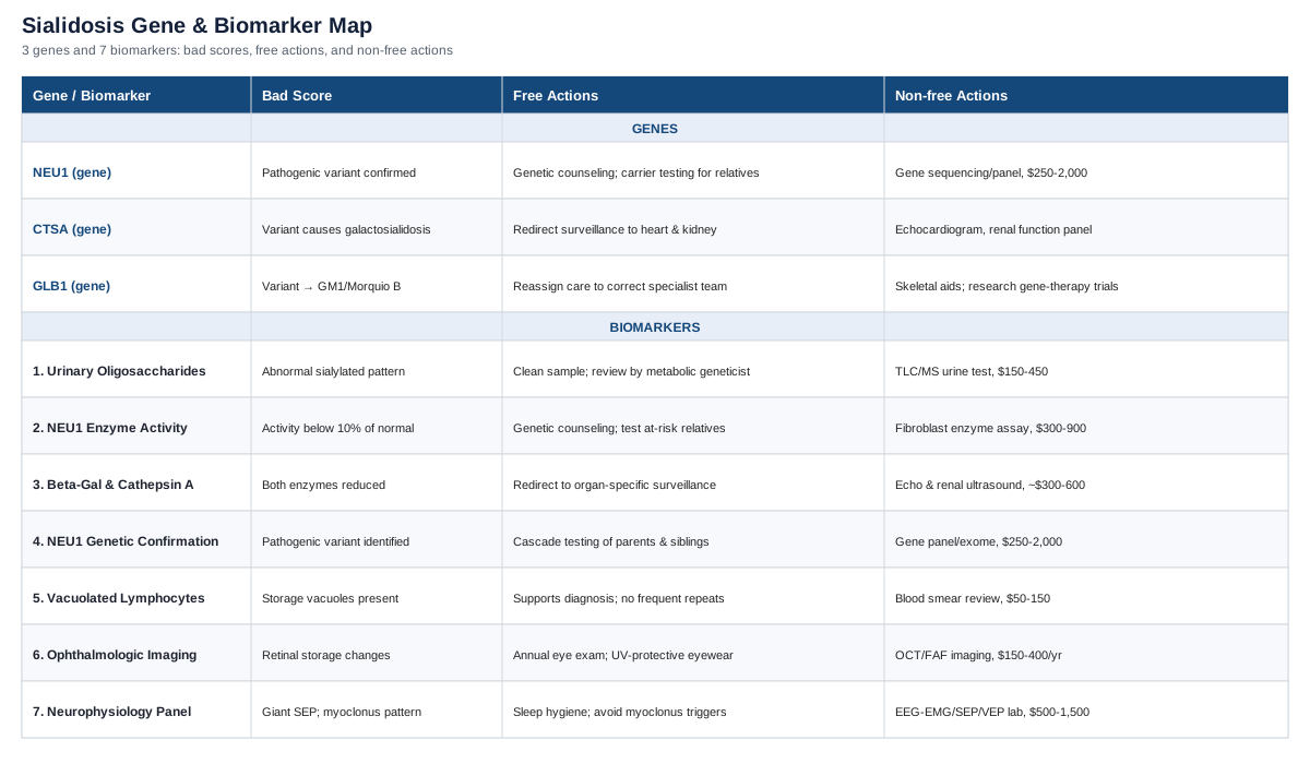
The Sialidosis Biomarker Panel: What to Track and Why
Sialidosis is caused by deficient activity of neuraminidase-1 (sialidase), the lysosomal enzyme that normally trims terminal sialic acid residues off glycoproteins and glycolipids. When it doesn't work, sialylated compounds accumulate inside lysosomes throughout the body, producing the two recognized clinical forms: type I, the milder, later-onset "cherry-red spot myoclonus syndrome," and type II, an earlier-onset, more systemic form with skeletal and facial involvement. Unlike biomarker panels built around modifiable cardiometabolic risk, most sialidosis biomarkers exist to answer three questions: is this sialidosis (versus a look-alike condition like galactosialidosis or GM1 gangliosidosis), how severe is the current disease burden, and is a specific symptom — usually myoclonus, seizures, or vision loss — getting better, worse, or staying stable under treatment. That reframes what "improving a biomarker" means here: for most of these markers, there is no lifestyle or supplement fix for the enzyme deficiency itself, and any claim otherwise should be treated with real skepticism. What is genuinely actionable is testing accuracy, monitoring cadence, symptom management, and knowing when a result should trigger a specialist referral.
1. Urinary Oligosaccharide Profile
A urine oligosaccharide screen looks for the abnormal pattern of sialylated oligosaccharides that spill into urine when neuraminidase-1 can't process them normally. It's usually the first biochemical clue that points a clinician toward sialidosis, well before an enzyme assay or genetic panel is ordered, and it remains useful afterward as a rough index of storage burden over time, as described in reviews of the condition's diagnostic workup (Khan & Sergi, Diagnostics, 2018).
How to measure it
A first-morning spot urine sample is analyzed by thin-layer chromatography as an initial screen, with mass spectrometry used at reference metabolic laboratories for more precise characterization. Cost typically runs from about 150 to 450 US dollars depending on the lab and whether MS confirmation is included; turnaround is usually one to three weeks. This is a specialized biochemical genetics test, not something available through routine bloodwork.If the score is bad, the plan without supplements
There is no behavioral fix for an abnormal oligosaccharide pattern — it reflects an enzyme deficiency, not diet or lifestyle. The practical plan is to make sure the sample was collected correctly (clean first-morning void, no contamination), to have it interpreted by a metabolic geneticist rather than a general lab, and to treat a single abnormal result as a starting point for further workup rather than a number to chase downward on repeat testing. Repeat testing every one to two years is reasonable for monitoring trends, not for detecting week-to-week change.If the score is bad, the plan with supplements or equipment
No supplement, enzyme cofactor, or piece of equipment lowers urinary oligosaccharide excretion in sialidosis — this is worth stating plainly, since storage-disease forums sometimes suggest otherwise. The only "equipment" that matters here is access to a reference laboratory experienced in lysosomal storage disorders, since community labs can produce false negatives on borderline cases.2. Neuraminidase (NEU1) Enzyme Activity Assay
This is the functional confirmation test: it directly measures how much sialidase activity remains in a patient's cells. Activity below roughly 10 percent of normal is typical in sialidosis, and the degree of residual activity correlates loosely with disease severity — patients with some residual function tend to have milder, later-onset disease (Genes, 2025).
How to measure it
Enzyme activity is measured in cultured skin fibroblasts (from a small punch biopsy) or in peripheral blood leukocytes, using a fluorogenic substrate assay at a specialized biochemical genetics lab. Fibroblast culture is more reliable but slower — plan on two to six weeks and a cost in the range of 300 to 900 US dollars, more if a skin biopsy procedure fee is included.If the score is bad, the plan without supplements
A confirmed low result is generally a one-time diagnostic test, not something to repeat routinely — once neuraminidase deficiency is documented, the practical next step is genetic counseling for the patient and testing of at-risk relatives, not repeat enzyme testing. Coordinating care through a lysosomal storage disease center avoids duplicate biopsies and ensures results are interpreted against the right reference ranges.If the score is bad, the plan with supplements or equipment
Nothing over-the-counter restores neuraminidase-1 activity. The genuinely promising angle here is pharmacological — small-molecule chaperones designed to stabilize misfolded but partially functional NEU1 protein are in early research stages, discussed as an emerging therapeutic direction in recent genetic reviews of sialidosis (Genes, 2025), but these are not yet available outside research settings and should not be confused with supplements.3. Beta-Galactosidase and Cathepsin A Activity Panel
Because NEU1 works inside a three-protein lysosomal complex alongside beta-galactosidase (GLB1) and cathepsin A (CTSA), a full workup usually adds these two enzyme activities to rule in or rule out galactosialidosis — a related but distinct disorder caused by primary cathepsin A deficiency, which secondarily drags both beta-galactosidase and neuraminidase activity down (Orphanet Journal of Rare Diseases, 2013). Getting this distinction right matters because galactosialidosis carries a different systemic surveillance plan, including cardiac and renal monitoring.
How to measure it
Run alongside the neuraminidase assay in the same fibroblast sample; adds roughly 300 to 600 US dollars to the panel. In true sialidosis, beta-galactosidase activity should be normal or near-normal; in galactosialidosis, both beta-galactosidase (roughly 15 percent of normal) and neuraminidase (under 1 percent) are markedly reduced.If the score is bad, the plan without supplements
If cathepsin A/beta-galactosidase results confirm galactosialidosis rather than isolated sialidosis, the useful action is redirecting surveillance: periodic echocardiogram, renal function testing, and skeletal survey, since these systems are more often involved in the combined deficiency. This is a diagnostic branch point, not something to "improve."If the score is bad, the plan with supplements or equipment
No supplement changes cathepsin A or beta-galactosidase activity. Equipment-wise, the relevant investment is in organ-specific screening tools (echocardiography, renal ultrasound) scheduled at the frequency your metabolic team recommends, typically annually, rather than in anything self-administered.4. NEU1 Genetic and Molecular Confirmation
Sequencing NEU1 (and, when indicated, CTSA) identifies the specific pathogenic variants responsible, which matters for prognosis, for distinguishing missense from null mutations, and for offering accurate carrier testing to siblings and future family planning (Genes, 2025).
How to measure it
Single-gene sequencing, a lysosomal storage disease gene panel, or whole-exome sequencing, depending on how the case presents. Costs vary widely — roughly 250 to 2,000 US dollars before insurance, with panels often cheaper than exome sequencing and turnaround of two to six weeks.If the score is bad, the plan without supplements
A confirmed pathogenic genotype is the trigger for genetic counseling and cascade testing of parents and siblings — this is where a diagnosis becomes actionable for the wider family, not just the patient. Variants of uncertain significance should be revisited periodically, since reclassification happens as more cases are published.If the score is bad, the plan with supplements or equipment
Genotype cannot be changed by supplementation. The one legitimate "equipment" step is checking clinical trial registries for studies matched to a specific NEU1 genotype, since some gene- and enzyme-targeted research programs recruit by variant type.5. Vacuolated Lymphocytes on Blood Smear
A simple peripheral blood smear can show vacuolated lymphocytes containing storage material — a low-cost, low-tech clue that supports a lysosomal storage disorder even before enzyme or genetic testing is complete (Diagnostics, 2018).
How to measure it
A routine blood draw with a request for morphology review, roughly 50 to 150 US dollars. Bone marrow aspirate can show similar storage granules but is now rarely necessary given how accessible enzyme and genetic testing have become.If the score is bad, the plan without supplements
This finding supports the diagnosis rather than something to correct; it doesn't need to be repeated frequently once storage disease is confirmed by enzyme or genetic testing.If the score is bad, the plan with supplements or equipment
Nothing clears intracellular storage material through diet or supplements — general nutritional adequacy supports overall cell health, but it does not resolve lysosomal accumulation.6. Ophthalmologic Imaging: Cherry-Red Spot, OCT, and Fundus Autofluorescence
The classic macular cherry-red spot is present in a meaningful share of type I patients but is far from universal — some cohort studies report it in under half of cases — so modern workups lean on optical coherence tomography (OCT) and fundus autofluorescence (FAF), which pick up increased reflectivity in the nerve fiber and ganglion cell layers even when no cherry-red spot is visible (Molecular Genetics & Genomic Medicine, 2020; Tremor and Other Hyperkinetic Movements, 2021).
How to measure it
A dilated fundus exam plus OCT and FAF imaging at an ophthalmology visit, roughly 150 to 400 US dollars depending on insurance coverage and country, done annually or when visual symptoms change.If the score is bad, the plan without supplements
Consistent annual ophthalmology follow-up, UV-protective eyewear, and prompt evaluation of visually significant cataracts (punctate lens changes are common in sialidosis) are the realistic non-pharmacological steps. Photosensitivity or myoclonus triggered by bright light can sometimes be reduced with tinted lenses.If the score is bad, the plan with supplements or equipment
No supplement reverses retinal storage deposits. General macular-support nutrients such as lutein and zeaxanthin (typically 10 mg and 2 mg daily) are reasonable for general retinal health, well-tolerated, and don't require cycling, but they should be framed as supportive, not disease-modifying. Low-vision optical aids (magnifiers, adaptive lighting) are the most practically useful "equipment" for patients with progressive visual decline.7. Neurophysiological Markers: EEG-EMG Polygraphy, SEP, and VEP
Because action myoclonus, giant somatosensory evoked potentials (SEP), and photosensitive seizures are central to sialidosis type I, neurophysiological testing functions as the closest thing to a severity and treatment-response biomarker available. Long-term case series show these measures tracking meaningfully with clinical course and medication response over decades of follow-up (Brain Sciences, 2020).
How to measure it
EEG with simultaneous EMG polygraphy, plus SEP and visual evoked potentials, performed in a neurophysiology lab. Cost ranges from about 500 to 1,500 US dollars depending on the setting, repeated when symptoms change or roughly annually to track medication response.If the score is bad, the plan without supplements
Sleep hygiene, avoidance of known myoclonus triggers (sudden stimuli, flashing lights, alcohol withdrawal patterns), and stress reduction all have plausible symptomatic value, since stimulus-sensitive and stress-aggravated myoclonus is well documented in this condition. Antiseizure medication management with a neurologist — not a supplement — is the primary lever for an abnormal EEG/EMG pattern.If the score is bad, the plan with supplements or equipment
Magnesium is sometimes used adjunctively for general neuromuscular excitability (typically 200 to 400 mg of elemental magnesium daily, no cycling required, main side effect being loose stools at higher doses, used cautiously in kidney disease). Wearable EEG or actigraphy-based movement trackers are increasingly used in research settings to quantify myoclonus frequency outside the clinic, and protective equipment (padded flooring, gait aids) is reasonable for patients with drop episodes. None of this substitutes for specialist-guided seizure management.Taken together, this panel tells a consistent story: most of these markers exist to diagnose accurately and monitor honestly, not to be "optimized" the way a lipid panel might be. That distinction matters for setting realistic expectations — and it's also the reason the underlying genetics are worth understanding directly, since they explain why these particular tests were chosen in the first place.
The Genes Behind Sialidosis: NEU1, CTSA, and GLB1
Sialidosis and its close relative, galactosialidosis, both trace back to a shared piece of lysosomal machinery: a three-protein complex made of neuraminidase-1, beta-galactosidase, and cathepsin A (also called the protective protein). Understanding how these three genes relate to each other explains both why sialidosis biomarkers look the way they do, and why "fixing" a bad gene score is a fundamentally different conversation than it is for common polygenic conditions.
NEU1
NEU1, on chromosome 6, encodes lysosomal sialidase, the enzyme that trims terminal sialic acid off glycoproteins and glycolipids (NCBI Gene: NEU1). More than 90 pathogenic variants have been catalogued, most of them missense mutations, and the specific combination a person carries drives most of the clinical variability between mild, late-onset type I disease and severe, early-onset type II disease (Genes, 2025). Notably, the same variant can behave differently across populations: one study found cherry-red spots and cognitive impairment were both less frequent in Asian cohorts than in Caucasian cohorts carrying comparable mutations, and a specific variant, c.544A>G, appears to be a founder mutation concentrated in Taiwanese patients.
If the gene is bad, the plan without supplements
Since sialidosis is autosomal recessive, the most consequential "non-supplement" action is genetic counseling and carrier testing for siblings and future children, since two carrier parents face a 25 percent recurrence risk per pregnancy. Working with a clinician experienced in lysosomal disease also helps map which mutations in your specific report tend to correlate with milder versus more progressive courses, since this varies considerably by variant.If the gene is bad, the plan with supplements or equipment
No supplement corrects a NEU1 mutation, and framing it that way would be misleading. What is genuinely on the horizon, still in early research, are small-molecule pharmacological chaperones intended to stabilize misfolded-but-partially-functional NEU1 protein enough to restore some enzyme activity, and enzyme replacement or gene therapy approaches being explored across the broader lysosomal storage disease field. None of these are available as consumer products; they belong in clinical trials, and current management is supportive (seizure control, physical therapy, vision aids) rather than gene-corrective.CTSA
CTSA encodes cathepsin A, a serine carboxypeptidase that acts as the scaffold holding the NEU1–GLB1 lysosomal complex together and ensuring both enzymes fold, traffic, and stay stable correctly (NCBI Gene: CTSA). When CTSA is mutated, the result is galactosialidosis — a secondary combined deficiency in which both beta-galactosidase and neuraminidase activity drop, even though neither of those genes is itself mutated (Orphanet Journal of Rare Diseases, 2013). This is a good illustration of how lysosomal disease genetics don't always map one gene to one enzyme — sometimes a single structural gene failure takes two enzymes down with it.
If the gene is bad, the plan without supplements
Confirming CTSA as the cause (rather than NEU1) redirects surveillance toward the systemic features more common in galactosialidosis — cardiac valve involvement, nephropathy, and skeletal dysplasia in the early-infantile form — so the practical step is ensuring your care team is screening the right organ systems rather than applying a generic sialidosis monitoring plan.If the gene is bad, the plan with supplements or equipment
As with NEU1, no supplement restores cathepsin A function. Equipment-wise, the useful investment is in the organ-specific monitoring tools appropriate to galactosialidosis (echocardiography, renal function panels, skeletal imaging) rather than anything self-directed.GLB1
GLB1 encodes beta-galactosidase, the third member of the complex, and is included here less because it's usually the primary cause of sialidosis and more because testing it is essential for differential diagnosis — mutations in GLB1 itself cause the distinct conditions GM1-gangliosidosis and Morquio B syndrome, not sialidosis, but a properly run enzyme panel needs to check it to rule these out (NCBI Gene: GLB1; GeneReviews: GLB1-Related Disorders).
If the gene is bad, the plan without supplements
If GLB1 itself turns out to carry the pathogenic mutation, the diagnosis shifts away from sialidosis toward GM1-gangliosidosis or Morquio B, each with its own specialist team and monitoring plan — which is exactly why running the full three-enzyme panel up front, rather than assuming sialidosis from symptoms alone, saves time.If the gene is bad, the plan with supplements or equipment
Again, no supplement changes GLB1 function. For Morquio B specifically, skeletal and mobility equipment (orthotics, mobility aids) often becomes clinically relevant, while GM1-gangliosidosis management is centered on neurological supportive care and, in some type 3 cases, research-stage gene therapy trials.The throughline across all three genes is the same: this is a structural, enzymatic problem, not a nutrient-deficiency or lifestyle problem, so the honest "fix" is accurate genetic diagnosis, informed family planning, and staying connected to the research pipeline — not a supplement stack. That distinction is also central to how genomic medicine researchers now think about interpreting any rare diagnosis, which is where the next section picks up.
What Genomic Medicine Research Reveals About Rare Genetic Risk
Ali Torkamani, director of genome informatics at Scripps Research, has spent much of his career studying how genetic information should actually be used in the clinic — work that shows up in interviews like his appearance on the Longevity by Design podcast and in his published research on genotype-first sequencing and polygenic risk. None of it was written about sialidosis specifically, but the underlying logic — how to interpret a genotype, how much weight to put on a single variant, and when genetics is and isn't destiny — applies directly to anyone trying to make sense of a rare monogenic diagnosis. Here are ten ideas from that body of work worth sitting with.
1. Residual function matters more than the diagnosis label
Two people with the same named condition can have very different outcomes depending on how much functional enzyme activity their specific mutations leave behind. Research on NEU1 genotype-phenotype correlation shows exactly this pattern: compound mutations combining one mild and one severe allele tend to produce milder disease than two severe alleles (Genes, 2025). The lesson generalizes — a diagnosis name is a starting point, not the whole picture.2. Sequencing first, symptoms second, flips the traditional order
Torkamani's "Wellderly" project sequenced disease-free elderly adults before looking for disease, rather than the usual symptom-first approach to genetic testing (Cell, 2016). This genotype-first logic is exactly what underlies newborn screening expansion and cascade testing in families with a known rare-disease variant — sequence relatives before symptoms appear, not after.3. Carrying a pathogenic variant doesn't guarantee a phenotype
One of the more counterintuitive Wellderly findings was that disease-free elderly participants carried rare pathogenic variants at similar rates to the general population — protection from disease wasn't simply the absence of risk variants (Cell, 2016). That's a useful caution against over-interpreting any single variant in isolation, including incidental findings unrelated to a primary diagnosis.4. Background genetics can modulate how a single-gene condition expresses itself
Torkamani's work on polygenic risk scores shows that most common disease risk is shaped by the combined, small effects of many variants rather than one gene (Nature Reviews Genetics, 2018). While sialidosis itself is monogenic, this framework helps explain why family members with identical NEU1 genotypes still show variable severity — modifier genes in the polygenic background likely play a role, even if they haven't been fully mapped for this specific disease yet.5. Rare disease diagnosis is still, too often, a years-long odyssey
Reviews of sialidosis note that diagnosis is frequently delayed because symptoms overlap with other progressive myoclonic and ataxic conditions (Diagnostics, 2018). Genotype-first thinking — sequencing earlier in the workup rather than as a last resort — is one of the more concrete ways this odyssey is shortening for newly diagnosed families.6. Ethnic background shapes both variant frequency and phenotype
The same NEU1 variant produces different clinical pictures across populations, and certain variants cluster geographically due to founder effects, as seen with c.544A>G in Taiwanese cohorts (Genes, 2025). Population-specific reference data matters when interpreting any genetic report, not just for common disease risk scores.7. Functional testing should always accompany a genetic result, not replace it
A genetic report naming a variant of uncertain significance isn't the end of the workup — pairing it with enzyme activity testing is how labs distinguish, for instance, true sialidosis from galactosialidosis when the biochemical complex shares components (GeneReviews: GLB1-Related Disorders). Genotype and phenotype-confirming biochemistry work best together.8. One diagnosis becomes prevention for an entire family
The genotype-first logic extends naturally to cascade testing: once a proband's variants are known, testing parents, siblings, and eventually reproductive partners turns a single diagnosis into actionable information for the whole family, well before another affected pregnancy occurs.9. Variant classifications change — revisit old genetic reports periodically
As sequencing databases grow, variants once labeled "uncertain significance" are regularly reclassified as pathogenic or benign. Anyone with an older genetic report, especially one that came back inconclusive, has good reason to ask their genetics team for a periodic reanalysis rather than assuming the original interpretation is final.10. The frontier is molecular, but quality of life is still shaped day to day
The most promising future interventions for monogenic lysosomal disease — chaperones, enzyme replacement, gene therapy — are molecular, not lifestyle-based. But while those mature, day-to-day symptom management, family planning, and stress around unpredictable myoclonus remain squarely in the domain of supportive care, which is exactly where evidence-based complementary approaches can genuinely help.That last point is the natural bridge to a smaller, more practical question: among the complementary and supportive approaches available today, which ones actually have human evidence behind them for the symptoms sialidosis produces?
Complementary Approaches Worth Considering
No complementary therapy changes NEU1 enzyme activity or reverses lysosomal storage — that needs to be said plainly before anything else. But sialidosis type I produces a specific, well-defined symptom cluster (cerebellar ataxia, stimulus-sensitive action myoclonus, and in some patients seizures), and a few approved modalities have real human evidence for exactly those symptoms in closely related neurological populations, even though condition-specific trials in sialidosis itself don't exist given how rare it is.
Biofeedback
Biofeedback uses real-time sensory feedback — visual, auditory, or vibrotactile — to help a person consciously correct posture, balance, or movement patterns that their damaged cerebellar circuits can no longer regulate automatically. Because gait ataxia is a core, often disabling feature of sialidosis type I, and it stems from the same cerebellar dysfunction studied in hereditary ataxia populations, this is one of the more directly relevant modalities on this list.
A crossover study using vibrotactile sensory augmentation during home-based balance training in people with hereditary cerebellar ataxia found statistically significant improvement in SARA ataxia scores and posture/gait subscores after the feedback-assisted training block, with no significant change during training without it (Sensors, 2022). The protocol involved five sessions per week of balance exercises over six-week blocks, using a smartphone-based inertial sensor worn on the lower back.
Realistically, this means working with a physical or occupational therapist familiar with ataxia rehabilitation to set up a structured, feedback-assisted balance program rather than expecting a single gadget to substitute for supervised training. Given that the supporting study was small and preliminary, expectations should stay modest — think incremental gait stability and fall-risk reduction, not reversal of underlying cerebellar disease, and progress should be reassessed every few months with a clinician.
Mindfulness Meditation and MBSR
Mindfulness-based stress reduction trains sustained, non-judgmental attention to present-moment experience, and it's relevant here because both seizures and stimulus-sensitive myoclonus in sialidosis can be aggravated by stress and anxiety, and because living with a rare, progressive diagnosis carries a real psychological burden of its own.
A randomized controlled trial in people with epilepsy found that a six-session, 2.5-hour weekly mindfulness-based intervention produced significant reductions in depression scores and significant improvements in quality of life (QOLIE-31) and mindfulness measures compared to a no-treatment control group, with benefits persisting at six-week follow-up (Epilepsy & Behavior, 2021). A broader systematic review of mindfulness interventions in epilepsy found consistent, if still preliminary, signals for reduced anxiety and depression and improved quality of life (Systematic review, 2017).
For someone managing sialidosis-related seizures or myoclonus, a structured six-to-eight week MBSR course — in person or via a validated app-based program — is a reasonable, low-risk addition to standard neurological care. It won't change seizure frequency on its own, but the evidence for improved mood and quality of life in a comparable seizure-prone population is genuine, and side effects are minimal beyond occasional early-session frustration with the practice itself.
Progressive Muscle Relaxation
Progressive muscle relaxation (PMR) involves systematically tensing and releasing muscle groups to reduce physiological arousal, and it's included here because stress and startle are recognized triggers for action myoclonus in sialidosis, even though PMR itself has not been studied in this condition specifically.
A 2024 systematic review covering 46 studies and over 3,400 adults confirmed PMR reliably reduces stress, anxiety, and depression, with effective protocols typically running 15 to 30 minutes per session, three to seven times weekly, over two to twelve weeks, and no meaningful difference in outcomes across that range of schedules (Psychology Research and Behavior Management, 2024).
Given the general (not sialidosis-specific) nature of this evidence, PMR is best framed as a low-cost, low-risk stress-management tool to layer alongside seizure and myoclonus management rather than a targeted therapy — a nightly ten-to-twenty-minute practice, guided by a free audio script or app, with essentially no side effects and no need for cycling on and off.
Conclusion
Sialidosis is, at its core, a single enzyme running short — but understanding it well means tracking the right seven signals (from urinary oligosaccharides to retinal imaging to neurophysiology), knowing how the three lysosomal genes NEU1, CTSA, and GLB1 relate to each other and to the look-alike conditions they help rule out, and being honest about what current science can and cannot change. No supplement fixes a recessive enzyme mutation, but accurate testing, informed family planning, symptom-focused supportive care, and a handful of evidence-backed complementary tools for stress and ataxia genuinely do improve day-to-day management. The next useful step is a practical one: bring this framework — the specific tests, the gene panel, and the realistic scope of supportive options — to your metabolic genetics or neurology team, and use it to ask which of these biomarkers are already being tracked, which aren't, and what a sensible monitoring schedule looks like from here.
Neurological: Movement Disorders Epilepsy & Seizures
Eye: Retinal Conditions