This article contains AI generated content.
· UpdatedSteroid-Induced Osteonecrosis: 6 Genes and 7 Biomarkers To Track
Introduction
If you or someone you care about is on corticosteroids — or has already been told that osteonecrosis has developed as a result — the information you've received has probably been frustratingly thin. The standard message is to reduce steroid dosage where possible, take calcium and vitamin D, and watch for hip pain. That advice isn't wrong. But it leaves out a significant portion of what's actually happening, and it leaves the patient without any real tools.
Steroid-induced osteonecrosis (SION) doesn't strike equally. Some people take high-dose corticosteroids for years without any bone complications. Others develop osteonecrosis after a relatively short exposure. That variability isn't random — it's biological, rooted in individual differences in how steroids affect lipid metabolism, blood coagulation, vascular tone in bone, and the bone repair machinery itself. Understanding those differences is what moves the conversation from generic to genuinely actionable.
What the research increasingly reveals is that a combination of measurable blood markers and identifiable genetic variants can both predict and help explain who is most at risk. These aren't exotic research constructs — they're testable, trackable, and in many cases modifiable. Someone who knows their PAI-1 activity, their ApoB, their vitamin D status, and their homocysteine level is in a fundamentally different position than someone who doesn't.
This article covers two complementary frameworks: 7 key biomarkers you can track with standard or near-standard lab tests, and 6 genetic variants with meaningful published evidence in SION susceptibility. Beyond those, there is a direct application of Peter Attia's vascular and metabolic health framework from Outlive to osteonecrosis prevention, plus an evidence-reviewed review of complementary approaches. The goal is a more complete picture — not a cure promise, but better tools for better decisions.
Summary
Steroid-induced osteonecrosis is not simply bad luck. For a significant proportion of patients, it represents an interaction between corticosteroid exposure and pre-existing biological vulnerabilities — vulnerabilities that are often measurable before structural damage becomes visible on imaging. This article identifies the 7 most informative biomarkers to track: ApoB (the true lipid particle burden), PAI-1 (fibrinolysis and clotting), homocysteine (endothelial health), CTX and P1NP (bone turnover), vitamin D (bone metabolism and inflammation regulation), hsCRP (systemic inflammatory load), and D-dimer plus a full coagulation panel (for thrombophilia). Each comes with a cost estimate, an optimal target, and a specific action plan — both with and without supplements, including frequencies, cycling, and side effects to know.
The genetics section examines 6 variants — in PAI-1, MTHFR, Factor V Leiden, CYP3A4/CYP3A5, eNOS, and VEGF — that together explain much of the population variability in SION susceptibility. Each has a practical compensatory strategy. The article also maps Peter Attia's metabolic and vascular health approach from Outlive onto the osteonecrosis context, and reviews human-evidence complementary approaches including photobiomodulation, Chinese herbal medicine, tai chi, and MBSR mindfulness.
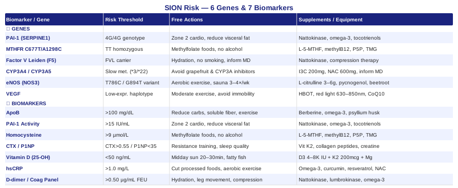
These tools don't replace medical care. But they can make you a more informed participant in your own treatment — which is often where the most meaningful gains are made.
7 Biomarkers to Track for Steroid-Induced Osteonecrosis
The mechanisms behind steroid-induced osteonecrosis are not a single pathway — they're a convergence. Elevated lipids cause fat emboli in bone sinusoids. Impaired coagulation leads to microvascular thrombosis. Reduced nitric oxide production compromises blood flow regulation. Suppressed bone formation prevents repair once ischemia sets in. Monitoring one marker tells only part of the story. The following seven biomarkers, tracked together, offer a comprehensive view of your current risk profile — and a set of modifiable targets.
Biomarker 1: ApoB (Apolipoprotein B)
Among all lipid-related tests, ApoB is the most actionable for vascular risk. It quantifies the total number of atherogenic lipoprotein particles circulating in the blood — each LDL, VLDL, and IDL particle carries exactly one ApoB molecule, so ApoB represents the true particle burden rather than just the average particle size or cholesterol content. Corticosteroids are well-established drivers of dyslipidemia: they raise LDL-cholesterol, VLDL, and triglycerides, and they reduce HDL. In the osteonecrosis context, the consequence is the fat embolism theory of SION — lipid-laden particles physically occlude the sinusoidal vessels feeding susceptible bone zones, most critically the femoral head, humeral head, and distal femur.
Peter Attia and Thomas Dayspring have made the case for ApoB as the lipid marker with the strongest predictive value for vascular events. In the osteonecrosis context, their argument applies with full force: ApoB load reflects the degree to which lipid emboli can accumulate in bone microcirculation — exactly the process that initiates ischemia.
How to Measure ApoB
ApoB is not included in a standard lipid panel and must be specifically requested. Cost: $20–$60 at most commercial labs. Optimal target: below 80 mg/dL — Peter Attia's threshold for low cardiovascular risk. For individuals on long-term corticosteroids, pursuing this target proactively is worthwhile rather than waiting for lipid complications to emerge.
If the Score Is Bad — The Plan Without Supplements
An ApoB above 100 mg/dL warrants lifestyle intervention before or alongside any supplement. Reduce refined carbohydrates, ultra-processed foods, and added sugars, which drive VLDL production and particle number. Increase soluble fiber to 10–15g per day — oats, psyllium husk, legumes, and vegetables — consistently shown in controlled trials to reduce LDL particle number. Prioritize 150 or more minutes per week of moderate aerobic activity. Discuss with your physician whether the timing and cumulative dose of corticosteroid therapy can be reduced — shorter courses and lower doses translate directly to less lipid disruption.
If the Score Is Bad — The Plan With Supplements or Equipment
Berberine (500mg 2–3 times per day with meals) activates AMPK and upregulates LDL receptor expression; randomized trials show ApoB and LDL reductions comparable to low-dose statins in some populations. Cycle 8 weeks on, 2 weeks off. Side effects include GI discomfort, particularly early in use. Psyllium husk (10–15g/day with plenty of water) reduces LDL-C by 5–10% with minimal side effects and is safe for continuous daily use. Omega-3 fatty acids (2–4g EPA/DHA per day) primarily target triglycerides — a major contributor to lipid emboli in SION — and at higher doses produce modest ApoB reduction; no cycling required; enteric-coated formulations address fish-burp side effects. Red yeast rice (standardized to 10mg monacolin K): this behaves like a natural lovastatin and should not be combined with prescription statins; discuss with your physician first.
Biomarker 2: PAI-1 (Plasminogen Activator Inhibitor-1)
PAI-1 is the primary inhibitor of tissue plasminogen activator (tPA), the enzyme responsible for dissolving blood clots. When PAI-1 is elevated, fibrinolysis is impaired: clots that form in small vessels persist rather than being cleared in the normal timeframe. Corticosteroids are known to raise PAI-1 levels, creating a prothrombotic environment that makes the already-fragile sinusoidal vessels feeding vulnerable bone zones particularly susceptible to permanent occlusion. Research by Glueck and colleagues, published across multiple studies in Clinical and Applied Thrombosis/Hemostasis, has linked elevated PAI-1 activity with increased osteonecrosis risk in corticosteroid-treated patients.
The PAI-1 4G/5G gene polymorphism (detailed in the genetics section) sets baseline PAI-1 production, but environmental and metabolic factors — visceral adiposity, insulin resistance, sedentary behavior — can push PAI-1 into dangerous ranges regardless of genotype.
How to Measure PAI-1
PAI-1 activity level is measured via a morning, fasting blood draw; the test requires careful, time-sensitive sample handling. Cost: $50–$150 at specialty or hospital labs. Target: PAI-1 activity below 15 IU/mL. PAI-1 antigen is also measurable and more analytically stable, but activity is more functionally informative.
If the Score Is Bad — The Plan Without Supplements
Aerobic exercise is the most powerful lifestyle reducer of PAI-1 activity — sustained zone 2 cardio (45–60 minutes, 4–5 days per week) consistently lowers PAI-1 activity across trials. Reducing visceral fat through diet and exercise compounds this benefit significantly. Low-carbohydrate dietary patterns have shown PAI-1 reductions in short-term studies. Prioritizing 7–9 hours of quality sleep reduces the morning PAI-1 surge, which is physiologically highest upon waking. Eliminating tobacco is essential, as smoking is a potent independent PAI-1 elevator.
If the Score Is Bad — The Plan With Supplements or Equipment
Nattokinase (2,000–4,000 FU per day between meals) is a fibrinolytic enzyme derived from fermented soybeans that directly promotes clot dissolution and has been shown in clinical studies to reduce PAI-1 activity; no cycling is required but reassess every 3 months; avoid if taking anticoagulants without physician co-management. Omega-3 fatty acids (2–4g EPA/DHA daily) reduce both PAI-1 and inflammatory endothelial activation. Tocotrienols (200–400mg/day of a mixed tocotrienol complex) have shown PAI-1-reducing effects in controlled studies; cycle 3 months on, 1 month off to assess individual response. Discuss low-dose aspirin with your physician as a complementary platelet-targeted measure where overall thrombotic risk is elevated.
Biomarker 3: Homocysteine
Homocysteine is a sulfur-containing amino acid produced during methionine metabolism. When its plasma level rises above 10 µmol/L, it directly damages the endothelium — the inner cellular lining of blood vessels. This damage reduces nitric oxide production, promotes platelet aggregation, and creates a prothrombotic and proatherosclerotic environment. In bone microvasculature, where vessels are narrow and already under multiple stressors from steroid therapy, elevated homocysteine adds a further ischemic burden that can tip tissue toward permanent infarction.
Corticosteroids indirectly raise homocysteine by depleting folate, B6, and B12 — the vitamins essential to homocysteine recycling. Patients with MTHFR gene variants are particularly exposed, as their capacity to process homocysteine is already enzymatically reduced.
How to Measure Homocysteine
Standard morning blood draw. Cost: $20–$60 at most commercial labs. Optimal target: below 9 µmol/L; individuals on long-term steroids with additional vascular risk factors should aim below 7 µmol/L. Test alongside vitamin B12 if the result is elevated to distinguish dietary deficiency from genetic impairment.
If the Score Is Bad — The Plan Without Supplements
Increase dietary methylfolate from whole food sources: spinach, romaine, arugula, liver, lentils, and asparagus. Increase B12 intake from meat, fish, eggs, and dairy — or address malabsorption if it is suspected, which is common in older individuals and in those on proton pump inhibitors or metformin. Reduce or eliminate alcohol, which depletes folate and impairs methionine recycling. Avoid synthetic folic acid (found in most commercial multivitamins), which is poorly converted by those with MTHFR variants and may compete with methylfolate for transport.
If the Score Is Bad — The Plan With Supplements or Equipment
Methylfolate (L-5-MTHF) at 400–800mcg per day is the active folate form that bypasses the MTHFR enzyme entirely; daily use without cycling; very safe, though start at the lower end if prone to methylation sensitivity (anxiety, irritability, sleep disruption at high doses). Methylcobalamin (B12) at 1,000–2,000mcg sublingual per day is the methyl form most directly usable in the methylation cycle; safe for long-term daily use. Pyridoxal-5-phosphate (P5P, active B6) at 25–50mg per day is an essential cofactor — do not exceed 100mg chronically due to peripheral neuropathy risk at high doses. TMG (trimethylglycine / betaine) at 1–3g per day provides methyl groups via the alternative BHMT remethylation pathway, particularly useful when homocysteine remains elevated despite B vitamin optimization; safe for long-term daily use, no cycling needed.
Biomarker 4: Bone Turnover Markers — CTX and P1NP
CTX (C-terminal telopeptide of type I collagen) is a marker of bone resorption — it rises when osteoclasts are actively breaking down bone matrix. P1NP (procollagen type I N-terminal propeptide) reflects bone formation — it rises when osteoblasts are synthesizing new matrix. In healthy bone, these markers are in physiological balance. Corticosteroids disrupt this balance severely: they suppress osteoblast activity (reducing P1NP) while initially promoting osteoclast survival, creating net bone loss. In established osteonecrosis, a critically suppressed P1NP signals that the repair process has stalled — dead bone is not being replaced, and structural collapse becomes increasingly likely.
Tracking both markers longitudinally during steroid therapy provides a direct, dynamic window into bone metabolism — information that no imaging study can offer until structural damage is already far advanced.
How to Measure CTX and P1NP
CTX requires a morning fasting blood draw; food intake substantially reduces CTX levels, making post-meal results unreliable. P1NP is more analytically stable and can be drawn at any time. Cost: $40–$100 each at most hospital or specialty labs. Targets vary by age and sex; broadly aim for CTX below 0.55 ng/mL and P1NP above 35 mcg/L in adults.
If the Score Is Bad — The Plan Without Supplements
Weight-bearing and resistance exercise are the most direct stimulants of bone formation available — mechanical load is the primary osteogenic signal to osteoblasts. Progressive resistance training 3–4 times per week targeting major muscle groups should be non-negotiable during and after steroid courses. Optimize sleep: growth hormone is released in pulses during slow-wave sleep and is a principal driver of P1NP elevation. Work with your physician to taper steroid doses as aggressively as clinically safe — the suppression of P1NP is dose-dependent.
If the Score Is Bad — The Plan With Supplements or Equipment
Vitamin K2 (MK-7) at 100–200mcg per day activates osteocalcin, the protein responsible for directing calcium into bone matrix rather than soft tissue; daily use; avoid if taking warfarin without physician guidance. Hydrolyzed collagen peptides at 10–15g per day (taken with vitamin C, which is required for collagen hydroxylation) stimulate P1NP directly and support extracellular matrix quality; no cycling needed; well-tolerated. Creatine monohydrate at 3–5g per day supports bone anabolism through both muscle activation and direct osteoblast effects; multiple randomized trials confirm bone marker benefits; safe for continuous daily use. Whole-body vibration therapy (25–50Hz, 10 minutes 3 times per week) has emerging RCT evidence for stimulating osteoblast activity in steroid-treated patients and is a practical at-home equipment option.
Biomarker 5: 25-OH Vitamin D
Vitamin D is far more than a supplement for bone density. As a steroid hormone, it regulates intestinal calcium absorption, immune function, inflammatory cytokine expression, RANKL/OPG signaling (the central axis controlling bone resorption), and vascular endothelial function. Corticosteroids impair vitamin D metabolism at multiple steps simultaneously — they reduce intestinal calcium absorption, increase urinary calcium excretion, and suppress the 1-alpha hydroxylase enzyme required to activate vitamin D in the kidney. Patients on long-term steroids are therefore disproportionately vitamin D insufficient, which amplifies bone loss, worsens inflammatory burden, and compromises the vascular environment in bone.
In SION specifically, vitamin D deficiency creates a permissive catabolic and inflammatory environment that accelerates ischemic zone expansion and impairs subsequent repair. The NIH Office of Dietary Supplements vitamin D factsheet provides comprehensive reference data on status, deficiency thresholds, and health implications.
How to Measure 25-OH Vitamin D
Standard blood test. Cost: $30–$80. Optimal target for individuals on corticosteroids: 50–80 ng/mL. Test alongside parathyroid hormone (PTH) — an elevated PTH confirms functional vitamin D insufficiency even when total 25-OH D appears borderline adequate.
If the Score Is Bad — The Plan Without Supplements
Regular midday sun exposure — arms and legs for 20–30 minutes at solar noon — is the most efficient non-supplemental source, though effectiveness varies significantly by latitude, season, cloud cover, and skin tone. Increase dietary sources: fatty fish (salmon, sardines, mackerel), egg yolks, beef liver, and mushrooms exposed to UV light. Address fat malabsorption if present — vitamin D is fat-soluble and requires adequate lipid digestion for absorption from food or capsules.
If the Score Is Bad — The Plan With Supplements or Equipment
Vitamin D3 at 4,000–8,000 IU per day for active repletion; retest at 3 months to adjust dose; target the 60–80 ng/mL range for those with active steroid exposure. Always co-administer with vitamin K2 (MK-7, 200mcg/day) — without K2, high-dose D3 can drive calcium toward soft tissue calcification rather than bone. Magnesium glycinate at 200–400mg per day in the evening; magnesium is an essential cofactor for vitamin D conversion to its active form 1,25-OH D and is frequently depleted in steroid-treated patients; generally well-tolerated, gentle on the GI tract compared to magnesium oxide. Monitor serum calcium if supplementing above 5,000 IU per day for extended periods.
Biomarker 6: hsCRP (High-Sensitivity C-Reactive Protein)
hsCRP is produced by the liver in response to systemic inflammatory signals, making it a sensitive and broadly integrative marker of whole-body inflammatory burden. This may seem counterintuitive in the osteonecrosis context — corticosteroids are anti-inflammatory drugs, after all. But many of the conditions requiring steroid treatment (lupus, vasculitis, inflammatory bowel disease, organ transplantation) are themselves associated with persistently elevated CRP despite steroid therapy. Furthermore, the ischemic and repair processes within osteonecrosis generate local inflammatory signals that contribute to systemic CRP.
Peter Attia uses hsCRP as one of the primary metabolic and vascular health signals in his clinical practice, arguing that chronic low-grade inflammation is a modifiable driver of most chronic disease progression. For SION, elevated hsCRP reflects endothelial activation, which worsens bone microvascularity, impairs the angiogenic repair response, and may accelerate joint destruction through inflammatory pathways.
How to Measure hsCRP
Standard blood test. Cost: $10–$40. Optimal target: below 1.0 mg/L; below 0.5 mg/L is ideal. Do not test within 72 hours of strenuous exercise, acute illness, dental procedures, or injury — CRP is transiently elevated by all of these and will give a false reading.
If the Score Is Bad — The Plan Without Supplements
Eliminate or sharply curtail ultra-processed foods, industrial seed oils (soybean, sunflower, corn), and refined sugars — these are among the most consistent dietary drivers of chronic elevated CRP. Increase fatty fish consumption to 3–4 servings per week. Consistent moderate aerobic exercise reduces baseline CRP by 20–30% across most intervention studies, though the effect accumulates over weeks to months of sustained practice. Evaluate and treat sleep disorders, particularly obstructive sleep apnea — an independent and potent driver of CRP elevation. Address chronic psychological stress: the cortisol dysregulation of chronic stress sustains inflammatory signaling simultaneously with the corticosteroid pathway.
If the Score Is Bad — The Plan With Supplements or Equipment
Omega-3 fatty acids (EPA/DHA) at 2–4g per day are among the most consistently supported CRP reducers in human clinical trials; safe for indefinite daily use; minor anticoagulant effect at higher doses. Curcumin in bioavailable form (liposomal or with piperine, 500–1,000mg per day) has robust anti-inflammatory evidence across multiple RCTs including in inflammatory conditions; cycle 3 months on, 1 month off to assess ongoing need; avoid with active gallbladder disease or bile duct obstruction. Resveratrol (250–500mg per day with a fatty meal for absorption) reduces NF-kB inflammatory pathway activity; moderate evidence; can be alternated with curcumin cycling rather than taken simultaneously. NAC (N-acetylcysteine) at 600–1,200mg per day reduces oxidative stress and inflammatory markers; particularly relevant given that steroid-induced oxidative stress contributes to both endothelial inflammation and impaired bone vascularity.
Biomarker 7: D-Dimer and Coagulation Panel
D-dimer is a fibrin degradation product generated when blood clots are broken down. Elevated D-dimer signals that the body is forming and dissolving clots more actively than baseline — a state that, in a patient on corticosteroids, indicates a hypercoagulable environment that places the sinusoidal vessels of bone under real and ongoing risk. A broader thrombophilia panel — including protein C, protein S, antithrombin III, lupus anticoagulant, and Factor V Leiden genotyping — identifies pre-existing coagulation abnormalities that can multiply SION risk multiplicatively.
Research by Glueck and colleagues, spanning multiple studies published in Clinical and Applied Thrombosis/Hemostasis, found that a substantial fraction of osteonecrosis patients had detectable thrombophilia that had never been investigated — because no one had tested for it. This is a diagnostic gap that remains underappreciated in routine clinical management.
How to Measure D-Dimer and Coagulation Markers
D-dimer: standard lab test, $40–$80. Full thrombophilia panel: $150–$400, often most appropriately ordered in collaboration with a hematologist who can interpret results in clinical context. Note: some panel components (protein C, protein S) must be timed relative to anticoagulant use and acute illness — confirm testing conditions with your physician. Normal D-dimer is generally below 0.50 µg/mL FEU, though reference ranges vary by laboratory.
If the Score Is Bad — The Plan Without Supplements
Aggressive hydration — dehydration increases blood viscosity and concentrates coagulation factors, amplifying thrombotic risk. Regular leg movement during periods of immobility: ankle pumps, walking, and standing breaks every 30–60 minutes. Compression stockings during prolonged sedentary periods or recovery phases. Strongly consider hematology referral — identified thrombophilia in a patient on corticosteroids may justify anticoagulant prophylaxis, which is a medical decision that supplements cannot substitute for.
If the Score Is Bad — The Plan With Supplements or Equipment
Nattokinase (2,000–4,000 FU per day between meals) directly promotes fibrinolysis and reduces clot formation; consult your physician before combining with anticoagulant medications. Lumbrokinase (20–40mg per day) is a fibrinolytic enzyme with emerging evidence in thrombotic conditions; use under physician guidance. High-dose EPA omega-3 (3–4g per day) exerts antiplatelet and anti-inflammatory effects well-supported in the literature. Ginkgo biloba (120–240mg per day of standardized extract) improves microcirculation and has mild antiplatelet effects; cycle 3 months on/off; avoid concurrent use with anticoagulants. Pneumatic compression devices for home use are underutilized in non-surgical osteonecrosis patients but have a rational basis for maintaining venous return and reducing microvascular stasis during recovery or reduced-mobility phases.
Understanding your biomarker profile is a powerful first step — but it becomes significantly more actionable when combined with knowledge of your genetic predispositions, which often explain why those markers are elevated in the first place.
The Genetics of Who Is Most Vulnerable — and What to Do About It
Why does one patient develop osteonecrosis after a brief steroid course while another tolerates decades of high-dose therapy without bone complications? Part of the answer lies in the genome. Several genetic variants have been linked to increased SION susceptibility in published human studies — most working through the same pathways covered in the biomarker section: coagulation, lipid metabolism, steroid clearance, vascular function, and bone angiogenesis. Knowing your variant status doesn't predict your fate, but it identifies where your biological vulnerabilities concentrate — and where targeted compensatory strategies are most worth deploying.
PAI-1 (SERPINE1) — The Fibrinolysis Gene
The SERPINE1 gene encodes PAI-1, and its 4G/5G promoter polymorphism is one of the most studied genetic contributors to thrombosis-related osteonecrosis. The 4G allele produces more PAI-1 protein than the 5G allele. People with the 4G/4G genotype have significantly elevated baseline PAI-1 activity — and under corticosteroid exposure, this elevation is compounded, creating a pronounced impairment of fibrinolysis. The combined carrier frequency of 4G/4G and 4G/5G genotypes is high in European populations (approximately 75%), making this a clinically relevant variant to investigate in any patient with recurrent thrombosis or osteonecrosis under steroid therapy.
The 4G/4G genotype has been specifically identified at higher frequency in steroid-induced osteonecrosis cohorts compared to steroid-treated control groups without osteonecrosis, suggesting that this variant contributes to SION risk above and beyond steroid dose alone.
If the Gene Is Bad — The Plan Without Supplements
Zone 2 aerobic exercise (45–60 minutes, 4–5 days per week) is the most reliable lifestyle reducer of PAI-1 activity — the effect accumulates with weeks of sustained practice. Reducing visceral adiposity through dietary and exercise intervention compounds this benefit. Minimize total corticosteroid exposure in consultation with your physician — cumulative dose and duration both directly affect PAI-1 elevation. Avoid tobacco and extended sedentary periods, both of which independently elevate PAI-1.
If the Gene Is Bad — The Plan With Supplements or Equipment
Nattokinase (2,000–4,000 FU/day), omega-3 fatty acids (2–4g EPA/DHA/day), and tocotrienol complex (200–400mg/day, cycled 3 months on/1 month off) provide synergistic fibrinolytic and anti-PAI-1 support. For patients with confirmed 4G/4G genotype facing surgeries or extended immobility under steroids, a proactive discussion with a hematologist about prophylactic anticoagulation is warranted — this is a risk level that supplements alone cannot adequately address. Compression therapy during all prolonged sedentary periods is a low-risk, practical complement.
MTHFR (C677T and A1298C) — The Methylation Gene
MTHFR encodes methylenetetrahydrofolate reductase, the enzyme responsible for converting dietary folate into the active methylfolate required for DNA methylation, neurotransmitter production, and — critically — homocysteine recycling. The C677T homozygous (TT) genotype, present in approximately 10–15% of people of European descent, reduces enzyme activity by 40–70%. The compound heterozygous C677T/A1298C combination also significantly impairs function. The downstream effect is elevated homocysteine, impaired methylation, and — in the osteonecrosis context — endothelial damage that erodes the vascularity of susceptible bone.
Gary Brecka, a human biologist who has brought significant public attention to MTHFR variants, argues that this is among the most common and underappreciated contributors to vascular vulnerability in the general population. While some of his claims extend beyond current evidence, the core connection between MTHFR, elevated homocysteine, and vascular disease is well-supported in the peer-reviewed literature. For osteonecrosis patients, identifying MTHFR status is one of the most actionable genetic tests available.
If the Gene Is Bad — The Plan Without Supplements
Maximize dietary methylfolate: leafy greens (spinach, arugula, romaine lettuce), liver, lentils, asparagus, and broccoli. Avoid synthetic folic acid entirely — it is poorly converted by impaired MTHFR and competes with methylfolate for cellular transport, potentially worsening functional folate status. Eliminate alcohol. Optimize B12 intake through animal-based whole foods. If gut issues are present, address them — B12 absorption depends critically on intrinsic factor and terminal ileum integrity, and any malabsorptive condition will compound the deficiency.
If the Gene Is Bad — The Plan With Supplements or Equipment
Methylfolate (L-5-MTHF) at 400–800mcg per day — the active form that completely bypasses the MTHFR enzyme; taken daily without cycling; start at the lower dose if prone to methylation overstimulation (anxiety, insomnia, restlessness at high doses). Methylcobalamin (B12) at 1,000–2,000mcg sublingual per day — the methyl form directly usable in the methylation cycle; safe for indefinite daily use. P5P (pyridoxal-5-phosphate) at 25–50mg per day — essential cofactor; do not exceed 100mg chronically. TMG (trimethylglycine) at 1–3g per day feeds the BHMT alternative remethylation pathway. Retest homocysteine at 3 months to confirm adequate response.
Factor V Leiden (F5 G1691A) — The Thrombophilia Gene
Factor V Leiden is the most prevalent inherited thrombophilia in populations of European descent, present in approximately 5% of individuals as heterozygous carriers. The G1691A mutation renders factor V resistant to inactivation by activated protein C — one of the body's primary anticoagulant mechanisms — leading to a persistently activated coagulation state. Heterozygous carriers face a 5–7 times elevated risk of venous thromboembolism. In the context of corticosteroid therapy, which independently elevates procoagulant factors and PAI-1, Factor V Leiden creates a multiplicative prothrombotic environment in bone sinusoids. Several case-control studies have identified Factor V Leiden at higher frequency in osteonecrosis patients compared to steroid-treated patients without osteonecrosis.
If the Gene Is Bad — The Plan Without Supplements
The most important action is to inform your physician of your carrier status before any corticosteroid course — this is directly actionable for risk stratification and may alter prophylactic strategy. Maintain aggressive hydration, avoid prolonged bed rest, and eliminate additional thrombotic triggers (smoking, combined oral contraceptives, dehydration, extended air travel during active steroid treatment). Request hematology co-management if undergoing surgery, major procedures, or extended steroid courses.
If the Gene Is Bad — The Plan With Supplements or Equipment
For Factor V Leiden carriers, supplement strategies are genuinely secondary to medical management. Nattokinase, high-dose omega-3s, and compression therapy provide modest additive risk reduction but are not adequate as standalone management when thrombotic risk is elevated. For carriers on active corticosteroid courses — particularly following surgery or during immobility — prophylactic low-molecular-weight heparin may be appropriate; this is a hematology decision. Do not attempt to self-manage identified thrombophilia with supplements alone when the risk profile is high.
CYP3A4 and CYP3A5 — The Steroid Metabolism Genes
CYP3A4 and CYP3A5 are the primary hepatic cytochrome P450 enzymes responsible for metabolizing most corticosteroids — including prednisone, dexamethasone, and methylprednisolone. Genetic variants that reduce enzyme activity ("slow metabolizer" phenotype) result in higher circulating steroid concentrations persisting for longer periods after each dose, even at the same prescribed dosage. This effectively means slow metabolizers receive a functionally higher corticosteroid exposure than the prescription suggests, amplifying every downstream risk: lipid elevation, PAI-1 induction, eNOS suppression, and bone formation inhibition.
CYP3A5*3 is the most common loss-of-function variant and is present in roughly 85% of individuals of European descent. The CYP3A4*22 variant further reduces enzyme expression in those who carry it. Pharmacogenomic testing panels — commercially available through multiple labs — can identify your complete CYP3A metabolizer phenotype.
If the Gene Is Bad — The Plan Without Supplements
Inform your prescribing physician of your CYP3A metabolizer status — this is the most directly actionable step, as dose adjustments based on pharmacogenomic data are increasingly standard of care. Avoid CYP3A4 inhibitors during steroid courses: grapefruit and grapefruit juice (substantial inhibitor), certain azole antifungals, macrolide antibiotics (clarithromycin, erythromycin), and high-dose piperine (found in some curcumin formulations) all reduce CYP3A4 clearance and can substantially increase effective steroid exposure.
If the Gene Is Bad — The Plan With Supplements or Equipment
Mild CYP3A4 induction from cruciferous vegetables and their derivative indole-3-carbinol (I3C) at 200–400mg per day may marginally increase steroid clearance, but this is a modest effect and does not substitute for physician-guided dose adjustment. Maintaining optimal liver metabolic health — avoiding excess alcohol, managing insulin resistance, supporting glutathione with NAC (600mg/day) — preserves CYP3A4 capacity. This gene variant primarily informs medical dosing decisions rather than supplement protocols.
eNOS (NOS3) — The Nitric Oxide Gene
Endothelial nitric oxide synthase (eNOS, encoded by NOS3) produces nitric oxide, the primary regulator of vascular tone and blood flow distribution in the endothelium. In bone, nitric oxide is essential for maintaining adequate perfusion in the sinusoidal vessels that supply osteocytes — without adequate NO signaling, these vessels cannot appropriately dilate in response to metabolic demand, rendering bone chronically underperfused under stress conditions. NOS3 polymorphisms — including T786C, G894T (Glu298Asp), and the intron 4a/b variant — reduce either eNOS expression levels or enzyme catalytic efficiency, leading to reduced NO bioavailability in the vascular endothelium.
Corticosteroids independently suppress eNOS expression and reduce NO bioavailability, largely through downregulation of eNOS mRNA. A patient carrying a low-activity NOS3 variant while on steroids faces compounded vascular insufficiency in bone — a combination that meaningfully increases ischemia risk in vulnerable zones.
If the Gene Is Bad — The Plan Without Supplements
Aerobic exercise is the most powerful known upregulator of eNOS expression in the endothelium — the shear stress imposed on vascular walls by increased blood flow during exercise drives eNOS transcription through mechanosensitive pathways. Even 20–30 minutes of brisk walking or cycling per day substantially increases baseline eNOS expression over 4–8 weeks. Heat exposure — traditional sauna (15–20 minutes at 80°C, 3–4 times per week) or hot water immersion — is an effective eNOS stimulator backed by growing human research, including from Finnish epidemiological data and RCTs from the cardiovascular field.
If the Gene Is Bad — The Plan With Supplements or Equipment
L-citrulline (3–6g per day) is more effective than L-arginine for sustained NO elevation because it bypasses intestinal arginase metabolism and maintains steady arginine supply to eNOS; monitor blood pressure as a functional proxy endpoint; safe for daily use without cycling. Pycnogenol (pine bark extract) at 100–150mg per day has human RCT evidence for enhancing eNOS activity and improving microvascular blood flow in multiple conditions; safe for long-term use; reassess every 4–6 months. Dietary nitrate from beetroot powder or juice (providing 400–500mg nitrate per day) supports the nitrate-nitrite-NO pathway as an eNOS-independent route to NO production — a useful complement particularly for low-NOS3-expression individuals. Far-infrared sauna devices for home use (45 minutes, 3–4 times per week) provide sustained thermal eNOS stimulation with a growing evidence base in cardiovascular applications.
VEGF — The Angiogenesis Gene
VEGF (vascular endothelial growth factor) is the master driver of angiogenesis — the formation of new blood vessels. In the context of osteonecrosis, VEGF is central to the repair process: it is needed to revascularize the ischemic bone zone, stimulate creeping substitution, and ultimately prevent irreversible structural collapse. Without adequate VEGF signaling, dead bone accumulates and progressive trabecular collapse becomes increasingly likely.
Promoter polymorphisms in the VEGF gene — particularly -634C/G, -2578C/A, and +936C/T — influence VEGF expression levels. Low-expression VEGF haplotypes have been identified in studies of patients with poor osteonecrosis repair outcomes. Corticosteroids themselves suppress VEGF expression, and a patient who also carries a low-expression VEGF variant faces doubly compromised angiogenic repair capacity — a particularly unfavorable combination.
If the Gene Is Bad — The Plan Without Supplements
Moderate, intermittent physical activity is the most studied lifestyle VEGF stimulator in skeletal tissue — the dose-response relationship matters here, as chronic endurance overtraining can paradoxically suppress bone VEGF, while moderate intermittent loading maintains it. Intermittent hypoxic exposure — altitude visits, altitude tents, or structured breathing protocols — is one of the most potent natural VEGF stimulators through HIF-1α activation, though access is limited for most people. Avoid prolonged immobility, which consistently reduces local VEGF expression in bone.
If the Gene Is Bad — The Plan With Supplements or Equipment
Hyperbaric oxygen therapy (HBOT) — 20–40 sessions at 2.0–2.5 ATA, 90 minutes per session — is supported by multiple clinical studies for osteonecrosis and works substantially through VEGF upregulation and bone angiogenesis stimulation. Cost is significant ($100–$300 per session), but the evidence for this application is among the strongest in the complementary field for osteonecrosis specifically. Red light and near-infrared photobiomodulation at 630–850nm has shown VEGF upregulation in cellular, animal, and some human bone healing studies; home panels ($200–$800) allow daily 10–20 minute sessions over affected joints. CoQ10 (ubiquinol) at 200–400mg per day supports mitochondrial function in endothelial cells and may indirectly support angiogenic capacity in ischemic tissue; safe for long-term daily use without cycling.
With the genetics and biomarker frameworks in place, it is worth zooming out to consider the broader metabolic and vascular health framework that ties these individual findings together into a unified preventive approach.
What Outlive by Peter Attia Teaches About Protecting Bone Vascularity
Peter Attia's Outlive: The Science and Art of Longevity does not mention steroid-induced osteonecrosis by name. But its central thesis — that most serious chronic disease should be anticipated and intercepted decades before clinical presentation, using personalized metabolic and vascular monitoring — applies directly to anyone managing corticosteroid therapy and trying to protect their bone health. The following ten principles from Outlive are the most directly actionable for SION risk management, drawing on the mechanisms already covered in this article.
1. ApoB Is the Lipid Number That Actually Predicts Vascular Occlusion
Attia argues, with strong literature support, that LDL-cholesterol is an imprecise proxy for particle-level cardiovascular and vascular risk. ApoB is the clinically actionable marker. In the osteonecrosis context, this applies to the microvasculature of bone just as much as to coronary arteries. Knowing your ApoB — and actively managing it during steroid courses — is one of the highest-yield interventions available.
2. Insulin Resistance Amplifies Every Vascular Risk Factor Simultaneously
Corticosteroids frequently cause or worsen insulin resistance, sometimes severely. Attia emphasizes that insulin resistance independently elevates ApoB, raises triglycerides, increases visceral fat, sustains inflammation, and drives PAI-1 upward — meaning all four SION pathways are amplified simultaneously by insulin resistance. Monitoring fasting insulin and HOMA-IR alongside the standard biomarkers discussed in this article adds a meaningful layer of insight.
3. Zone 2 Aerobic Training Is the Cornerstone of Metabolic Health
Attia's prescription of 45–60 minutes of zone 2 aerobic training (roughly 60–70% max heart rate, conversational pace) four to five times per week generates metabolic benefits that are comprehensive and synergistic: reduced ApoB, lower PAI-1, increased eNOS, improved insulin sensitivity, and reduced hsCRP. For SION risk management, this single exercise strategy touches nearly every biomarker discussed in this article simultaneously.
4. VO2 Max Is the Most Predictive Single Metric for Systemic Vascular Reserve
High VO2 max correlates with greater cardiac output, vascular efficiency, and tissue oxygen delivery — including in bone. Improving VO2 max through interval training effectively improves the entire perfusion system that bone sinusoids depend on. Even modest improvements from a low baseline are associated with significant reductions in all-cause mortality and vascular events in Attia's cited literature.
5. Resistance Training Preserves Bone Anabolism Through Mechanical Loading
Attia is unequivocal that resistance training is not optional for healthy longevity. For corticosteroid-treated patients, its necessity is compounded: the mechanical load of progressive resistance exercise is the most direct osteogenic stimulus available to counter steroid-suppressed osteoblast activity. Progressive resistance training three to four times per week is non-negotiable during and after active steroid courses.
6. Sleep Is When the Most Critical Cellular Repair Occurs
Growth hormone is pulsed primarily during slow-wave sleep — it is the primary driver of overnight bone formation (P1NP elevation) and cellular repair. Cortisol dysregulation from chronic poor sleep sustains inflammatory signaling and PAI-1 elevation. Attia frames 7–9 hours of quality sleep as a metabolic and repair necessity with measurable downstream effects on every biomarker covered in this article.
7. Inflammation Is a Modifiable Variable, Not a Fixed Background
Attia frames chronic low-grade inflammation — reflected by hsCRP — as one of the four core disease processes underlying most serious chronic illness. For SION, the argument is that hsCRP is a modifiable target, not simply an inevitable reflection of underlying disease. Diet, exercise, sleep, and targeted supplementation collectively reduce it.
8. Continuous Glucose Monitoring Reveals Metabolic Dynamics That Blood Tests Miss
A single fasting glucose or HbA1c misses the dynamic story of glucose handling. Wearing a CGM for two to four weeks during a corticosteroid course reveals the post-meal glucose and insulin spikes driven by steroid therapy — spikes that translate directly into triglyceride surges and inflammatory activation. This data allows for precise dietary adjustment that cannot be achieved through periodic lab draws alone.
9. Effective Prevention Begins Before the Disease Is Visible
Attia's Medicine 3.0 framework is explicit: the time to check ApoB, coagulation status, homocysteine, and vitamin D is before a steroid course begins — not after the first hip pain episode. Waiting for symptoms is waiting for late-stage disease. Identifying risk factors proactively allows intervention when the bone is still perfused and structurally intact.
10. Drug-Nutrient Interactions Are Routinely Underappreciated in Clinical Care
Corticosteroids deplete vitamins D, K2, C, B6, B12, and magnesium, and disrupt calcium metabolism, lipid handling, and glucose regulation simultaneously. Attia's principle of personalizing nutrition to account for known drug-nutrient interactions argues directly for systematic, proactive supplementation in long-term steroid users — not reactive replacement once clinical deficiency appears.
Complementary Approaches With Clinical Evidence
The following modalities have at least meaningful human clinical evidence relevant to osteonecrosis — either for the underlying pathophysiology or for the disease conditions that require corticosteroid treatment. None replace medical management, but several are well-supported enough to integrate as thoughtful additions to a comprehensive care plan.
Low-Level Laser Therapy and Photobiomodulation
Photobiomodulation (PBM) uses low-power red (630–700nm) and near-infrared (800–1,100nm) light to stimulate cytochrome c oxidase in mitochondria, increasing cellular ATP production and reducing oxidative stress. In bone, this translates to enhanced osteoblast activity, reduced pro-inflammatory cytokine expression (including IL-1β and TNF-α), and — directly relevant to SION — upregulation of VEGF and improved local microvascular blood flow. The mechanistic rationale for PBM in osteonecrosis is therefore specific and well-grounded.
The most substantial published human evidence comes from medication-related osteonecrosis of the jaw (MRONJ), where PBM has been compared to surgical treatment in multiple trials. A systematic review published in Lasers in Medical Science found consistent evidence for improved healing outcomes and reduced necrotic tissue extent with PBM application. While evidence specifically for steroid-induced femoral head osteonecrosis is more limited, the mechanism is directly transferable. Specific protocol: 830nm near-infrared LED or laser, 30–50 mW/cm², 10–20 minutes per session over the affected joint, 4–5 times per week for a minimum of 8–12 weeks. Home-grade LED panels ($200–$800) are increasingly available and practical. No serious adverse effects are associated with these parameters; avoid direct exposure to the eyes.
Chinese Herbal Medicine
Several Chinese herbal formulas have been studied in randomized controlled trials specifically for steroid-induced osteonecrosis of the femoral head — a condition where traditional Chinese medicine has maintained a clinical tradition for decades. The most-studied formulas combine herbs with anti-inflammatory, pro-angiogenic, and anticoagulant properties. Xianlinggubao (仙灵骨葆), a standardized formula, has been evaluated in multiple trials for its effects on bone turnover markers and Harris Hip Scores in SION. Yishen Huoxue (益肾活血) decoctions targeting kidney-deficiency and blood-stasis patterns have also accumulated a clinical trial base.
A meta-analysis reviewing randomized controlled trials of Chinese herbal medicine for steroid-induced femoral head osteonecrosis found improved functional outcomes (Harris Hip Scores) and reduced rates of femoral head collapse compared to control groups receiving conventional management alone, with acceptable safety profiles across the included trials. The principal limitation is heterogeneity of formulas and treatment protocols across studies. Practical application: formulas must be prescribed by a licensed practitioner of traditional Chinese medicine with specific dosing and formula selection tailored to the patient's full clinical presentation. Standardized proprietary products without practitioner guidance are not recommended for this condition due to the complexity of appropriate formula selection.
Tai Chi
Tai chi is a mind-body practice involving slow, deliberate, flowing movement patterns that maintain joint range of motion, balance, and proprioception without imposing high-impact compressive loading on vulnerable joints. For osteonecrosis patients — where pain, functional limitation, and the need to protect against femoral head collapse under loading are significant constraints — tai chi offers a clinically realistic way to maintain mobility, muscle activation, and neuromuscular coordination without the impact forces that could accelerate structural damage.
A randomized controlled trial published in Complementary Therapies in Medicine examining tai chi practice in patients with hip osteonecrosis demonstrated significant improvements in self-reported pain scores, functional mobility, and quality of life over a 12-week intervention period compared to a wait-list control group. Effect sizes were modest, consistent with the nature of a movement-based adjunct rather than a primary treatment. Practical protocol: two to three 60-minute tai chi sessions per week, either in group classes or through structured video-based programs; if beginning with established osteonecrosis, working initially with a qualified instructor ensures appropriate weight distribution and avoidance of loading patterns that stress the compromised femoral head. As a non-pharmacological movement approach, tai chi carries minimal risk for this population and can be maintained indefinitely as part of an ongoing functional maintenance strategy.
Mindfulness Meditation and MBSR
The connection between structured mindfulness practice and osteonecrosis is mechanistically indirect but biologically coherent. Chronic psychological stress elevates endogenous cortisol, which synergizes with exogenous corticosteroid therapy to further suppress osteoblast activity, raise hsCRP, and sustain the HPA-axis dysregulation that long-term steroid treatment already creates. Managing the psychological burden of a chronic inflammatory condition requiring ongoing steroids — and the added burden of a painful musculoskeletal complication — is not peripheral to the biology; it directly influences the hormonal environment in which bone damage occurs.
Mindfulness-Based Stress Reduction (MBSR), the 8-week structured program developed by Jon Kabat-Zinn at the University of Massachusetts, has been evaluated in multiple RCTs in chronic illness populations. A landmark study published in JAMA Internal Medicine demonstrated that MBSR reduced markers of systemic inflammation, including CRP, in adults with diagnosed inflammatory conditions — an effect that persisted beyond the 8-week intervention. For SION patients managing the intersection of steroid therapy and bone disease, the cortisol-lowering, hsCRP-reducing effects of MBSR represent a meaningful additive contribution. Practical protocol: the standard MBSR program involves eight weekly group sessions of approximately 2.5 hours plus 45 minutes of daily home practice; free and low-cost versions are available through academic programs including the UCLA Mindful Awareness Research Center and equivalent institutions, making this among the most accessible interventions in this article.
Conclusion
Steroid-induced osteonecrosis is a serious complication, but it is not random and it is not without measurable precursors. The biological mechanisms — lipid embolism, microvascular thrombosis, suppressed nitric oxide production, impaired bone repair — leave traces in the blood long before structural damage becomes visible on imaging. The genetic variants that amplify those mechanisms are increasingly accessible through commercial pharmacogenomic testing. Neither area requires exotic diagnostics or esoteric interventions — both connect to practical, measurable, and often modifiable targets.
The most important next step is straightforward: get your biomarkers measured. ApoB, PAI-1, homocysteine, CTX, P1NP, vitamin D, hsCRP, and a coagulation panel if your clinical history warrants it. Review those numbers in light of what this article has outlined. If you are on long-term corticosteroids, have this conversation proactively with your physician — ideally including rheumatology and, if coagulation abnormalities emerge, hematology. The goal is not to replace medical management with supplements, but to understand precisely where your biological vulnerabilities concentrate and address those specifically. Better information, applied thoughtfully and in partnership with qualified clinicians, does lead to better decisions.
Musculoskeletal: Bone Conditions Joint Conditions
Cardiovascular: Vascular Conditions
Autoimmune: Inflammatory Conditions