This article contains AI generated content.
Traumatic Neuroma of the Knee: 4 Genes and 6 Biomarkers to Track
Introduction
A neuroma after knee surgery or trauma doesn't behave like ordinary post-operative soreness. It burns, it buzzes, it flares when you least expect it, and it often sits in a small, specific spot — over the infrapatellar branch of the saphenous nerve, along an old incision, or near a portal scar — while the rest of the knee has otherwise healed. If you've been told to "give it more time" or to just "manage the pain," you already know that advice like this rarely matches what you're actually feeling.
Generic guidance struggles here because a traumatic neuroma isn't one thing. It's the result of a nerve that was cut, stretched, or compressed, followed by a regeneration process that went slightly wrong — axons trying to reconnect to their distal stump, failing, and tangling into a small, disorganized, pain-generating mass. What determines whether that regrowth resolves quietly or turns into years of localized neuropathic pain isn't fully within your control, but it also isn't a mystery. It involves measurable biology: inflammatory signaling, nerve growth factor activity, micronutrient status, and to a lesser extent, inherited differences in how your nervous system processes pain.
This article looks at that biology directly. Instead of repeating "avoid pressure on the area" and "try physical therapy," it walks through the blood tests and functional markers that can tell you whether your body's healing environment is working for you or against you, and it explains the genetic variants that may shape how intensely you experience nerve pain in the first place. Neither is a diagnostic shortcut — nerve conduction studies, ultrasound, and a skilled clinician's exam remain central to actually diagnosing and managing a neuroma. But the data underneath those exams is where informed decisions get made.
None of this promises a cure, and anyone offering one for a structural nerve problem should be treated with skepticism. What better information does offer is a narrower, more useful set of questions to bring to your next appointment. The sections ahead cover the biomarkers most worth tracking, the genetic variants with the clearest human evidence, a research-backed perspective on pain from outside conventional orthopedics, and a short review of complementary approaches with real trial data behind them — each one meant to replace vague hope with a more grounded, workable plan.
Summary
Before you dive in: a traumatic knee neuroma is driven less by "damage that hasn't healed" and more by an active biological process — elevated nerve growth factor at the injury site, ongoing low-grade inflammation, and (in some people) a nervous system that's genetically wired to amplify pain signals more than average. This article breaks down the six blood-based and functional markers worth checking first — from a $15 inflammation test to a specialized nerve-damage marker most doctors have never ordered — and explains exactly what a "bad" result means and how to respond to it, with and without supplements. It then covers four gene variants (including the sodium-channel gene behind some of the most extreme pain disorders ever studied) that may explain why two people with an identical surgical injury end up with very different pain experiences. Beyond that, you'll find a research-backed pain-science perspective drawn from clinical pain specialists, and a review of the complementary therapies — including one with a documented "number needed to treat" of roughly 2 — that have actual clinical trial evidence behind them for nerve pain, not just general wellness claims.
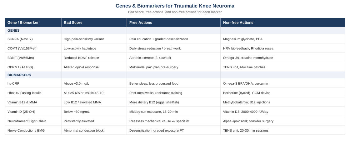
6 Biomarkers Worth Tracking After a Knee Nerve Injury
Before looking at genetics, it helps to look at what's actually measurable right now, because biomarkers reflect your current biological state rather than a fixed inherited risk. Peter Attia, Thomas Dayspring, and Allan Sniderman have each argued, in the context of cardiometabolic disease, that generic reference ranges often miss the person in front of you — the same logic applies to nerve injury recovery. A "normal" lab value on a standard panel can still be suboptimal for someone actively trying to quiet an irritated nerve. The six markers below combine affordable, widely available tests with a couple of more specialized ones, in roughly the order most clinicians would prioritize.
1. High-Sensitivity C-Reactive Protein (hs-CRP)
hs-CRP is a general marker of systemic low-grade inflammation, produced by the liver in response to interleukin-6 signaling. It doesn't diagnose a neuroma, but chronically elevated hs-CRP indicates a body-wide inflammatory tone that can keep an irritated nerve ending sensitized longer than it otherwise would be. This is the same marker Peter Attia frequently references as underused outside of cardiology, and its relevance extends to soft-tissue and nerve healing environments, not just vascular risk.
How to Measure It
A simple blood draw, widely available through primary care or direct-to-consumer lab services. Cost typically runs $15 to $40 without insurance, and it's often bundled into routine metabolic panels at no extra charge.
If the Score Is Bad: The Plan Without Supplements
An hs-CRP above roughly 3.0 mg/L warrants a look at sleep debt, visceral fat, alcohol intake, and unaddressed dental or gum inflammation, all of which independently raise CRP. Prioritizing 7 to 9 hours of consistent sleep and reducing ultra-processed food intake for 8 to 12 weeks, then retesting, is a reasonable first step with no cost and no side effects beyond the general effort of habit change.
If the Score Is Bad: The Plan With Supplements or Equipment
Omega-3 fatty acids (EPA/DHA, 2 to 3 grams daily with food) have the best human evidence for modestly lowering hs-CRP over 8 to 12 weeks; cycling isn't necessary, but doses above 3 g/day can increase bleeding risk and should be discussed with a physician if you're on anticoagulants. Curcumin with piperine (500 to 1000 mg daily) has more modest, mixed evidence for inflammation reduction and can cause GI upset or interact with blood thinners at higher doses.
2. HbA1c and Fasting Insulin
Glycemic control matters for nerve healing well below the threshold of diagnosed diabetes. Elevated HbA1c and fasting insulin are associated with slower peripheral nerve conduction and higher neuropathy risk even in people classified as "pre-diabetic," which is directly relevant when a traumatized nerve is trying to regenerate a healthy myelin sheath.
How to Measure It
Standard blood draw; HbA1c costs $15 to $40, fasting insulin $20 to $50. Both are frequently included in comprehensive metabolic and diabetes-screening panels.
If the Score Is Bad: The Plan Without Supplements
An HbA1c above 5.6% or fasting insulin above roughly 8 to 10 μIU/mL responds reliably to a 20 to 30 minute daily walk after meals, resistance training twice weekly, and reducing refined carbohydrate load at the meal closest to inactivity (usually dinner). This is a slow lever — recheck at 12 weeks, not sooner.
If the Score Is Bad: The Plan With Supplements or Equipment
Berberine (500 mg, two to three times daily with meals) has trial evidence comparable to metformin for insulin sensitivity, but should be cycled — commonly 8 to 12 weeks on, then a break — and can cause GI cramping or interact with other glucose-lowering medications. A continuous glucose monitor (roughly $50 to $100 per month) gives real-time feedback on which specific foods spike your glucose, which is often more actionable than the supplement itself.
3. Vitamin B12 and Methylmalonic Acid (MMA)
Vitamin B12 supports myelin maintenance and Schwann cell function, and human and animal data both show that B12 promotes remyelination and improves nerve conduction velocity after injury, as documented in research on vitamin B12 and peripheral nerve regeneration. Serum B12 alone can be misleading, since levels can appear normal while functional deficiency is present — pairing it with methylmalonic acid (MMA), which rises specifically when B12 is functionally low, gives a clearer picture.
How to Measure It
Blood draw; B12 costs $30 to $50, MMA adds another $40 to $80. Order them together if you have any history of reduced meat intake, metformin use, or GI absorption issues.
If the Score Is Bad: The Plan Without Supplements
Increasing dietary sources — eggs, shellfish, organ meats, dairy — over 8 to 12 weeks can correct mild insufficiency in people without an absorption problem. This won't help if the issue is absorption (common after certain GI surgeries or with long-term PPI use), which is why retesting matters.
If the Score Is Bad: The Plan With Supplements or Equipment
Methylcobalamin, 1000 mcg daily sublingually, is well tolerated with essentially no toxicity ceiling since excess is renally cleared; for documented malabsorption, monthly intramuscular B12 injections are more reliable. No cycling is needed — this is a nutrient replacement, not a hormetic stressor. Side effects are rare but can include mild acne in sensitive individuals at high doses.
4. Vitamin D (25-OH)
Sensory neurons express vitamin D receptors, and clinical trial data shows vitamin D3 supplementation reduces neuropathic pain scores compared to placebo, likely through modulation of oxidative and inflammatory pathways in pain-signaling pathways. This is one of the more actionable markers on this list given how common insufficiency is.
How to Measure It
25-hydroxyvitamin D blood test, $40 to $80, widely available and often covered by insurance when ordered alongside a metabolic panel.
If the Score Is Bad: The Plan Without Supplements
15 to 20 minutes of midday sun exposure on bare arms and legs, several times weekly, can meaningfully raise levels in lighter-skinned individuals over 2 to 3 months, though this is seasonal and latitude-dependent and unreliable as a sole strategy in winter months or northern regions.
If the Score Is Bad: The Plan With Supplements or Equipment
Vitamin D3, 2000 to 4000 IU daily with a fat-containing meal, taken continuously rather than cycled, with retesting at 3 months to confirm you're in range (commonly targeted at 40 to 60 ng/mL) rather than overshooting. Doses above 10,000 IU/day sustained for months can cause hypercalcemia, so self-escalating beyond the standard range without retesting is not advisable.
5. Neurofilament Light Chain (NfL)
This is the most specialized marker on the list and the one most clinicians won't think to order. NfL is a structural protein released into blood when large myelinated axons are actively damaged, and it's increasingly used as a biomarker of ongoing neuroaxonal injury in peripheral nerve conditions, detected via ultra-sensitive single-molecule array (Simoa) assays. For a knee neuroma, a persistently elevated NfL could suggest ongoing axonal injury rather than a settled, stable scar — information that may support a more assertive treatment conversation with a surgeon or pain specialist.
How to Measure It
Not yet a routine order; available through specialty and research-oriented labs (such as those used in multiple sclerosis and neurodegeneration monitoring) for roughly $100 to $300. Ask a neurologist or physiatrist whether it's accessible in your region — this is genuinely a frontier test, not a mainstream one.
If the Score Is Bad: The Plan Without Supplements
There's no lifestyle lever that directly lowers NfL — an elevated result is a signal to revisit the mechanical situation itself: whether the nerve is still being compressed by scar tissue, hardware, or a brace, and whether imaging or a repeat nerve conduction study is warranted. This is a "go back to the specialist" marker more than a "change your habits" marker.
If the Score Is Bad: The Plan With Supplements or Equipment
Because NfL reflects active axonal damage rather than a modifiable metabolic state, the most relevant intervention is typically procedural (neuroma excision with nerve capping or targeted muscle reinnervation, desensitization protocols, or a nerve block) rather than a supplement. Alpha-lipoic acid (600 mg daily) has separate evidence for reducing neuropathic symptoms and may be a reasonable adjunct, generally well tolerated, though it can occasionally cause mild GI discomfort and should be used cautiously alongside diabetes medications due to added glucose-lowering effect.
6. Nerve Conduction Study / EMG
Not a blood biomarker, but a functional one, and arguably the single most clinically useful data point for a suspected neuroma. It measures how well electrical signals travel along the affected nerve and can help localize the injury, distinguish a neuroma from more diffuse neuropathy, and provide an objective baseline to track against future testing.
How to Measure It
Performed by a neurologist or physiatrist; cost ranges from roughly $300 to $1,500 depending on region and insurance coverage, and it's frequently covered when ordered for a documented post-surgical or post-traumatic complaint.
If the Score Is Bad: The Plan Without Supplements
An abnormal result pointing to a localized conduction block supports pursuing desensitization therapy, activity modification around the affected area, and a structured graded exposure program with a hand or physical therapist trained in peripheral nerve injuries — not a passive "wait and see" approach.
If the Score Is Bad: The Plan With Supplements or Equipment
A TENS (transcutaneous electrical nerve stimulation) unit, used 20 to 30 minutes per session, up to twice daily, can meaningfully reduce localized neuropathic pain for many patients with minimal side effects (occasional skin irritation at electrode sites); it should be trialed for 4 to 6 weeks to judge effect before deciding whether it's worth keeping in a long-term routine.
Blood work and electrophysiology tell you about your current biological terrain, but they don't explain why two people with an almost identical surgical injury can end up with completely different pain trajectories. That gap is where genetics offers a partial, still-developing explanation.
What Your Genes May Be Telling You About Nerve Pain Sensitivity
Genetic research on traumatic neuromas specifically is thin — there isn't a "neuroma gene" with strong human trial data. What does have solid human evidence is a small set of genes that shape how intensely the nervous system processes and amplifies pain signals in general, which indirectly affects how a knee neuroma is experienced. Researchers like Ali Torkamani, who has written extensively on using genomic data to inform personalized health decisions, and clinicians like Gary Brecka, who popularized accessible genetic and biomarker panels for identifying modifiable health risks, are a reasonable starting point if you want to explore your own raw genetic data rather than relying on population averages.
SCN9A (Nav1.7 Sodium Channel)
SCN9A encodes the Nav1.7 sodium channel, and its role in human pain perception is unusually well established — loss-of-function mutations cause congenital insensitivity to pain, while gain-of-function variants cause some of the most severe inherited pain disorders known to medicine, including erythromelalgia and paroxysmal extreme pain disorder. Common variants short of these rare disease-causing mutations are associated with differences in everyday pain sensitivity, which is relevant to how intensely a neuroma's discharge is perceived, though this is generalized pain-sensitivity evidence rather than neuroma-specific data.
If the Gene Variant Suggests High Pain Sensitivity: The Plan Without Supplements
Pain neuroscience education — understanding that heightened nerve signaling doesn't equal ongoing tissue damage — combined with graded, structured exposure to light touch and pressure over the affected area (desensitization) has direct evidence for retraining an oversensitized system. This costs nothing beyond a referral to a physical or occupational therapist familiar with nerve injuries, done in short daily sessions of a few minutes, building gradually over 4 to 8 weeks.
If the Gene Variant Suggests High Pain Sensitivity: The Plan With Supplements or Equipment
Magnesium glycinate (200 to 400 mg nightly) has a plausible mechanism for reducing neuronal excitability and is generally well tolerated, with loose stools being the main side effect at higher doses; no strict cycling is required, though periodic breaks (one week off every couple of months) are a reasonable precaution against dependency on any single input. Palmitoylethanolamide (PEA), 300 to 600 mg twice daily, has emerging human trial evidence in neuropathic pain conditions and a favorable safety profile, typically trialed for 8 weeks before assessing benefit.
COMT (Catechol-O-Methyltransferase)
COMT breaks down catecholamines involved in pain modulation, and specific low-activity haplotypes (built around the Val158Met variant) are consistently associated with higher experimental pain sensitivity and higher risk of developing chronic musculoskeletal pain, with the clearest evidence coming from fibromyalgia and postoperative pain cohorts. This is a moderately well-replicated finding, though it reflects general pain amplification tendency rather than anything specific to neuroma tissue itself.
If the Gene Variant Suggests Low COMT Activity: The Plan Without Supplements
Because low COMT activity interacts strongly with psychological stress to amplify pain, structured stress-reduction practice (even 10 minutes of daily breathwork or a short walk without a phone) has a plausible, low-cost effect on the pathway this gene affects. Consistency matters more than intensity — daily practice for 8 weeks before judging results.
If the Gene Variant Suggests Low COMT Activity: The Plan With Supplements or Equipment
There's no supplement that reliably raises COMT activity, so the more relevant tool here is behavioral: a biofeedback or heart-rate-variability device (many wearables now include this) to build objective awareness of your stress response in real time, used for a few minutes daily. Rhodiola rosea (200 to 400 mg in the morning) has some evidence for stress-response modulation and is generally well tolerated, though it should be cycled — commonly 6 to 8 weeks on, 2 weeks off — since tolerance can build, and it may cause mild jitteriness in sensitive individuals.
BDNF Val66Met
Brain-derived neurotrophic factor supports neuroplasticity and peripheral nerve regeneration. The Val66Met variant reduces activity-dependent BDNF release, and while most of the strongest human data comes from mood and cognitive research rather than peripheral nerve injury specifically, the mechanistic link to slower or less complete neural remodeling after injury is biologically plausible and an area of early, ongoing investigation rather than settled fact.
If the Gene Variant Suggests Reduced BDNF Signaling: The Plan Without Supplements
Aerobic exercise is the single most consistent, well-evidenced way to raise BDNF levels in humans. Three to four sessions of 30 minutes at a moderate-to-vigorous intensity per week, sustained over months, is the dose most associated with meaningful increases — and it's free.
If the Gene Variant Suggests Reduced BDNF Signaling: The Plan With Supplements or Equipment
Omega-3s (already discussed above) have secondary evidence for supporting BDNF signaling alongside their anti-inflammatory effect. Creatine monohydrate (3 to 5 g daily, no cycling needed) has preliminary evidence for supporting neural energy metabolism broadly; it's one of the more extensively safety-tested supplements available, with mild water retention being the most common side effect.
OPRM1 (Mu-Opioid Receptor Gene)
The A118G variant in OPRM1 affects opioid receptor binding and is associated with differences in opioid analgesic requirements and pain response after surgery. This matters less for neuroma formation itself and more for how postoperative pain management should be approached — some carriers may need higher opioid doses for equivalent relief, which has implications for a more deliberate, non-opioid-forward pain plan from the outset.
If the Gene Variant Suggests Altered Opioid Response: The Plan Without Supplements
Discussing multimodal pain control with a surgeon or pain specialist before any procedure — nerve blocks, scheduled non-opioid analgesics, and desensitization started early — reduces reliance on opioid dose escalation regardless of genetic variant, and costs nothing beyond that conversation.
If the Gene Variant Suggests Altered Opioid Response: The Plan With Supplements or Equipment
A TENS unit (described above) and topical lidocaine patches (available over the counter in low concentrations, prescription-strength through a physician) provide localized analgesia without systemic opioid exposure, used as needed with minimal systemic side effects — occasional local skin irritation being the main concern.
Genetics and lab values describe the biological substrate, but a growing body of pain-science research argues that the story doesn't end there — how the brain interprets nerve signals matters just as much, which is where the next perspective comes in.
A Pain-Science Perspective Worth Understanding
Andrew Huberman's podcast conversation with Stanford pain specialist Dr. Sean Mackey, Understanding and Conquering Pain, is one of the more useful long-form resources on chronic and neuropathic pain outside of a formal medical textbook, precisely because it challenges the simple "damage equals pain" model that still dominates a lot of orthopedic conversation. It's especially relevant to a traumatic neuroma, where structural healing and pain intensity frequently diverge — some people with clear nerve injury on imaging have modest pain, others with subtle findings have severe pain. Below are ten of the most useful, evidence-grounded points from that body of work.
1. Pain Is Constructed by the Brain, Not Just Transmitted by It
Nociceptive signals from an injured nerve are real, but the brain integrates them with expectation, mood, prior experience, and context before producing the sensation of pain. This isn't a claim that pain is "in your head" in a dismissive sense — it's a claim that the brain is an active participant in how much a given signal hurts, which is why identical injuries produce different pain experiences.
2. Chronic Pain Can Persist After Tissue Has Healed
Once a pain pathway has been active long enough, the nervous system can become sensitized independent of ongoing tissue damage — a phenomenon called central sensitization. For a neuroma, this means pain intensity months out doesn't always track cleanly with how "damaged" the nerve currently is, which is part of why purely structural fixes don't always resolve pain on their own.
3. Attention Amplifies Pain Signals
The more attention directed at a painful area, the larger the perceived signal tends to become — this is a documented feature of the attentional gating of pain, not a character flaw. Practical implication: engagement in absorbing, meaningful activity is a legitimate pain management tool, not a distraction trick.
4. Fear of Movement Can Worsen Long-Term Outcomes
Avoiding all activity near an injured nerve out of fear of triggering pain (kinesiophobia) is associated with worse long-term function. Controlled, graded reintroduction of movement — exactly what desensitization protocols are built around — tends to outperform strict avoidance.
5. Sleep Deprivation Directly Lowers Pain Thresholds
A single night of poor sleep measurably increases next-day pain sensitivity. For anyone managing a neuroma, sleep isn't a peripheral wellness recommendation — it's a direct lever on how the pain will feel the next day.
6. Deep, Slow Breathing Has a Measurable Analgesic Effect
Structured slow-breathing practices engage the parasympathetic nervous system and have been shown to reduce pain ratings in controlled settings. It's a zero-cost, zero-side-effect tool that's easy to underrate.
7. Cold and Heat Exposure Modulate Pain Through Distinct Pathways
Cold tends to blunt acute nociceptive signaling locally, while heat can reduce muscle guarding around a painful area — both are legitimate, low-cost adjuncts, though neither addresses the underlying nerve injury itself.
8. Belief and Expectation Change Measurable Pain Outcomes
Expectation of relief activates real, measurable descending pain-inhibition pathways in the brain (part of the mechanism behind placebo analgesia). This isn't an argument for false hope — it's a reason why working with a clinician you trust, who explains what's happening clearly, has a documented physiological benefit beyond the specific treatment offered.
9. Exercise Produces Its Own Analgesic Effect
Moderate aerobic exercise triggers exercise-induced hypoalgesia, a short-term reduction in pain sensitivity through endogenous opioid and endocannabinoid pathways. For a knee neuroma, this supports maintaining general fitness (cycling, swimming, walking) even while local desensitization work continues.
10. Pain Education Alone Changes Outcomes
Simply understanding the biology of pain — that heightened signaling doesn't always mean worsening damage — has been shown in clinical trials to reduce pain and disability scores on its own. This is arguably the single most underused, zero-risk intervention available to anyone dealing with a persistent nerve pain problem.
These pain-science principles pair naturally with a set of hands-on, body-based therapies that have their own direct clinical evidence in nerve pain populations.
Complementary Approaches With Real Evidence Behind Them
The following four approaches were chosen because each has direct or closely related human clinical evidence for neuropathic or post-surgical pain, not just general relaxation claims. None should replace a structured medical evaluation of the neuroma itself — think of them as evidence-supported additions to, not substitutes for, that care.
Low-Level Laser Therapy / Photobiomodulation
Photobiomodulation uses low-intensity red or near-infrared light to reduce inflammation and modulate nerve signaling at the cellular level, and it's one of the more mechanistically direct options for a localized neuroma given its documented effects on glial cell activity and pro-inflammatory cytokine reduction in neuropathic pain models.
A commonly studied protocol uses near-infrared wavelengths delivered at higher irradiance for rapid pain-signal blocking, followed by lower-irradiance sessions for sustained relief; the mechanistic basis for this near-infrared approach to neuropathic pain has been described in detail in peer-reviewed research, as summarized in this analysis of photobiomodulation therapy for neuropathic pain.
In practice, this means seeking out a physical therapist or pain clinic with a class III or IV therapeutic laser device (home-use low-power devices exist but have far less direct evidence at effective doses), applying it directly over the site of the neuroma, typically two to three times weekly for several weeks, then reassessing — treating it as a trial rather than an assumed fix, since study designs and outcomes in this area are still notably heterogeneous.
Guided Imagery and Graded Motor Imagery
Guided imagery, and specifically the graded motor imagery (GMI) protocol, is directly relevant to nerve injury pain because it retrains the brain's distorted representation of the affected limb without requiring direct physical contact with a hypersensitive area — a useful feature when the neuroma site is too painful to touch early in recovery.
The original and most influential trial, led by Lorimer Moseley, tested a sequential program of limb laterality recognition, imagined movements, and mirror therapy in patients with complex regional pain syndrome and related neuropathic conditions, finding a roughly 25-point improvement on the Neuropathic Pain Scale and a number needed to treat of about 2 for a 50% pain reduction — a notably strong effect size for a non-invasive intervention, as reported in this randomized controlled trial of graded motor imagery for pathologic pain.
For a knee neuroma, this can be adapted with the help of a physical or occupational therapist trained in the protocol: roughly two weeks each of laterality recognition drills, imagined knee movements, and finally mirror-assisted movement, done in short daily sessions before progressing to direct desensitization work on the area itself.
Mindfulness Meditation / MBSR
Mindfulness-Based Stress Reduction doesn't reduce nerve signaling directly, but it changes the relationship between attention, stress reactivity, and perceived pain intensity — directly relevant given how strongly attention and stress amplify neuropathic pain, as discussed above.
The most rigorous test of this came from a large randomized trial in chronic low back pain, where an 8-week MBSR program produced improvements in pain and function comparable to cognitive behavioral therapy and superior to usual care; a detailed companion analysis of that same trial cohort is available in this study on MBSR and CBT effects on pain-related catastrophizing and self-efficacy. Evidence specific to post-surgical knee neuroma pain is limited, so this should be understood as strong general chronic-pain evidence applied by extension.
A standard 8-week MBSR course (widely available in person and online, often $200 to $500, though some hospital systems offer it at low or no cost) with daily home practice of 20 to 30 minutes is the format used in most supporting trials — shorter, informal "mindfulness app" use has much weaker direct evidence behind it.
Massage Therapy
Manual scar and soft-tissue massage around a neuroma site is one of the most commonly recommended conservative first-line measures in the surgical literature itself, used specifically for desensitization and to reduce local tissue adhesion around the injured nerve.
A published case report describes meaningful pain relief in Morton's neuroma (a closely related peripheral nerve entrapment condition) using structured therapeutic massage, detailed in this case report on therapeutic massage for neuroma-related pain, and standard surgical guidance on painful neuromas explicitly includes local massage as part of an initial conservative treatment trial before considering surgery.
Realistically, this means daily, gentle, progressively firmer massage or scar mobilization directly over and around the neuroma site, ideally taught first by a hand or physical therapist to avoid overly aggressive pressure early on, continued for a six-month conservative trial period before more invasive options are considered — consistent with standard neuroma management guidance.
Conclusion
A traumatic knee neuroma sits at the intersection of mechanical injury, active inflammatory biology, and a nervous system that processes pain signals somewhat differently in every person. No single test, gene, or therapy explains the whole picture, but together — inflammatory and glycemic markers, nerve-specific lab values, a small set of well-studied pain-related genes, and a handful of complementary approaches with genuine trial evidence — they give you a far more specific set of questions than "wait and see."
The most useful next step isn't dramatic. It's practical: get hs-CRP, HbA1c, vitamin B12 with MMA, and vitamin D checked if you haven't recently, ask your physician whether a nerve conduction study makes sense for your specific case, and bring this information into your next conversation with the surgeon, neurologist, or physical therapist managing your recovery. Better information doesn't replace their expertise — it makes the conversation with them sharper and the decisions that follow more grounded.
Musculoskeletal: Joint Conditions Sports Injuries